. 2015 Apr 28;112(17):5533-8.
doi: 10.1073/pnas.1504154112. Epub 2015 Apr 13.

Plant immunity triggered by engineered in vivo release of oligogalacturonides, damage-associated molecular patterns

Affiliations

Plant immunity triggered by engineered in vivo release of oligogalacturonides, damage-associated molecular patterns

Manuel Benedetti et al. Proc Natl Acad Sci U S A. .

Abstract

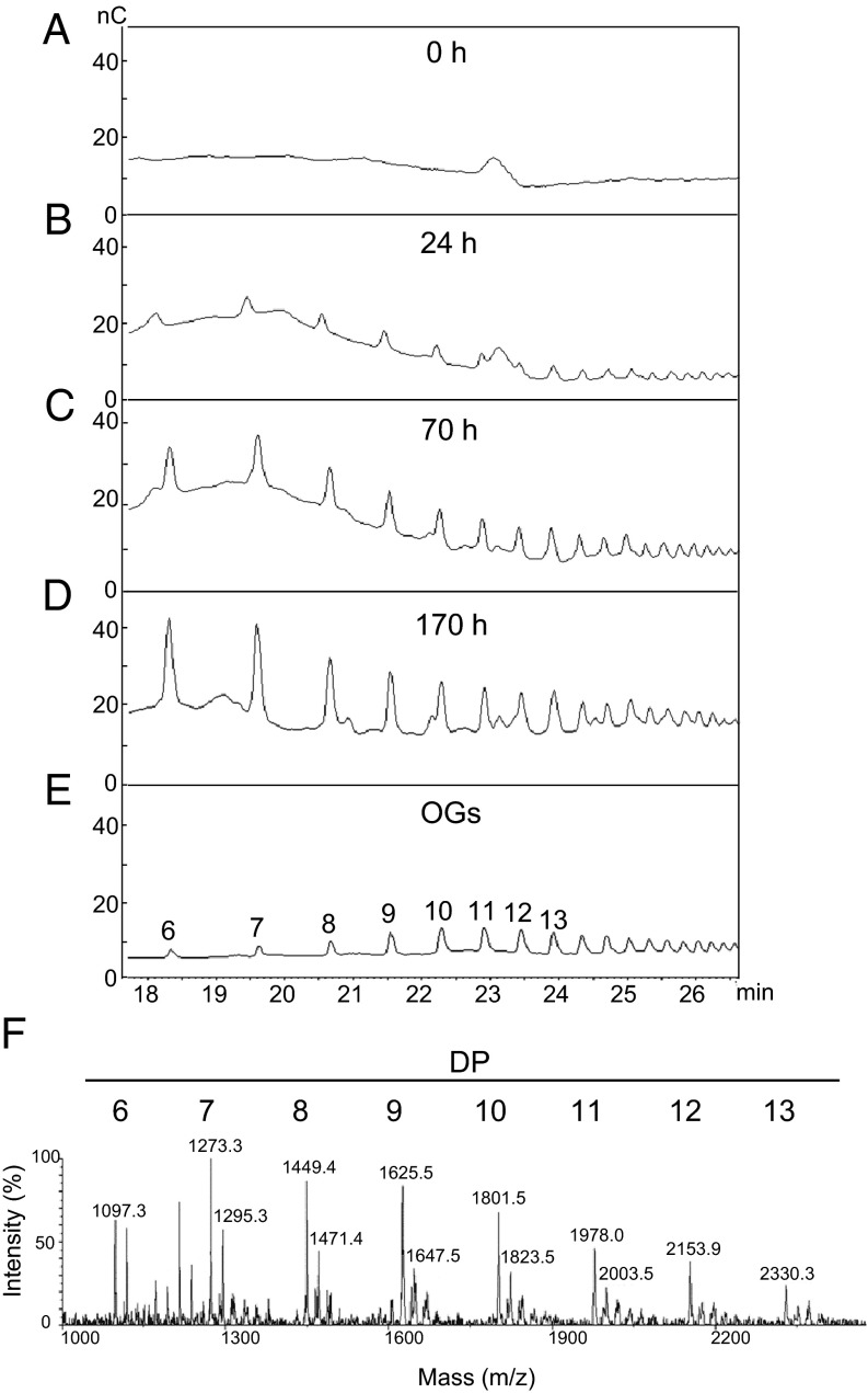
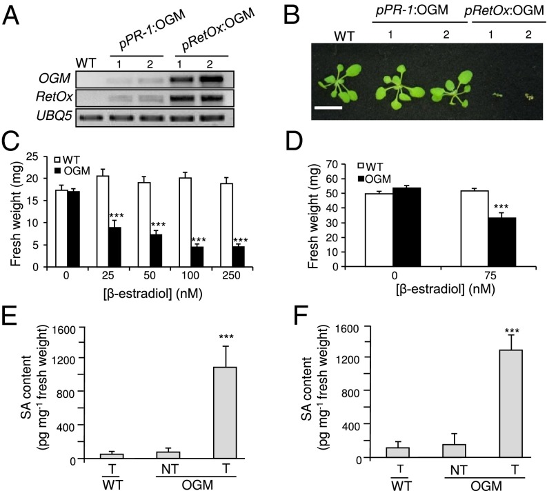
Oligogalacturonides (OGs) are fragments of pectin that activate plant innate immunity by functioning as damage-associated molecular patterns (DAMPs). We set out to test the hypothesis that OGs are generated in planta by partial inhibition of pathogen-encoded polygalacturonases (PGs). A gene encoding a fungal PG was fused with a gene encoding a plant polygalacturonase-inhibiting protein (PGIP) and expressed in transgenic Arabidopsis plants. We show that expression of the PGIP-PG chimera results in the in vivo production of OGs that can be detected by mass spectrometric analysis. Transgenic plants expressing the chimera under control of a pathogen-inducible promoter are more resistant to the phytopathogens Botrytis cinerea, Pectobacterium carotovorum, and Pseudomonas syringae. These data provide strong evidence for the hypothesis that OGs released in vivo act as a DAMP signal to trigger plant immunity and suggest that controlled release of these molecules upon infection may be a valuable tool to protect plants against infectious diseases. On the other hand, elevated levels of expression of the chimera cause the accumulation of salicylic acid, reduced growth, and eventually lead to plant death, consistent with the current notion that trade-off occurs between growth and defense.

Keywords: DAMPs; PGIP; oligogalacturonides; plant immunity; polygalacturonase.


Conflict of interest statement

The authors declare no conflict of interest.

Figures

Fig. 1.
Fig. 1.
Biochemical characterization of the PGIP–PG chimera expressed in P. pastoris. (A) Representation of two PGIP–PG chimeric proteins forming a homodimer. PvPGIP2 (in green), FpPG (in purple), and the (Ala)3 linker are indicated. (B) (Upper) PG activity of purified PGIP–PG (220 ng) and FpPG (1 ng) evaluated by agar diffusion assay; (Lower) immunoblot analysis of PGIP–PG (220 ng) and FpPG (1 ng) samples using an antibody against FpPG. The expected molecular mass of PGIP–PG (80 kDa) and FpPG (37 kDa) are indicated. (C) SDS/PAGE analysis of purified PGIP-PG eluted from a PGAII affinity column and after chemical cross-linking by formaldehyde (OGM-CL). The molecular weight marker (M, Amersham High Molecular Weight Calibration Kit) and the calculated molecular mass of PGIP-PG monomer and multimers are indicated.
Fig. 2.
Fig. 2.
Expression of the PGIP–PG chimera induces defense responses. (A) PGIP–PG chimera levels determined by immunoblot upon induction with 50 µM β-estradiol. (B) PG activity evaluated by agar diffusion assay in protein extracts of Arabidopsis nontreated (−) and treated (+) with 50 µM β-estradiol. (C) A representative PGIP–PG transgenic plant without treatment or after 170 h of induction. (D) Callose staining after 140 h of induction. Both images are at the same scale. (Magnification: 10×.) (E) Defense gene induction upon treatment with 50 µM β-estradiol. The photographs shown are representative of typical results.
Fig. 3.
Fig. 3.
Inducible release of OGs in Arabidopsis expressing the PGIP–PG chimera. (A–D) Plants expressing the inducible PGIP–PG chimera were treated for the indicated times with 50 µM β-estradiol and oligosaccharides in the cell wall pectin fraction were analyzed by HPAEC-PAD. Chromatograms indicate signal intensity (nC) at each retention time (min). (E) Representative chromatogram of purified OGs; the numbers indicate the DP. (F) MALDI-TOF analysis of the fraction in D. Numbers above peaks indicate their mass (m/z). Mass values correspond to OG sodium adducts. Numbers above mass peaks indicate the calculated DP of the corresponding oligomer.
Fig. 4.
Fig. 4.
Increased resistance of plants expressing a pathogen-inducible OGM (PGIP–PG chimera). (A–D) pPR-1:OGM lines 1 and 2 were inoculated with B. cinerea. OGM transcript (A) and protein (B) levels were analyzed at 0 (−) and 48 h postinfection (hpi) (+). Bars, average ± SD, n = 3. (C) Percentage of spreading lesions (n > 60 in four combined independent experiments) and (D) average lesion area ± SE (n > 12) at 72 hpi. (E) Average lesion area ± SE, n > 12, after P. carotovorum infection. (F) P. syringae pv. tomato DC3000 growth (average ± SE, n > 6) in wild-type (WT) and transgenic plants at 72 hpi. *P < 0.05; ***P < 0.01, Fischer’s exact test (C) or Student’s t test (D–F). All experiments were repeated at least twice with similar results.
Fig. 5.
Fig. 5.
High level of expression of OGM (PGIP–PG chimera) reduces plant growth and promotes SA accumulation. (A) Levels of OGM and RetOx transcripts in 2-wk-old wild-type (WT) plants, pPR-1:OGM 1 and 2 plants, and in two representative pRetOx:OGM T1 plants. UBQ5 was used as reference. (B) Representative photo of the same plants used in A. (Scale bar, 2 cm.) (C and D) Plants expressing the inducible OGM were germinated in the presence of β-estradiol (C) or were treated with β-estradiol 5 d after germination (D), and fresh weight was measured after 10 d (C) or 5 d (D). Bars: average (n > 10) ± SE; ***P < 0.001, statistical difference between control- and estradiol-treated seedlings (Student’s t test). (E and F) Accumulation of SA in plants expressing the inducible OGM. Four-week-old plants (E) or 10-d-old seedlings (F) were treated with β-estradiol (T) or DMSO (NT) and SA levels were determined after 24 h. Asterisks indicate statistically significant differences with β-estradiol–treated wild-type (WT) plants, according to Student’s t test (P < 0.01).

Similar articles

Cited by

References

    1. Vance RE, Isberg RR, Portnoy DA. Patterns of pathogenesis: Discrimination of pathogenic and nonpathogenic microbes by the innate immune system. Cell Host Microbe. 2009;6(1):10–21. - PMC - PubMed
    1. Jones JD, Dangl JL. The plant immune system. Nature. 2006;444(7117):323–329. - PubMed
    1. Boyer L, et al. Pathogen-derived effectors trigger protective immunity via activation of the Rac2 enzyme and the IMD or Rip kinase signaling pathway. Immunity. 2011;35(4):536–549. - PMC - PubMed
    1. McEwan DL, Kirienko NV, Ausubel FM. Host translational inhibition by Pseudomonas aeruginosa exotoxin A triggers an immune response in Caenorhabditis elegans. Cell Host Microbe. 2012;11(4):364–374. - PMC - PubMed
    1. Dunbar TL, Yan Z, Balla KM, Smelkinson MG, Troemel ER. C. elegans detects pathogen-induced translational inhibition to activate immune signaling. Cell Host Microbe. 2012;11(4):375–386. - PMC - PubMed

Publication types

MeSH terms