. 2015 Apr 28;112(17):5431-6.
doi: 10.1073/pnas.1500541112. Epub 2015 Apr 13.

Microgrooves and fluid flows provide preferential passageways for sperm over pathogen Tritrichomonas foetus

Affiliations

Microgrooves and fluid flows provide preferential passageways for sperm over pathogen Tritrichomonas foetus

Chih-kuan Tung et al. Proc Natl Acad Sci U S A. .

Abstract

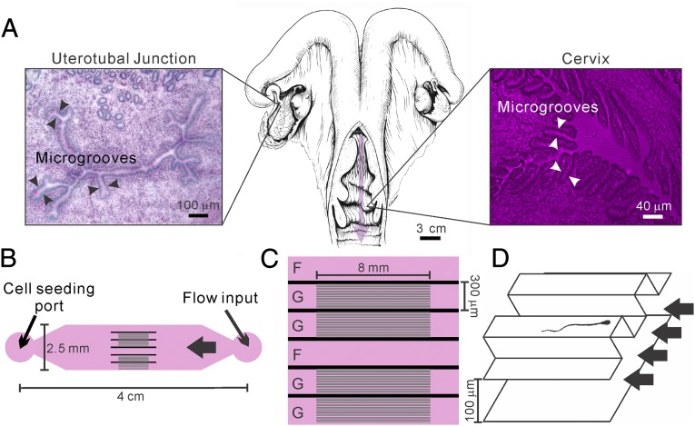
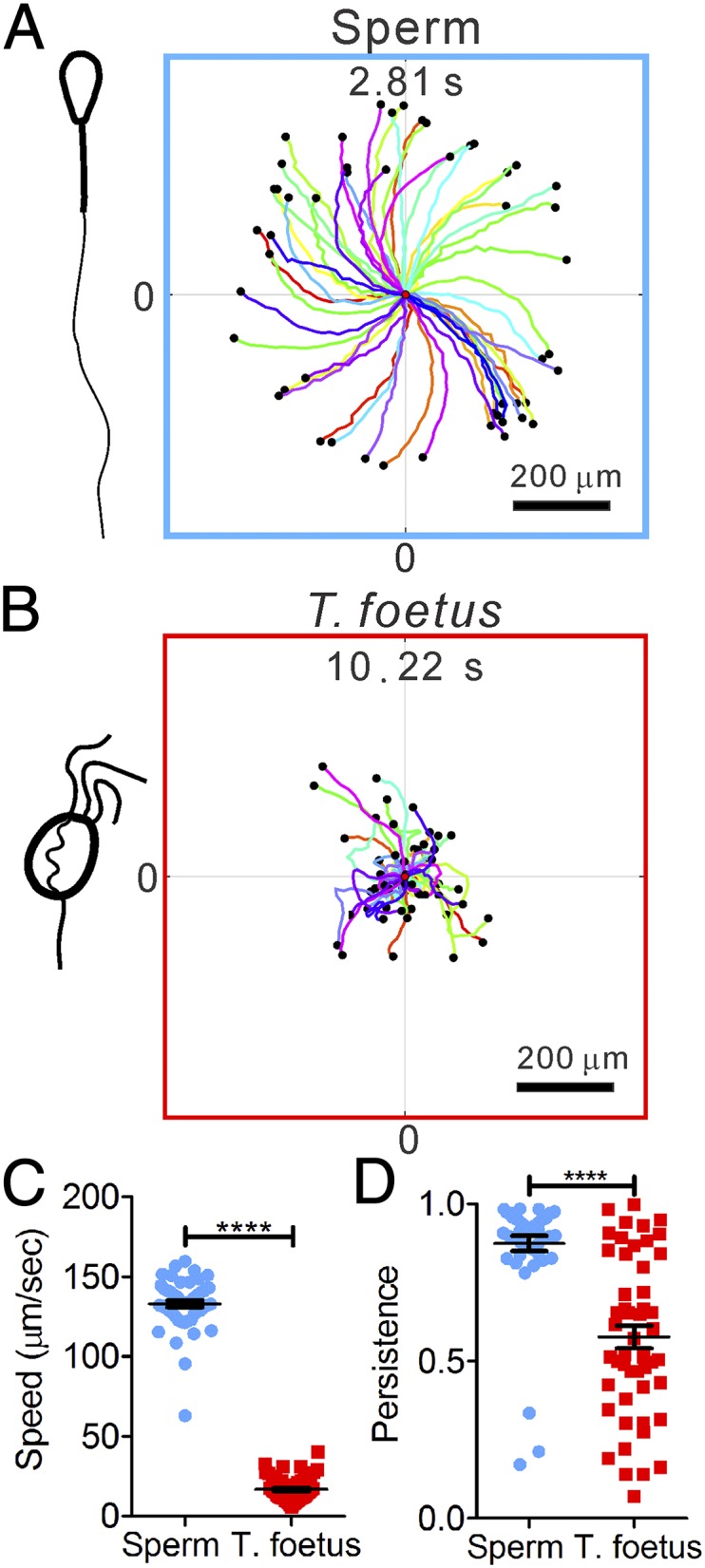
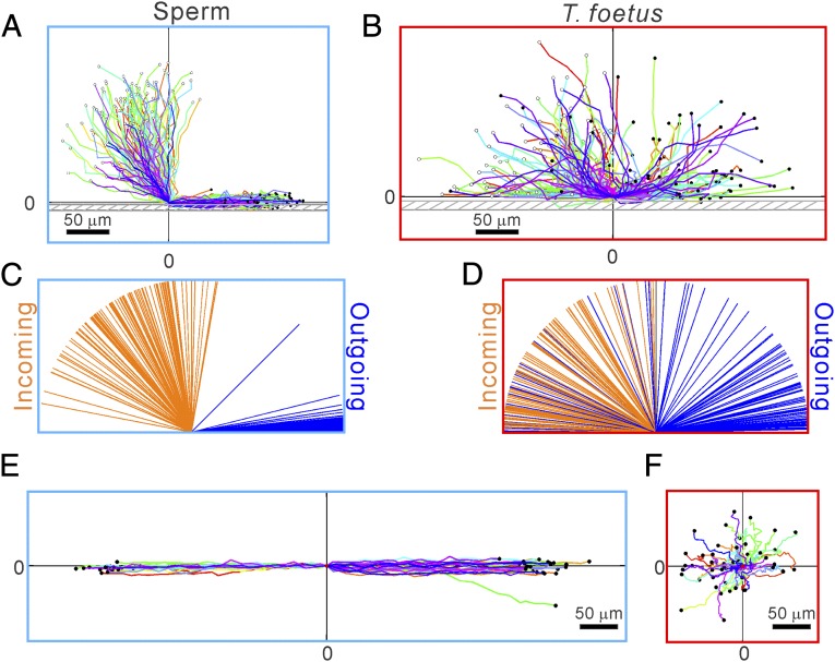
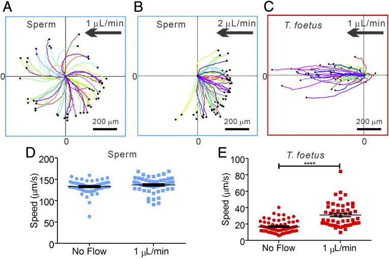
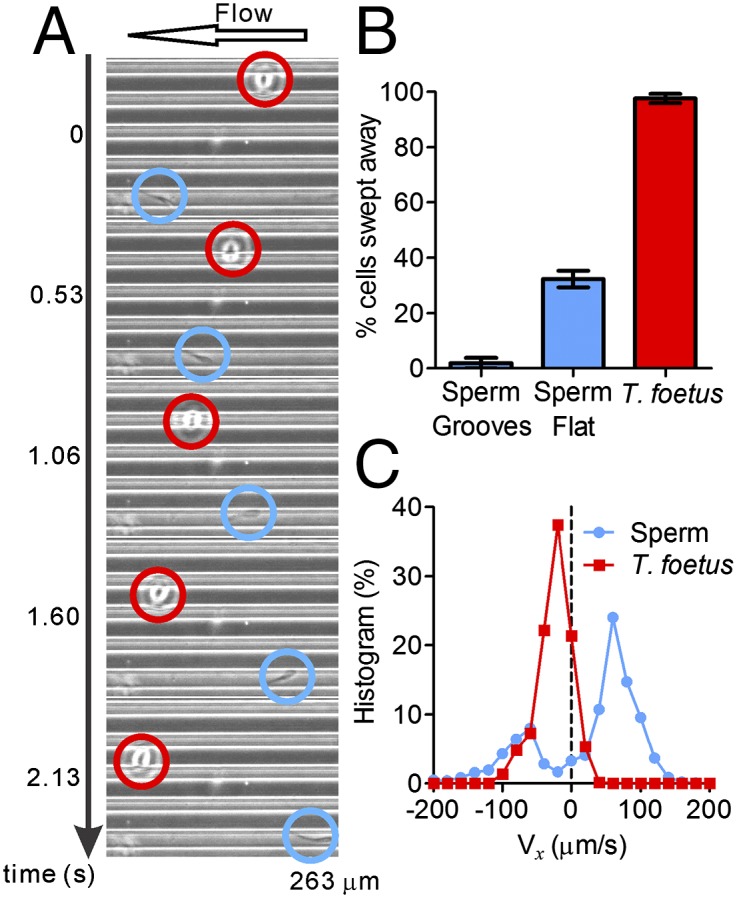
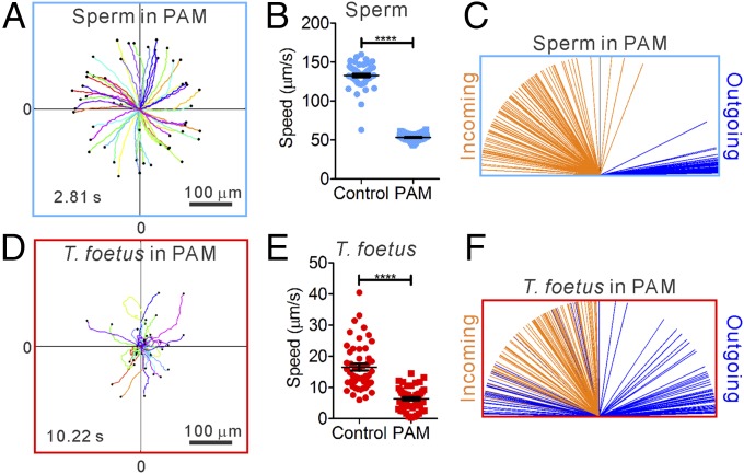
Successful mammalian reproduction requires that sperm migrate through a long and convoluted female reproductive tract before reaching oocytes. For many years, fertility studies have focused on biochemical and physiological requirements of sperm. Here we show that the biophysical environment of the female reproductive tract critically guides sperm migration, while at the same time preventing the invasion of sexually transmitted pathogens. Using a microfluidic model, we demonstrate that a gentle fluid flow and microgrooves, typically found in the female reproductive tract, synergistically facilitate bull sperm migration toward the site of fertilization. In contrast, a flagellated sexually transmitted bovine pathogen, Tritrichomonas foetus, is swept downstream under the same conditions. We attribute the differential ability of sperm and T. foetus to swim against flow to the distinct motility types of sperm and T. foetus; specifically, sperm swim using a posterior flagellum and are near-surface swimmers, whereas T. foetus swims primarily via three anterior flagella and demonstrates much lower attraction to surfaces. This work highlights the importance of biophysical cues within the female reproductive tract in the reproductive process and provides insight into coevolution of males and females to promote fertilization while suppressing infection. Furthermore, the results provide previously unidentified directions for the development of in vitro fertilization devices and contraceptives.

Keywords: cell motility; cervix; microfluidics; microswimmer; trichomoniasis.


Conflict of interest statement

The authors declare no conflict of interest.

Figures

Fig. 1.
Fig. 1.
A microfluidic device modeling fluid flows and microgrooves within the female reproductive tract. (A) Illustration (Center) of a bovine reproductive tract (adapted from ref. 36). The pink arrow points in the direction of fluid flow through the cervix. Microgrooves are seen in periodic acid-Schiff stain/hematoxylin-stained frozen sections of the uterotubal junction (Left) and the cervix (Right). Detailed methods are in ref. . (B) Illustration of the microfluidic device that recreates the microgrooves and the fluid flows that are in the female reproductive tract. Here, the cell seeding port is on one side and the flow inlet on the other, connected by channels with and without microgrooves. (C) Details of the channel design in the middle of the device. There are six channels for parallel experimentation; G denotes a channel with microgrooves in the upper surface, and F denotes a control channel lacking microgrooves. (D) A 3D drawing illustrates the details within a grooved channel. Here the main channel is 120 μm in height, and the microgrooves have a sectional area of 20 × 20 μm. Drawing is not to scale.
Fig. 2.
Fig. 2.
Differential swimming behavior of bovine sperm and T. foetus. (A and B) Clockwise (CW) chiral trajectories of sperm (each 2.81 s long) (A) and random motion trajectories of T. foetus (each 10.22 s) (B). Each colored line is a swimming trajectory, and all of the trajectories start at (0,0) and end at the black dots. A total of 50 trajectories are shown in each plot. (C) Speeds of T. foetus are significantly slower than sperm. ****P < 0.001 (nonparametric Mann– Whitney test). (D) Sperm trajectories are more persistent than T. foetus. Persistence is defined as the ratio between the vector length of the displacement to the contour length of a trajectory. Error bars show SEM.
Fig. 3.
Fig. 3.
Differential swimming behavior of sperm and T. foetus near a channel side wall. (A and B) Trajectories of sperm (A) and T. foetus (B) when they encounter a sidewall. Here, (0,0) is defined as the center position of the cell head at the time when the cell head hits the wall, and the wall is parallel to the x axis. Most sperm swim parallel to the wall after hitting it; in contrast, most T. foetus leave the wall in a random direction, whereas some of them swim along the wall for a few seconds before leaving the wall. Each trajectory is ∼1.6 s (A) or 5.9 s (B) long. (C and D) Line diagram showing the incoming and outgoing angles of the sperm (C) and T. foetus (D) before and after they hit a sidewall. Cell locations 0.4 s (C) or 1.48 s (D) away from (0,0) were used to calculate the angles. n = 100. (E and F) Swimming trajectories of sperm (E) and T. foetus (F) near a surface patterned with microgrooves. When sperm encountered the microgrooves, they entered and followed the grooves. When T. foetus encountered the grooves, they did not swim into the grooves; instead, they executed random motion in all directions. n = 50.
Fig. 4.
Fig. 4.
Sperm swim upstream and T. foetus are swept away by a gentle flow. (A) At 1 μL/min flow, sperm trajectories are similar to the case in which there is no flow. (B) At 2 μL/min flow, sperm swim against the flow. (C) Almost all (but one) T. foetus are swept downstream by a 1 μL/min flow. (D) A gentle flow of 1 μL/min has very limited impact on the motility of sperm, because the difference in speeds is not statistically significant (P = 0.25). (E) Motility of T. foetus is significantly altered by the presence of a flow. The higher speed in the presence of the flow is due to the fact that T. foetus were passively swept away by the flow. Flow speed in the channel has a Poiseuille profile across the vertical direction and ranges from 6 to 86 μm/s. T. foetus is distributed along the vertical direction in the channel. Number of cells analyzed is 50. Error bars show SEM.
Fig. 5.
Fig. 5.
Microgrooves facilitate upstream swimming in sperm, but have no impact on T. foetus. (A) A montage of micrographs shows sperm (blue circles) swimming against the flow in a microgroove, whereas a T. foetus (red circles) does not enter the microgroove and is brought downstream. (B) Percentage of cells swept away by a 3 μL/min flow in three different situations: sperm swimming within microgrooves, sperm on a flat surface, and T. foetus on a flat surface. n = 50. Error bars show SEM. (C) Histogram of instantaneous x-velocity shows an upstream bias for sperm at a 3 μL/min flow and a downstream bias for T. foetus at 1 μL/min flow. Statistics were acquired from 50 tracks of sperm (2.81 s long) and 50 tracks of T. foetus (10.22 s long).
Fig. 6.
Fig. 6.
Swimming behaviors of sperm and T. foetus in viscoelastic fluid. (A) CCW chiral trajectories of swimming sperm. (B) Sperm swim significantly slower in the presence of PAM than in control medium. n = 50. (C) Sperm swim along a wall after it hits the wall in the presence of PAM. n = 100. (D) Random motion of T. foetus in the presence of PAM. (E) T. foetus swim significantly slower in the presence of PAM than in control medium. n = 50. Error bars show SEM. (F) T. foetus does not show a tendency to swim along the wall after it hits the wall. n = 100.

References

    1. Denissenko P, Kantsler V, Smith DJ, Kirkman-Brown J. Human spermatozoa migration in microchannels reveals boundary-following navigation. Proc Natl Acad Sci USA. 2012;109(21):8007–8010. - PMC - PubMed
    1. Tung C-K, Ardon F, Fiore AG, Suarez SS, Wu M. Cooperative roles of biological flow and surface topography in guiding sperm migration revealed by a microfluidic model. Lab Chip. 2014;14(7):1348–1356. - PMC - PubMed
    1. Miki K, Clapham DE. Rheotaxis guides mammalian sperm. Curr Biol. 2013;23(6):443–452. - PMC - PubMed
    1. Kantsler V, Dunkel J, Blayney M, Goldstein RE. Rheotaxis facilitates upstream navigation of mammalian sperm cells. eLife. 2014;3:e02403. - PMC - PubMed
    1. Suarez SS, Brockman K, Lefebvre R. Distribution of mucus and sperm in bovine oviducts after artificial insemination: The physical environment of the oviductal sperm reservoir. Biol Reprod. 1997;56(2):447–453. - PubMed

Publication types