. 2015 May 19;112(20):E2725-34.
doi: 10.1073/pnas.1504393112. Epub 2015 Apr 13.

Neuronal medium that supports basic synaptic functions and activity of human neurons in vitro

Affiliations

Neuronal medium that supports basic synaptic functions and activity of human neurons in vitro

Cedric Bardy et al. Proc Natl Acad Sci U S A. .

Erratum in

Abstract

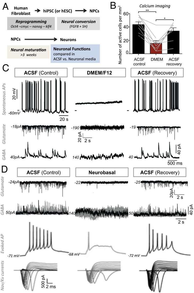
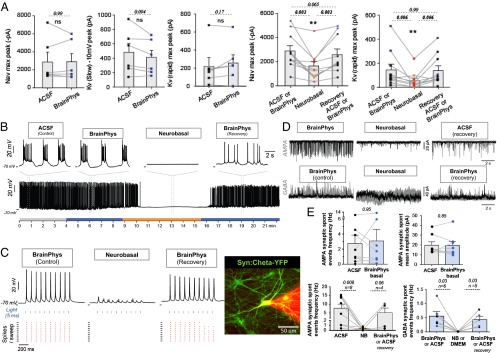
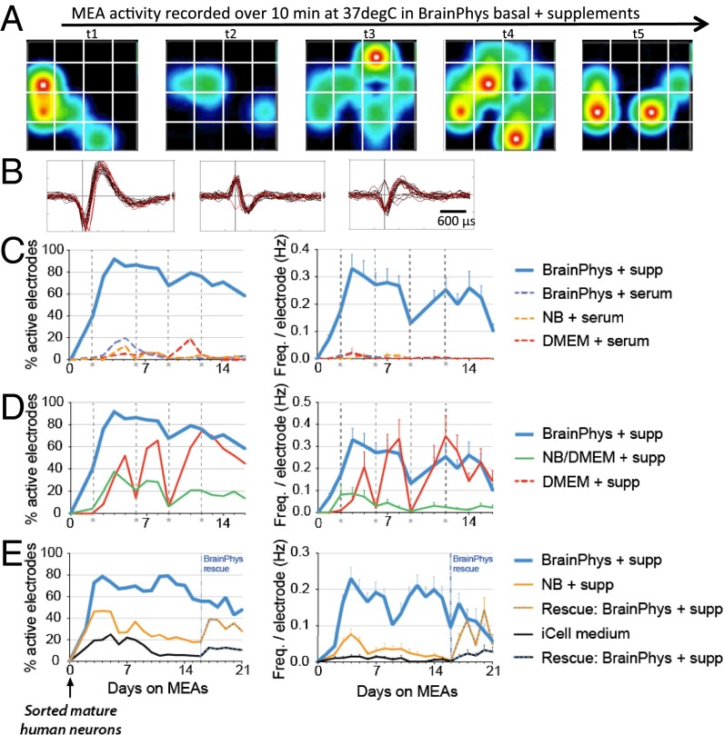
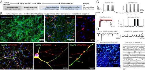
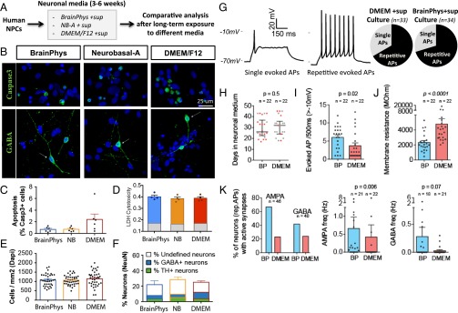
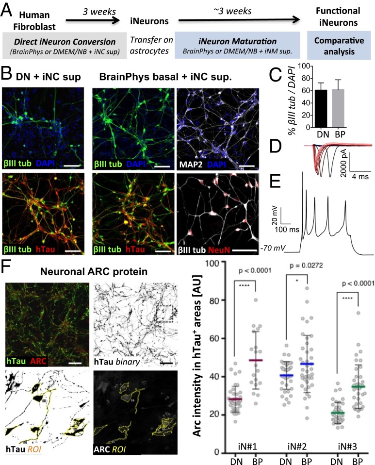
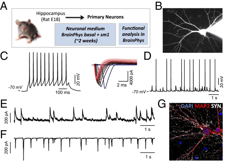
Human cell reprogramming technologies offer access to live human neurons from patients and provide a new alternative for modeling neurological disorders in vitro. Neural electrical activity is the essence of nervous system function in vivo. Therefore, we examined neuronal activity in media widely used to culture neurons. We found that classic basal media, as well as serum, impair action potential generation and synaptic communication. To overcome this problem, we designed a new neuronal medium (BrainPhys basal + serum-free supplements) in which we adjusted the concentrations of inorganic salts, neuroactive amino acids, and energetic substrates. We then tested that this medium adequately supports neuronal activity and survival of human neurons in culture. Long-term exposure to this physiological medium also improved the proportion of neurons that were synaptically active. The medium was designed to culture human neurons but also proved adequate for rodent neurons. The improvement in BrainPhys basal medium to support neurophysiological activity is an important step toward reducing the gap between brain physiological conditions in vivo and neuronal models in vitro.

Keywords: BrainPhys; induced pluripotent stem cells; neurobasal DMEM; neuromedium; tissue culture milieu.


Conflict of interest statement

Conflict of interest statement: The Salk Institute, C.B., and F.H.G. have filed a patent for the new BrainPhys medium described in this paper.

Figures

Fig. 1.
Fig. 1.
DMEM/F12 basal and Neurobasal media impair neuronal and synaptic activity. (A) Human neurons derived from iPSCs or ESCs were plated directly on glass coverslips. The basic neuronal functions of single mature neurons were tested and compared in two classic extracellular basal media (DMEM/F12 or Neurobasal-A). The perfusion of extracellular media was switched from ACSF to DMEM/F12 or Neurobasal-A and back to ACSF for recovery. (B) Calcium imaging analysis showing the number of spontaneously active cells in 10-min movies of the same fields of view recorded in ACSF, DMEM/F12 (n = 9), and back to ACSF (n = 8). Inorganic salt concentration and the osmolarity of ACSF were matched to DMEM. The gray lines represent individual fields of view. Statistics were performed with Wilcoxon test two-tailed, **P = 0.004; *P = 0.016). See also detailed calcium spikes analysis in SI Appendix, Fig. S1A. (C) Top traces: Current-clamp recordings reveal that spontaneous APs observed in ACSF quickly disappear in DMEM, together with a large increase in resting membrane potential (SI Appendix, Fig. S1). Middle traces: Voltage-clamp recordings (at −70 mV, Cl reversal potential) reveal that DMEM-based medium induces a large Na+ inward current and completely blocks spontaneous AMPA synaptic activity. Bottom traces: Voltage-clamp recordings (at 0 mV, Na+ reversal potential) reveal that DMEM-based medium induces large Cl currents and completely blocks spontaneous GABA synaptic activity (SI Appendix, Fig. S2). (D) Upper traces (synaptic activity): Voltage-clamp recordings reveal that Neurobasal-A based medium strongly reduces spontaneous AMPA synaptic activity. In addition, it induces large tonic Cl currents and completely blocks spontaneous GABA synaptic activity (SI Appendix, Fig. S2). Lower traces (APs): Neurobasal-A does not affect the resting membrane potential but it strongly impairs evoked APs (500-ms step of current), voltage-dependent Nav/Kv currents (I-V traces, clamp −70 mV, voltage steps 5 mV), and spontaneous APs.
Fig. 2.
Fig. 2.
BrainPhys basal medium supports optimal APs and synaptic activity. (A) Voltage-activated (VA) sodium and potassium currents (measured from I-V curves, voltage clamp −70 mV, steps 5 mV) were similar in BrainPhys and ACSF. In the three panels on the left, each neuron (n = 6) was tested both in ACSF and BrainPhys (black vs. blue dots). The maximum amplitudes of voltage-activated sodium and potassium currents of single neurons (n = 10) were significantly reduced in Neurobasal media (red dots, Neurobasal-A; orange dots, Neurobasal) compared with BrainPhys (blue dots) or ACSF (black dots). Nonparametric paired Wilcoxon test P values are shown in italics. Histograms represent the mean ± SE. (B) The resting membrane potential, spontaneous and evoked APs were the same in ACSF and BrainPhys. (C) Typical mature patched neuron expressing an optogene (synapsin: ChETA-YFP, green) and filled with rhodamine from the patch pipette (red shadow on the right). Optogenetic control of neuronal activity can be reliably achieved in BrainPhys. Recordings from single mature neurons tested with the same parameters in BrainPhys or Neurobasal-A illustrate that optogenetic control is dramatically improved in BrainPhys. Bottom part of the graphs in C are the corresponding raster plots. See also related results in SI Appendix, Fig. S3. (D) Both excitatory (AMPA) and inhibitory (GABA) synaptic activities were clearly apparent in BrainPhys medium. The perfusion of different media while recording the spontaneous synaptic activity of single neurons revealed that BrainPhys better supported synaptic function compared with other media such as Neurobasal-A. Twelve sweeps are superimposed in gray and one of them is highlighted in black for clarity. (E) Patched neurons (n = 22) tested in different basal media are represented by the paired dots. Quantification of AMPA receptor-mediated spontaneous synaptic activity shows similar properties in ACSF and BrainPhys basal (n = 8). Neurobasal significantly reduces AMPA synaptic events (n = 8). Both Neurobasal and DMEM completely block GABA synaptic events (n = 4 + 2). All tests comparing spontaneous activity were performed without any synaptic antagonists or voltage-gated sodium channel blockers. Voltage clamp at respective reversal potential of chloride and sodium was used to distinguish glutamatergic from gabaergic events. Synaptic blockers (NBQX or Gabazine) were used in a subset of cells (n = 15) only to confirm the nature of the receptors mediating the observed synaptic activity. Wilcoxon P values are shown in italics. Two-tailed tests were used except for recovery from 0 Hz; then a one-tailed test was used.
Fig. 3.
Fig. 3.
Long-term exposure of mature human neurons to BrainPhys basal (+supplements without serum) supports optimal and stable neuronal activity on MEAs. (A–E) Electrical activity of purified mature human iPSC-derived neurons (iCell neurons from Cellular Dynamics) was recorded in 48-well multielectrode array (MEAs) plates (Axion) in different neuronal maturation media at 37 °C every day for 2–3 wk. (A) Example illustrating the heatmap activity of seven snapshots of the same array in a well. Recordings (10 min) from neurons in BrainPhys with supplements (for 5 d) (see logg.salk.edu/files/BrainPhys_movies.pptx). (B) Examples of spikes detected by individual electrodes in BrainPhys+sup medium. (C–E) Percentage of active electrodes (>0.005 Hz) and frequency of detected spikes per electrode averaged from daily 10-min recordings. Each value represents the average of at least 96–192 electrodes spread in four to eight wells per conditions. The same batches of cells were compared in the 48-well plates with different media. Dashed lines in C and D mark the feeding dates. Recordings were performed before feeding. Each experiment was repeated at least twice independently with different set of cells and combined in the figures. For clarity, the experiment in C and D was split into two graphs, but the same control (BrainPhys+sup) is represented twice. See analysis of spikes properties in SI Appendix, Table S1.
Fig. 4.
Fig. 4.
Characterization of human neurons cultured for several weeks in BrainPhys basal + supplements. (A) Human NPCs derived from iPSCs or ESCs were plated directly on glass coverslips. Human neurons were matured in neuronal medium (BrainPhys + sup) for 3–6 wk. Analysis of electrophysiological properties was performed in the same neuronal medium: BrainPhys +sup. (B) Immunostaining of typical human IPSC-derived neuronal cultures grown in BrainPhys with supplements for 4 wk. (C and D) Electrophysiological activity of typical functional neurons after 3–6 wk in BrainPhys-based neuronal medium. Patch-clamp recordings were also performed in BrainPhys medium with supplements. (C) From left to right: Train of APs evoked by a small 500-ms depolarizing step of current or brief flashes of light (syn: ChETA-YFP). I-V traces (clamp −70 mV, steps 5 mV) showing typical voltage-activated Na+ and potassium currents. Spontaneous APs recorded in current clamp. (D) Spontaneous synaptic events mediated by AMPA receptors (sensitive to NBQX, voltage clamp at −70 mV) and GABA receptors (sensitive to Gabazine, voltage clamp at 0 mV). (E) Calcium imaging showing typical activity of neuronal cultures grown and recorded in BrainPhys + supplements. Time series analysis is plotted for the active regions of interest (white circles) (see logg.salk.edu/files/BrainPhys_movies.pptx); 91% of the active ROIs showed clear calcium spikes; the remaining showed slow calcium waves. Statistics for the calcium spikes of 41 active ROIs (mean ± SE): 8 ± 1 calcium spikes per 10 min, df/f = 1.1 ± 0.1; rise 10–90% = 3 ± 0.3 s; decay 37% = 11 ± 1s; half-width = 12 ± 1s.
Fig. 5.
Fig. 5.
Comparison of human neurons cultured in BrainPhys vs. classic media based on DMEM or Neurobasal. (A) In these experiments, we seeded human NPCs on coated glass coverlips (no feeder layer) in neural progenitor medium (SI Appendix, Methods). We then gradually switched to three different media: BrainPhys basal, DMEM/F12, or Neurobasal-A with the same serum-free supplements (SI Appendix, Methods). Half-media changes were performed every 3 d for 3–6 wk before analysis. (B–F) Representative immunostaining and analysis of cells cultured in the same plate with three different conditions. Nuclei were stained with DAPI (blue). Despite some variability, no clear differences in cell survival or proportion of neuronal subtypes (e.g., dopaminergic, GABAergic) were found between the three conditions. Quantification on all FOV with 20× objective (tiles 2 × 2) (for TH, n = 23 FOV; for NeuN, n = 20 FOV; for GABA, n = 23 FOV). (G) To characterize the influence of BrainPhys basal on functional properties, we compared the neurons matured in BrainPhys+sup with those in DMEM/F12+sup for 2–6 wk. Alternative blind patch clamping of the two groups was performed in ACSF for unbiased comparison. Electrophysiological types of neurons were defined based on their optimal firing patterns in response to 500-ms depolarizing steps of currents in ACSF. Similar proportion of cells with single or repetitive APs was found in the two media cultures. (H–K) For better sample homogeneity, the analysis was focused on neurons with repetitive APs. (H) The cells with repetitive APs spanned throughout the time windows of recording in both groups. Red dots represent neurons with evoked firing >10H (threshold −10 mV). Median and interquartile ranges are shown. (I) The evoked firing frequency (threshold −10 mV) of neurons with repetitive APs was significantly higher in the BrainPhys+sup cultures. (J) The membrane resistance of neurons with repetitive APs was significantly lower in the BrainPhys+sup cultures. (K) BrainPhys+sup culture also showed higher proportions of neurons receiving active AMPA synaptic inputs (NBQX sensitive) and GABA synaptic inputs (Gabazine sensitive). The presence of active synapses was determined by detecting at least four spontaneous synaptic events with distinctive kinetics over a 4-min recording in voltage clamp (−70 or 0 mV, respectively).
Fig. 6.
Fig. 6.
BrainPhys basal with the appropriate supplements permits efficient direct neuronal conversion, as well as development of human iNs, and increases ARC protein expression. (A) Experimental design: iN-competent human dermal fibroblasts were converted in neuronal conversion (NC) medium for 3 wk. For maturation, iN cells were relocated onto astrocytes and further cultured in neuronal maturation (NM) medium (see Methods for more details). (B) Immunofluorescence staining for β-III-tubulin, human Tau (hTau), MAP2ab, and NeuN following 3-wk conversion in media based on DMEM/F12:Neurobasal (Left) or BrainPhys (Right) with the same supplements. (Scale bar, 50 µm.) (C) The percentage of TUJ1+/DAPI was counted after 3 wk of conversion in different basal media with the same supplements. No significant difference was observed in the efficiency of neuronal conversion when either basal medium was used. (D and E) Following 3 wk of conversion, the iNs were replated on mouse astrocytes for 2 wk in BrainPhys basal+sup before patching and showed strong sodium/potassium currents and repetitive evoked APS. (F) Representative image of immunofluorescence staining for hTau and ARC in iN cells after maturation on astrocytes (3-wk conversion + 3-wk maturation). Quantification using ROI selection and measurement in ImageJ. (Scale bar, 100 µm.) Plot illustrating the quantification of neuronal ARC protein levels in iN cultures derived from three donors. Gray points represent individual cells (average n = 38 per group, minimum n = 19 per group). Bar show means ± SD of each group. Unpaired t test: *P < 0.05, **P < 0.01, ***P < 0.001, ****P < 0.0001.
Fig. 7.
Fig. 7.
Differentiation of healthy and active rat primary hippocampal neurons in BrainPhys basal+sm1. (A) Rat hippocampal neurons were plated on coated glass coverslips (no feeder layer, no serum) in BrainPhys basal medium (with sm1) for ∼2 wk before characterization. (B) Images of a typical patch-clamped neuron filled with Rhodamine. On the right, the shadow is the Rhodamine-filled patch pipette. Electrophysiological characterization was performed in BrainPhys. (C) Train of APs evoked by a small 500-ms depolarizing step of current. I-V traces (clamp −70 mV, steps 5 mV) showing typical voltage-dependent Na+ and K+ currents. Colored traces correspond to increasing voltage steps. (D) Spontaneous APs recorded in current clamp (0 pA). (E) Spontaneous GABA synaptic events recorded in voltage clamp at (0 mV). (F) Spontaneous AMPA synaptic events recorded in voltage clamp (−70 mV). (G) Coverslips were fixed after patching and processed for immunohistochemistry. Dendrites were stained with MAP2, presynaptic terminals with Synapsin, and nuclei with DAPI.

Comment in

  • Reconstructing the neuronal milieu intérieur.
    Livesey FJ. Livesey FJ. Proc Natl Acad Sci U S A. 2015 May 19;112(20):6250-1. doi: 10.1073/pnas.1506303112. Epub 2015 May 11. Proc Natl Acad Sci U S A. 2015. PMID: 25964319 Free PMC article. No abstract available.

Similar articles

Cited by

References

    1. Takahashi K, et al. Induction of pluripotent stem cells from adult human fibroblasts by defined factors. Cell. 2007;131(5):861–872. - PubMed
    1. Marchetto MC, Winner B, Gage FH. Pluripotent stem cells in neurodegenerative and neurodevelopmental diseases. Hum Mol Genet. 2010;19(R1):R71–R76. - PMC - PubMed
    1. Bellin M, Marchetto MC, Gage FH, Mummery CL. Induced pluripotent stem cells: The new patient? Nat Rev Mol Cell Biol. 2012;13(11):713–726. - PubMed
    1. Brennand KJ, et al. Modelling schizophrenia using human induced pluripotent stem cells. Nature. 2011;473(7346):221–225. - PMC - PubMed
    1. Israel MA, et al. Probing sporadic and familial Alzheimer’s disease using induced pluripotent stem cells. Nature. 2012;482(7384):216–220. - PMC - PubMed

Publication types