. 2015 Apr 28;112(17):5503-8.
doi: 10.1073/pnas.1422986112. Epub 2015 Apr 13.

Host attachment and fluid shear are integrated into a mechanical signal regulating virulence in Escherichia coli O157:H7

Affiliations

Host attachment and fluid shear are integrated into a mechanical signal regulating virulence in Escherichia coli O157:H7

Ghadah Alsharif et al. Proc Natl Acad Sci U S A. .

Abstract

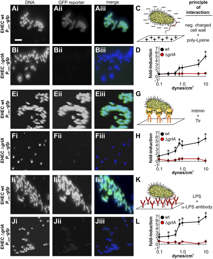
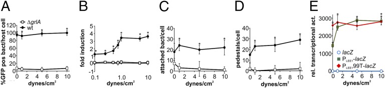
Enterohemorrhagic Escherichia coli (EHEC) is a foodborne pathogen causing hemorrhagic colitis and hemolytic uremic syndrome. EHEC colonizes the intestinal tract through a range of virulence factors encoded by the locus of enterocyte effacement (LEE), as well as Shiga toxin. Although the factors involved in colonization and disease are well characterized, how EHEC regulates its expression in response to a host encounter is not well understood. Here, we report that EHEC perceives attachment to host cells as a mechanical cue that leads to expression of LEE-encoded virulence genes. This signal is transduced via the LEE-encoded global regulator of LEE-encoded regulator (Ler) and global regulator of Ler and is further enhanced by levels of shear force similar to peristaltic forces in the intestinal tract. Our data suggest that, in addition to a range of chemical environmental signals, EHEC is capable of sensing and responding to mechanical cues to adapt to its host's physiology.

Keywords: attaching/effacing pathogens; enterohemorrhagic Escherichia coli; gastrointestinal infection; locus of enterocyte effacement; mechanosensing.


Conflict of interest statement

The authors declare no conflict of interest.

Figures

Fig. 1.
Fig. 1.
Attachment to host cells triggers LEE1 promoter activation in a GrlA-dependent manner. EHEC wild-type (A) or EHEC ΔgrlA (B) harboring a PLEE1-gfp transcriptional fusion as reporter were used to infect HeLa cells [multiplicity of infection (MOI) 10, 4 h]. Samples were fixed and DNA (Hoechst), reporter activation (GFP), and F-actin (rhodamine-phalloidin) were visualized by fluorescence microscopy. Several actin pedestals caused by EHEC attachment are marked by arrows. Example of an EHEC bacterium adsorbed to the glass slide, rather than attached to host cells, is marked by an asterisk. (Scale bar, 10 μm.) Percentage GFP-positive bacteria (C), average GFP intensity per bacterium (for GFP-positive cells) (D), number of attached bacteria per host cell (E), and number of pedestals per host cell (F) were determined from these experiments. Data are representative of three independent experiments (>100 HeLa cells each). The asterisk denotes significant differences between samples based on Student’s t test (P < 0.05). ns, not significant (P ≥ 0.05).
Fig. 2.
Fig. 2.
Induction of LEE1 is bacteria-driven, and a host response to infection is not required for signal transmission to GrlA. HeLa cells were infected with EHEC harboring a PLEE1-gfp transcriptional fusion (MOI 10, 4 h), following pretreatment with DMSO as control (A), 10 μg/mL cycloheximide (B), or 1 μg/mL cytochalasin D (C) for 1 h. Samples were fixed and DNA (Hoechst), reporter activation (GFP), and F-actin (rhodamine-phalloidin) were visualized by fluorescence microscopy. (Scale bar, 10 μm.) Percentage GFP-positive bacteria (D), average GFP intensity per bacterium (for GFP-positive cells) (E), number of attached bacteria per host cell (F), and number of pedestals per host cell (G) were determined for untreated (U), cycloheximide-treated (CHX), and cytochalasin d-treated (CD) cells. Data are representative of three independent experiments (>100 HeLa cells each). The asterisk denotes significant differences between samples based on Student’s t test (P < 0.05). ns, not significant (P ≥ 0.05); NA, not analyzed (no pedestals formed in CD-treated cells).
Fig. 3.
Fig. 3.
Population-level analysis of LEE1 induction rates in EHEC wild-type and mutant strains. Fluorescence intensity (AFU) was measured as a read-out for promoter activation, using promoterless gfp (blue), PLEE1-gfp (red), or PLEE199T-gfp (green) reporter constructs in EHEC wild-type cells grown in the presence (A) or absence (B) of host cells. Fluorescence was also measured in EHEC ΔgrlA (C), Δtir (D), and Δeae (E) strains incubated in the presence of HeLa cells for 1, 2, 3, or 4 h. Data are representative of three independent experiments done in triplicate. Asterisks denote significant differences between samples based on Student’s t test (P < 0.05). ns, not significant (P ≥ 0.05).
Fig. 4.
Fig. 4.
Bacterial attachment over time in EHEC wild-type and deletion strains. HeLa cells were infected with EHEC wild-type or deletion strains (MOI of 10), and bacterial attachment to host cells was determined after 1 h (gray bars) or 4 h (black bars) of infection by dilution plating. Data are representative of three independent experiments done in triplicate. The asterisk denotes significant differences between wild type and deletion strains at the respective time, based on Student’s t test (P < 0.05). ns, not significant (P ≥ 0.05).
Fig. 5.
Fig. 5.
LEE1 induction is independent of the mode of attachment, but the shape of the force response curve is substrate-dependent. EHEC wild-type (A, E, I) or ΔgrlA (B, F, J) strains containing a PLEE1-gfp reporter were introduced into substrate-coated flow cells and incubated for 1 h under static conditions, followed by 3 h of flow to give a defined fluid shear force ranging from 0 to 10 dynes/cm2. Substrates included poly-l-lysine (A–D), Tir-peptide (E–H), and α-LPS antibody (I–L) and were chosen to represent different modes of bacterial attachment. Images are representative of bacteria incubated under static conditions (0 dynes/cm2). (Scale bar, 5 μm.) After the experiment, average fluorescence intensity (AFU) per bacterium was determined from image analysis, and values were blotted as fold-change compared with wild-type EHEC on poly-K under static conditions (D, H, L). Data are representative of three independent experiments (>100 cells each). The asterisk denotes significant differences between samples based on Student’s t test (P < 0.05).
Fig. 6.
Fig. 6.
Fluid shear exacerbates LEE1 activation in host-attached bacteria. EHEC wild-type (black circles) or ΔgrlA strains (white squares) containing a PLEE1-gfp reporter were used to infect HeLa cells grown in glass flow cells and incubated for 1 h under static conditions, followed by 3 h of flow to give a defined fluid shear force ranging from 0 to 10 dynes/cm2. After the experiment, percentage GFP-positive bacteria per cell (A), fold-change in average GFP intensity per bacterium compared with static conditions (B), attached bacteria per cell (C), and pedestals per cell (D) were determined from image analysis. Data are representative of three independent experiments (>100 HeLa cells each). HeLa cells grown in glass flow cells were also infected with EHEC wild-type strain containing promoter-less lacZ (blue), PLEE1-lacZ (green), or PLEE199T-lacZ (red) reporters, as described earlier. After the experiment, cells were detached from the flow cells, using Triton-X100, and samples were used to determine relative transcriptional activities (E). Data are representative of three independent experiments performed in triplicate. The asterisk denotes significant differences between samples based on Student’s t test (P < 0.05).
Fig. 7.
Fig. 7.
Only free, but not GrlR-bound, GrlA is competent for attachment-mediated LEE1 induction; attachment does not relieve GrlR-mediated repression of GrlA. EHEC wild-type strain harboring a PLEE1-gfp transcriptional fusion as reporter and GrlR (A), GrlRA (B), or GrlA (C) expression vectors were used to infect HeLa cells (MOI 10, 4 h). Samples were fixed and DNA (Hoechst), reporter activation (GFP), and F-actin (rhodamine-phalloidin) were visualized by fluorescence microscopy. (Scale bar, 10 μm.) Percentage GFP-positive bacteria (D), average GFP intensity per bacterium (for GFP-positive cells) (E), number of attached bacteria per host cell (F), and number of pedestals per host cell (G) were determined from these experiments. Data are representative of three independent experiments (>100 HeLa cells each). HeLa cells were also infected (MOI 10, 4 h) with EHEC wild-type strain harboring a PLEE1-lacZ transcriptional fusion as reporter and empty vector (cont), GrlR, GrlRA, or GrlA expression constructs (H). Nonadherent bacteria (red) were recovered from the supernatant. Host cells were then washed and Triton-X100 lysed to recover adherent bacteria (green). Both fractions were used to determine β-galactosidase activity, and results were normalized to cfu/mL and are shown as relative transcriptional activity. Relative transcriptional activity was also determined for bacteria grown in planktonic LB cultures (blue). The asterisk denotes significant differences between bacteria harboring empty vector and expression constructs, based on Student’s t test (P < 0.05; n = 3). ns, not significant (P ≥ 0.05).

References

    1. Nataro JP, Kaper JB. Diarrheagenic Escherichia coli. Clin Microbiol Rev. 1998;11(1):142–201. - PMC - PubMed
    1. McDaniel TK, Kaper JB. A cloned pathogenicity island from enteropathogenic Escherichia coli confers the attaching and effacing phenotype on E. coli K-12. Mol Microbiol. 1997;23(2):399–407. - PubMed
    1. Jerse AE, Yu J, Tall BD, Kaper JB. A genetic locus of enteropathogenic Escherichia coli necessary for the production of attaching and effacing lesions on tissue culture cells. Proc Natl Acad Sci USA. 1990;87(20):7839–7843. - PMC - PubMed
    1. McDaniel TK, Jarvis KG, Donnenberg MS, Kaper JB. A genetic locus of enterocyte effacement conserved among diverse enterobacterial pathogens. Proc Natl Acad Sci USA. 1995;92(5):1664–1668. - PMC - PubMed
    1. Elliott SJ, et al. The locus of enterocyte effacement (LEE)-encoded regulator controls expression of both LEE- and non-LEE-encoded virulence factors in enteropathogenic and enterohemorrhagic Escherichia coli. Infect Immun. 2000;68(11):6115–6126. - PMC - PubMed

Publication types

MeSH terms