. 2015 Jul;88(1):1-11.
doi: 10.1124/mol.115.098913. Epub 2015 Apr 13.

Desformylflustrabromine (dFBr) and [3H]dFBr-Labeled Binding Sites in a Nicotinic Acetylcholine Receptor

Affiliations

Desformylflustrabromine (dFBr) and [3H]dFBr-Labeled Binding Sites in a Nicotinic Acetylcholine Receptor

Ayman K Hamouda et al. Mol Pharmacol. 2015 Jul.

Erratum in

Abstract

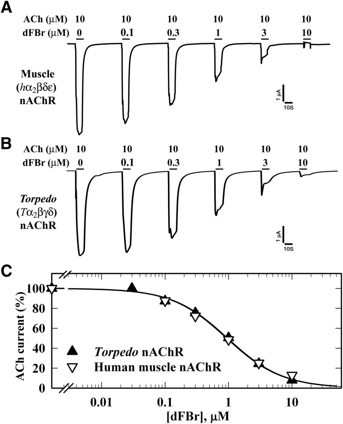
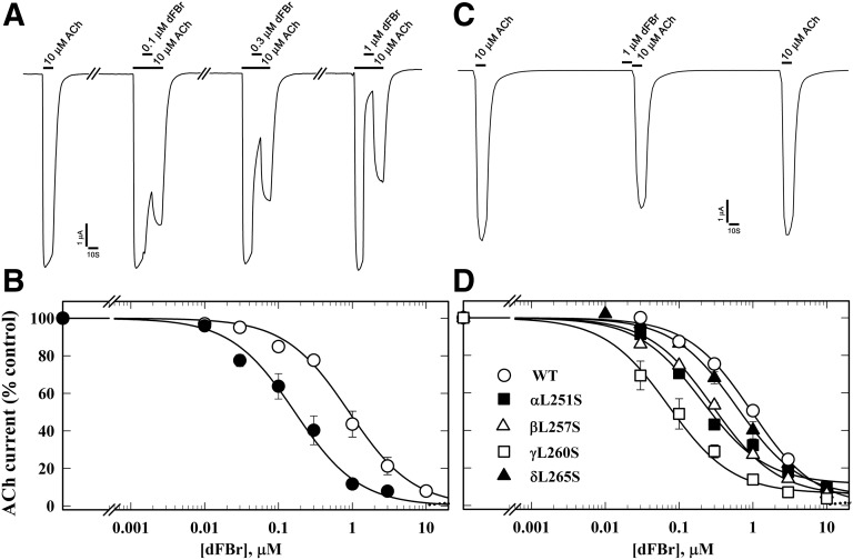
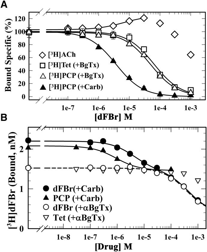
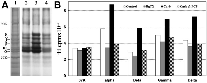
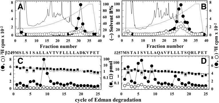
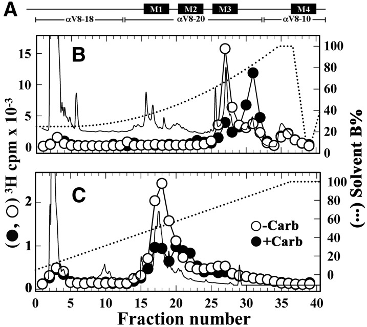
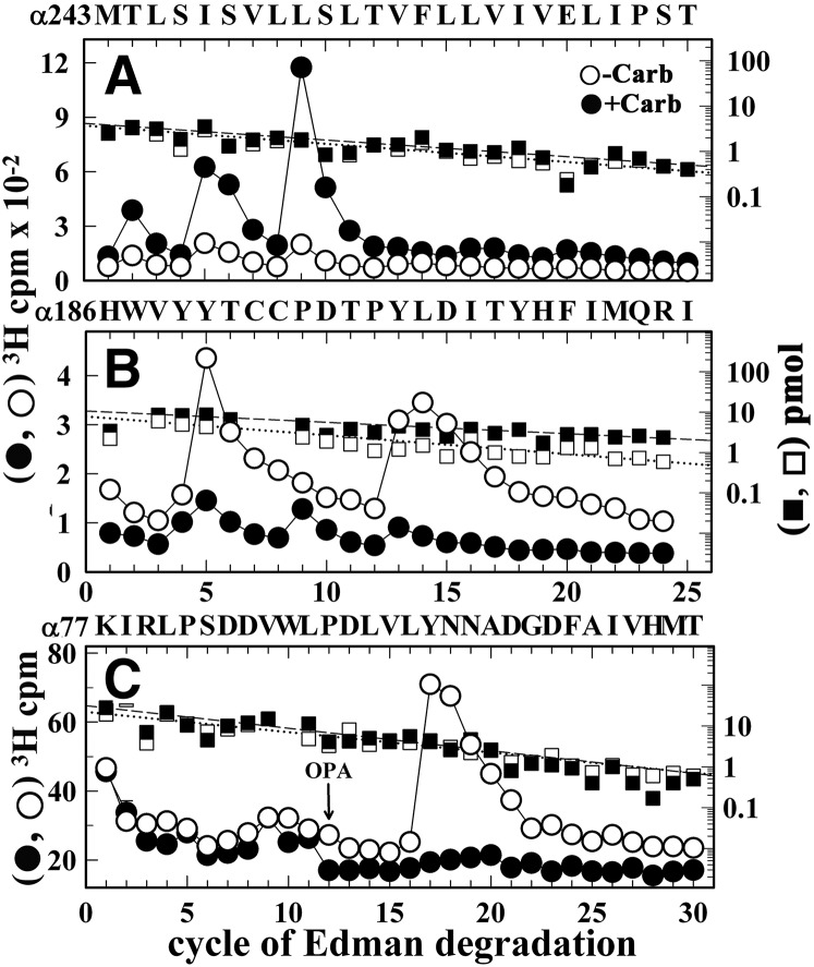
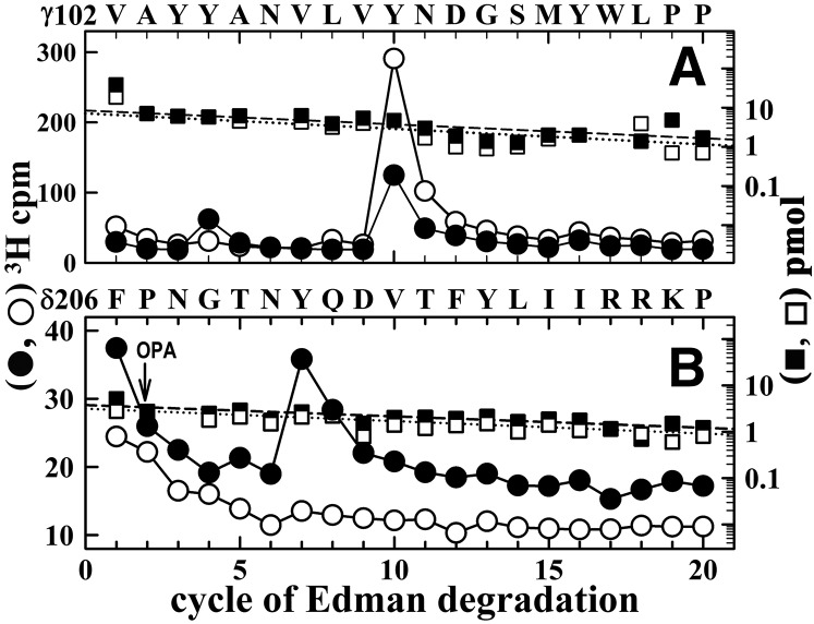
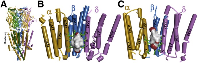
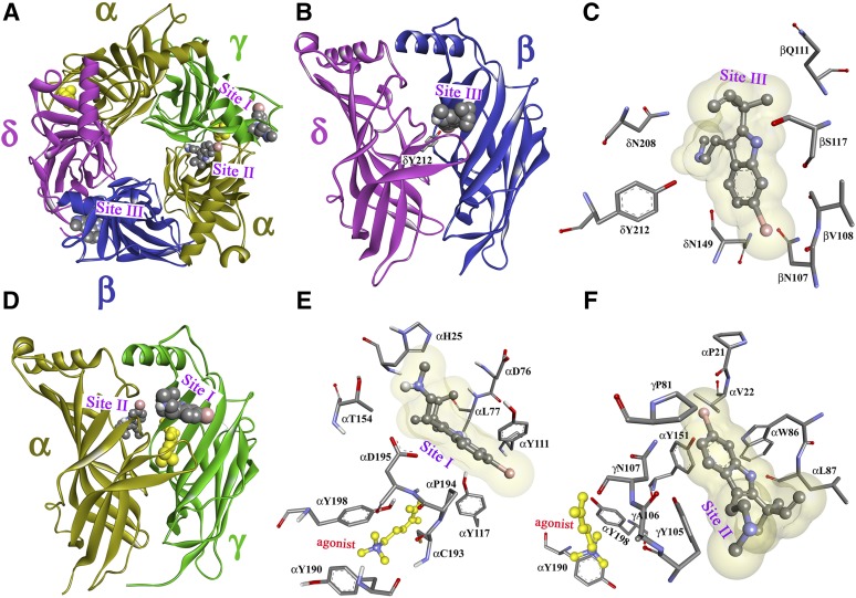
Desformylflustrabromine (dFBr) is a positive allosteric modulator (PAM) of α4β2 and α2β2 nAChRs that, at concentrations >1 µM, also inhibits these receptors and α7 nAChRs. However, its interactions with muscle-type nAChRs have not been characterized, and the locations of its binding site(s) in any nAChR are not known. We report here that dFBr inhibits human muscle (αβεδ) and Torpedo (αβγδ) nAChR expressed in Xenopus oocytes with IC50 values of ∼ 1 μM. dFBr also inhibited the equilibrium binding of ion channel blockers to Torpedo nAChRs with higher affinity in the nAChR desensitized state ([(3)H]phencyclidine; IC50 = 4 μM) than in the resting state ([(3)H]tetracaine; IC50 = 60 μM), whereas it bound with only very low affinity to the ACh binding sites ([(3)H]ACh, IC50 = 1 mM). Upon irradiation at 312 nm, [(3)H]dFBr photoincorporated into amino acids within the Torpedo nAChR ion channel with the efficiency of photoincorporation enhanced in the presence of agonist and the agonist-enhanced photolabeling inhibitable by phencyclidine. In the presence of agonist, [(3)H]dFBr also photolabeled amino acids in the nAChR extracellular domain within binding pockets identified previously for the nonselective nAChR PAMs galantamine and physostigmine at the canonical α-γ interface containing the transmitter binding sites and at the noncanonical δ-β subunit interface. These results establish that dFBr inhibits muscle-type nAChR by binding in the ion channel and that [(3)H]dFBr is a photoaffinity probe with broad amino acid side chain reactivity.


Figures

Fig. 1.
Fig. 1.
Structure of dFBr.
Fig. 2.
Fig. 2.
dFBr inhibition of muscle-type nAChRs. Xenopus oocytes expressing wild-type (A) human (2α:1β:1δ:1ε; ▽) or (B) Torpedo (2α:1β:1γ:1δ;▼) nAChR were voltage clamped at −50 mV, and currents elicited by 10 second applications of 10 µM ACh were recorded in the absence or presence of increasing concentrations of dFBr. Each drug concentration was tested at least three times and peak currents were normalized to the peak current elicited by 10 µM ACh alone. (C) Data (average ± S.E.) from at least three oocytes were plotted and fit to a single-site model (eq. 3); human nAChR, IC50 = 1.0 ± 0.08 µM; Torpedo nAChR, IC50 = 1.0 ± 0.06 µM.
Fig. 3.
Fig. 3.
dFBr inhibition of Torpedo nAChR is enhanced in the presence of agonist and by mutations in the ion channel favoring the open state. (A) Representative traces showing the effect of dFBr on ACh-induced current responses when 10 µM ACh was applied for 10 seconds, followed by co-application of 10 µM ACh and dFBr for 10 seconds, and then 10 seconds of 10 µM ACh alone. (B) The concentration dependence of dFBr inhibition when co-applied with ACh as in (A), with the current measured at the end of dFBr co-application normalized to the current just before (⬤, IC50 = 0.12 ± 0.01 µM), compared with a parallel experiment quantifying dFBr inhibition of peak ACh responses without prior ACh exposure (○, dashed line, fit from Fig. 2 for data from 10 oocytes). (C) Representative traces showing the effect of preapplication of 1 µM dFBr for 10 seconds on nAChR sensitivity to ACh. The ACh responses were inhibited by ∼15 ± 3% (N = 6). (D) dFBr inhibition of mutant nAChRs containing leucine to serine substitutions at position M2-9′ within the ion channel, quantified from the peak current elicited by 10 µM ACh with co-application of dFBr normalized to the current in the absence of dFBr. IC50 values for wild type, α, β, γ, and δ M2-9′ mutants were 1.0 ± 0.07, 0.2 ± .04, 0.3 ± 0.03, 0.1 ± 0.02, and 0.5 ± 0.06 µM, respectively. For each dFBr concentration in (B) and (D), average ± S.E. of several replicas from at least three oocytes were plotted and fit to a single-site model.
Fig. 4.
Fig. 4.
dFBr binds with high affinity to a site in the nAChR ion channel (desensitized state). The effects of dFBr on the equilibrium binding of [3H]ACh, [3H]PCP, and [3H]tetracaine (A) and the effect of dFBr, PCP, and tetracaine on the equilibrium binding of [3H]dFBr (B) to the Torpedo nAChR in the presence of α-BgTx (□, ▽, △) or Carb (⬤, ▴) were determined using centrifugation assays. (A) For each dFBr concentration, the specific binding of the radioligand was calculated from the total and nonspecific binding, normalized to the specific binding in the absence of dFBr, and fit to a single-site model using eq. 3. dFBr enhanced [3H]ACh binding (125% at 30 µM), inhibited [3H]PCP binding (+Carb) with IC50 = 3.6 ± 0.2 µM, and inhibited [3H]PCP and [3H]tetracaine binding in the presence of α-BgTx with IC50 values of 49 ± 6 and 63 ± 11 µM, respectively. (B) For each drug concentration, bound [3H]dFBr (in cpm/ml) was converted to nM [3H]dFBr. Inhibition curves were fit to a single-site model (eq. 1) or for inhibition by nonradioactive dFBr (+Carb) to a two-site model (eq. 2). For PCP (+Carb), IC50 = 0.9 ± 0.1 µM and Bns = 1.4 ± 0.1 nM, respectively. For dFBr (+α-BgTX), IC50 = 380 ± 70 µM, Bspec = 1.1 ± 0.08 nM, and Bns = 0.36 ± 0.09 nM. For inhibition by dFBr (+Carb), the high-affinity component of binding was fixed as the total binding [(+Carb) − (+α-BgTx)], the low-affinity component was equal to Bspec(+α-BgTx), and Bns was the same as in the presence of α-BgTx. With these assumptions, the calculated IC50 values for the high- and low-affinity components of binding were 4.1 ± 0.4 and 460 ± 30 µM, respectively.
Fig. 5.
Fig. 5.
Photoincorporation of [3H]dFBr into Torpedo nAChR-rich membranes. Membranes were photolabeled on an analytical scale with 0.5 µM [3H]dFBr in the absence and presence of nAChR ligands. Polypeptides were resolved by SDS-PAGE on duplicate gels that were stained with Coomassie blue (A, lane 1). One gel was prepared for fluorography (A, lanes 2–4), and polypeptide bands were excised from the second gel for 3H determination by liquid scintillation counting (B). Drug additions for the fluorograph in (A): lane 2, no other ligand added; lane 3, +1 mM Carb; lane 4, +1 mM Carb and 100 µM PCP. The electrophoretic mobilities of the nAChR α, β, γ, and δ subunits, rapsyn (Rn), the Na+/K+-ATPase α subunit (90K) and calectrin (37K) are indicated on the left.
Fig. 6.
Fig. 6.
Agonist-enhanced [3H]dFBr photolabeling in βM2, and δM2. Torpedo nAChR-rich membranes were photolabeled at preparative scale with 0.5 µM [3H]dFBr in the absence (○, ⬜) or presence (⬤, ▪) of 1 mM Carb. (A and B) Elution of peptides (solid line) and 3H (○, ⬤) during rpHPLC purifications of an ∼8 kDa β subunit fragment isolated by from a trypsin digest (A) and an ∼14 kDa δ subunit fragment isolated from an EndoLys-C digest (B). (C and D) 3H (○, ⬤) and phenylthiohydantoin amino acids (⬜, ▪) released during sequence analyses of rpHPLC fractions 30 to 31 from (A) and fractions 27–29 from (B), respectively. (C) The only sequence detected began at the N-terminus of βM2 (βMet249; 5 pmol each condition), and the major peak of 3H release in cycle 9 indicates photolabeling of βLeu257 (βM2-9; −Carb/+Carb, 2/50 cpm/pmol). (D) The only sequence detected began at the N-terminus of δM2 (δMet257; 10 pmol each condition). The peaks of 3H release in cycles 2, 5, 6, 9, and 16 indicate photolabeling (−Carb/+Carb, in cpm/pmol) at δSer258 (δM2-2; 1/7), δIle261 (δM2-5; 1/14), δSer262 (δM2-6; 2/17), δLeu265 (δM2-9; 0.5/14), and δLeu272 (δM2-16; 1/12).
Fig. 7.
Fig. 7.
Isolation of [3H]dFBr-photolabeled α subunit peptides. The band for nAChR α subunit from the photolabeling experiment described in Fig. 6 was excised and transferred to a 15% acrylamide mapping gel and subjected to in-gel digestion with V8 protease to produce large nAChR α subunit fragments shown schematically in (A). rpHPLC fractionations of EndoLys-C digests of (B) αV8-20, which begins at αSer173, and (C) αV8-18, which begins at αThr52. Elution of peptides (solid line) and 3H (○-Carb, ⬤+Carb) were monitored. rpHPLC fractions 27–29 and 30–32 from the V8-20 digest and fractions 17–19 from the V8-18 digest were pooled for sequence analysis (Fig. 8).
Fig. 8.
Fig. 8.
[3H]dFBr photolabeling within α subunit. 3H (○, ⬤) and phenylthiohydantoin amino acids (⬜, ▪) released during sequence analyses of rpHPLC fractions 30–32 (A) and 27–29 (B) from the fractionation of Endolys-C digests of αV8-20 (Fig. 7B) and fractions 17–19 (C) from the purification of the Endolys-C digest of αV8-18 (Fig. 7C). (A) The primary sequence began at the N-terminus of αM2 (αMet243; 4 pmol each condition) with a contaminating peptide beginning at βM249 (∼0.5 pmol each condition). The peaks of Carb-enhanced 3H release in cycles 2, 5, and 9 indicate [3H]dFBr photolabeling −Carb/+Carb, in cpm/pmol) at αThr244 (αM2-2, 4/15), αIle247 (αM2-5, 11/36), αSer248 (αM2-6, 7/31), and αIle251 (αM2-9, 15/101). (B) The only sequence began at αHis186 (⬜−Carb/▪+Carb, 8/10 pmol), with Carb-inhibitable peaks of 3H release at cycles 5 and 13 indicating [3H]dFBr photolabeling at αTyr190 and αTyr198 (−Carb/+Carb, 12/1 and 19/2 cpm/pmol, respectively). (C) The pool of material in rpHPLC fractions 17–19 from Fig. 7C was sequenced with the sequencing filter treated before cycle 12 with o-phthalaldehyde (OPA), which prevents sequencing of peptides not containing a proline at that cycle (Middleton and Cohen, 1991). After OPA treatment, sequencing continued only for the fragment beginning at αLys77 (⬜-Carb/▪+Carb, 22/31 pmol) that contains αPro88 in cycle 12. The peak of 3H release in cycle 17 indicated [3H]dFBr photolabeling of αTyr93 (−Carb/+Carb, 3/0.1 cpm/pmol).
Fig. 9.
Fig. 9.
[3H]dFBr photolabels γTyr105, γTyr111, and δTyr212 in the presence of agonist. 3H (○, ⬤) and phenylthiohydantoin amino acids (⬜, ▪) released during sequence analyses of a γ subunit fragment beginning at γVal102 (∼8 pmol each condition) (A) and a δ subunit fragment beginning at δPhe206 (∼4 pmol each condition) (B), each isolated as described in Materials and Methods from the nAChR photolabeling of Fig. 6 (−Carb (○, ⬜), +Carb (⬤, ▪)). (A) The major peak of 3H release at cycle 10 indicates [3H]dFBr photolabeling of γTyr111 (−Carb/+Carb, 18/6 cpm/pmol). The small peak of Carb-enhanced 3H release at cycle 4 indicates photolabeling of γTyr105 (−Carb/+Carb, 0.2/1.4 cpm/pmol). (B) During sequencing of the fragment beginning at δPhe206, recovered from rpHPLC fractions 23–25 in Fig. 6B, the peak of Carb-enhanced 3H release at cycle 7 indicates photolabeling of δTyr212 (-Carb/+Carb, 0.2/1.4 cpm/pmol). OPA, o-phthalaldehyde.
Fig. 10.
Fig. 10.
dFBr binding site in the Torpedo muscle-type nAChR ion channel. (A and B) Side view (A) and view from within the ion channel toward the α-β-δ subunits (B) of a nAChR homology model based on the X-ray structure of the mouse serotonin 5-HT3R (PDB ID 4PIR) (Hassaine et al., 2014) and (C) a homology model based on the cryo-electron microscopy structure of Torpedo marmorata nAChR (PDB ID 2BG9) (Unwin, 2005). The nAChR α, β, γ, and δ subunits are colored in gold, blue, green, and violet, respectively. Amino acids photolabeled by [3H]dFBr within the α/β/δM2 helices are colored in green and identified by their position numbers from the N-terminus of the M2 helices. In each model, the ensemble of the 100 lowest energy dFBr docking solutions within the channel is shown as a Connolly surface representation colored by atom (C, black; H, white; Br, red), defining volumes of 905 Å3 (B) and 821 Å3 (C) compared with the dFBr molecular volume of 241 Å3.
Fig. 11.
Fig. 11.
dFBr binding sites in the Torpedo nAChR ECD. Views of the homology model, the Torpedo nAChR ECD based on the x-ray structure of the L-AChBP in the presence of Carb (PDB ID 1UV6) (Celie et al., 2004) with the α, β, γ, and δ subunits colored in gold, blue, green, and violet, respectively. A view from the top (A) and side views from the exterior (B and D) are shown with dFBr, in space-filling representation, docked at three distinct sites in the presence of the agonist Carb (yellow). Sites I and II are at the agonist-binding canonical interface near entry to the ACh binding site (Site I; A, D, and E) and in the vestibule of the ion channel (Site II; A, D, and F). Site III (A, B, and C) is at the δ-β subunit interface. Views from the exterior (C and E) and a view from the ion channel vestibule (F) showing dFBr docked in its predicted lowest energy orientations in Site I (E), Site II (F), and Site III (C).

References

    1. Arevalo E, Chiara DC, Forman SA, Cohen JB, Miller KW. (2005) Gating-enhanced accessibility of hydrophobic sites within the transmembrane region of the nicotinic acetylcholine receptor’s δ-subunit. A time-resolved photolabeling study. J Biol Chem 280:13631–13640. - PubMed
    1. Arias HR. (2010) Positive and negative modulation of nicotinic receptors. Adv Protein Chem Struct Biol 80:153–203. - PubMed
    1. Bertrand D, Bertrand S, Cassar S, Gubbins E, Li J, Gopalakrishnan M. (2008) Positive allosteric modulation of the α7 nicotinic acetylcholine receptor: ligand interactions with distinct binding sites and evidence for a prominent role of the M2-M3 segment. Mol Pharmacol 74:1407–1416. - PubMed
    1. Bertrand D, Gopalakrishnan M. (2007) Allosteric modulation of nicotinic acetylcholine receptors. Biochem Pharmacol 74:1155–1163. - PubMed
    1. Blanton MP, Cohen JB. (1994) Identifying the lipid-protein interface of the Torpedo nicotinic acetylcholine receptor: secondary structure implications. Biochemistry 33:2859–2872. - PubMed

Publication types

MeSH terms

Associated data

LinkOut - more resources