. 2015 Mar 27:9:71.
doi: 10.3389/fnbeh.2015.00071. eCollection 2015.

Conditional targeting of medium spiny neurons in the striatal matrix

Affiliations

Conditional targeting of medium spiny neurons in the striatal matrix

Björn Reinius et al. Front Behav Neurosci. .

Abstract

The striatum serves as the main input to the basal ganglia, and is key for the regulation of motor behaviors, compulsion, addiction, and various cognitive and emotional states. Its deterioration is associated with degenerative disorders such as Huntington's disease. Despite its apparent anatomical uniformity, it consists of intermingled cell populations, which have precluded straightforward anatomical sub-classifications adhering to functional dissections. Approximately 95% of the striatal neurons are inhibitory projection neurons termed medium spiny neurons (MSNs). They are commonly classified according to their expression of either dopamine receptor D1 or D2, which also determines their axonal projection patterns constituting the direct and indirect pathway in the basal ganglia. Immunohistochemical patterns have further indicated compartmentalization of the striatum to the striosomes and the surrounding matrix, which integrate MSNs of both the D1 and D2 type. Here, we present a transgenic mouse line, Gpr101-Cre, with Cre recombinase activity localized to matrix D1 and D2 MSNs. Using two Gpr101-Cre founder lines with different degrees of expression in the striatum, we conditionally deleted the vesicular inhibitory amino acid transporter (VIAAT), responsible for storage of GABA and glycine in synaptic vesicles. Partial ablation of VIAAT (in ~36% of MSNs) resulted in elevated locomotor activity compared to control mice, when provoked with the monoamine reuptake inhibitor cocaine. Near complete targeting of matrix MSNs led to profoundly changed motor behaviors, which increased in severity as the mice aged. Moreover, these mice had exaggerated muscle rigidity, retarded growth, increased rate of spontaneous deaths, and defective memory. Therefore, our data provide a link between dysfunctional GABA signaling of matrix MSNs to specific behavioral alterations, which are similar to the symptoms of Huntington's disease.

Keywords: GABA; SLC32A1; behavior; huntington's disease; matrix; medium spiny neuron; patches; striosome.


Figures

Figure 1
Figure 1
Gpr101-Cre expression in the brain. Fluorescence microscopy images showing coronal brain sections of (A) Gpr101-Cre-Atg/wt:dtTomatotg/wt and (B) Gpr101-Cre-Btg/wt:dtTomatotg/wt reporter mice. Numbers denote approximate bregma coordinates.
Figure 2
Figure 2
Gpr101-Cre-B expression and projections. Fluorescence microscopy images showing (A) a sagital brain section of Gpr101-Cre-Btg/wt:dtTomatotg/wt (RFP) combined with immunohistochemistry for Tyrosine hydroxylase (TH). (B) Coronal section at the vertical mark intersecting SN in (A) of Gpr101-Cre-Btg/wt:dtTomatotg/wt combined with immunohistochemistry for TH. (C) High resolution confocal microscopy image of Gpr101-Cre-Btg/wt:dtTomatotg/wt combined with immunohistochemistry for VIAAT, showing RFP/VIAAT positive terminals in the SNr.
Figure 3
Figure 3
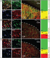
Gpr101-Cre demarks medium spiny neurons of both D1 and D2 type. Confocal microscopy images, showing brain sections of Gpr101-Cre-Btg/wt:dtTomatotg/wt combined with staining for either DARPP32 protein, D1 mRNA, or D2 mRNA. The leftmost panels show high resolution images in the striatum. The middle panels show wide-field images, CP: caudate putamen, Ctx: cortex. The rightmost panels show the percentage of cells with dual labeling (yellow) and single labeling (green or red) in cell counting experiments. #individuals = 2 and #sections = 10 per staining. (A) Immunohistchemical staining of the medium spiny neuron marker DARPP32 in Gpr101-Cre-Btg/wt:dtTomatotg/wt. (1) DARPP32pos:RFPneg cell, (2) DARPP32pos:RFPpos cell. (B) In situ hybridization of dopamine receptor 1 in Gpr101-Cre-Btg/wt:dtTomatotg/wt. (1) RFPpos:D1neg cell, (2) RFPneg:D1pos cell, (3) RFPpos:D1pos cell. (C) In situ hybridization of dopamine receptor 2 in Gpr101-Cre-Btg/wt:dtTomatotg/wt. (1) RFPpos:D2neg cell, (2) RFPneg:D2pos cell, (3) RFPpos:D2pos cell.
Figure 4
Figure 4
Gpr101-Cre delineates the striatal matrix. Confocal microscopy images showing (A) Gpr101-Cre-Btg/wt:dtTomatotg/wt combined with immunohistochemistry for DARPP32. The arrows point to patches in CP lacking RFP staining. Red: RFP, green: DARPP32, blue: DAPI. (B) Gpr101-Cre-Btg/wt:dtTomatotg/wt combined with immunohistochemistry for the matrix marker calbindin (CALB). Red: RFP, green: CALB, blue: DAPI.
Figure 5
Figure 5
Behavioral phenotypes upon partial matrix MSN VIAAT deletion. Results from behavioral tests using Gpr101-Cre-Btg/wt:Viaatlx/lx (KOB, black) and Gpr101-Cre-Bwt/wt:Viaatlx/lx (Ctrl, white). (A) Body weight at 6 and 26 wks. (B) Forelimb grip strength in three consecutive trials. (C) Dominance tube test. Proportion of wins. (D) Marble burying test. Number of marbles buried out of 18, shown as violin plots with the medians marked by circles. (E) Elevated plus maze. Proportion of time spent in areas during a 10 min trial, and number of open arm entries, shown as violin plots with the medians marked by circles. (F) Forced swim test. Average swim speed during 30 s segments of time over a 5 min trial. Barplots showing the percent time spent floating, swimming, and escaping (climbing against the cylinder wall), during the three first 30 s segments of time in the trial. (G) Rotarod test. Rotations per minute (rpm) at which the mice fell off the rotor as measured in three consecutive trials. (H) Basal locomotion. Number of dual beam breaks during 5 min segments of time over a 50 min trial. (I) Cocaine-induced locomotion. The basal locomotion was first monitored during 25 min, and at the time-point marked by the arrow 15 μg/g body weight of cocaine hydrochloride was injected. (J) Injection control. The mice were monitored during 25 min and injected with physiological saline solution at the time-point marked by the arrow (#KOBinj−ctrl = 6, #Ctrlinj−ctrl = 6). Bars and boxes show the group averages ±SEM. #KOB = 11, #Ctrl = 11 if not stated otherwise. P-values: *P < 0.05 and **P < 0.01, according to two-sided t-tests [A, B, E(time), F–J]; two-sided binomial test (C); or two-sided Wilcoxon signed-rank test [D, E(entries)].
Figure 6
Figure 6
Behavioral phenotypes upon broad matrix MSN VIAAT deletion. Results from CALB staining and behavioral tests using Gpr101-Cre-Atg/wt:Viaatlx/lx (KOA, black) and Gpr101-Cre-Awt/wt:Viaatlx/lx (Ctrl, white). (A) Confocal microscopy images showing Gpr101-Cre-Atg/wt:dtTomatotg/wt combined with immunohistochemistry for CALB. (B) Body weights up to adult age. (C) Survivability during 200 days (#KOA = 22, #Ctrl = 24), P-value according to Fischer's exact test at day 200. (D) Forelimb grip strength in 10 consecutive trials. (E) Basal locomotion. Number of dual beam breaks during 10 min segments of time over a 60 min trial. (F) Rearing activity. Number of beam breaks during 10 min segments of time over a 60 min trial. (G) Gait analysis. Footprints recorded from KOA and Ctrl mice during locomotion (hind paws: green, fore paws: red) and summary statistics. (H) Object recognition test. A schematic illustration of the paradigm, and the proportion of time spent interacting with objects. Bars and boxes show the group averages ±SEM. #KOA = 5, #Ctrl = 6. P-values: *P < 0.05, **P < 0.01, ***P < 0.001, according to two-sided t-tests if not stated otherwise.

References

    1. Albin R. L., Young A. B., Penney J. B. (1989). The functional-anatomy of basal ganglia disorders. Trends Neurosci. 12, 366–375. 10.1016/0166-2236(89)90074-X - DOI - PubMed
    1. Arango-Lievano M., Schwarz J. T., Vernov M., Wilkinson M. B., Bradbury K., Feliz A., et al. . (2014). Cell-type specific expression of p11 controls cocaine reward. Biol. Psychiatry 76, 794–801. 10.1016/j.biopsych.2014.02.012 - DOI - PMC - PubMed
    1. Aziz N. A., Van Der Marck M. A., Pijl H., Rikkert M. G. M. O., Bloem B. R., Roos R. A. C. (2008). Weight loss in neurodegenerative disorders. J. Neurol. 255, 1872–1880. 10.1007/s00415-009-0062-8 - DOI - PubMed
    1. Bateup H. S., Santini E., Shen W. X., Birnbaum S., Valjent E., Surmeier D. J., et al. . (2010). Distinct subclasses of medium spiny neurons differentially regulate striatal motor behaviors. Proc. Natl. Acad. Sci. U.S.A. 107, 14845–14850. 10.1073/pnas.1009874107 - DOI - PMC - PubMed
    1. Bolam J. P., Izzo P. N., Graybiel A. M. (1988). Cellular substrate of the histochemically defined striosome matrix system of the caudate-nucleus - a combined golgi and immunocytochemical study in cat and ferret. Neuroscience 24, 853–875. 10.1016/0306-4522(88)90073-5 - DOI - PubMed