Review
. 2015 Mar;4(2):182-90.
doi: 10.3978/j.issn.2225-319X.2015.03.08.

Mini-Bentall procedure

Affiliations
Review

Mini-Bentall procedure

Tristan D Yan. Ann Cardiothorac Surg. 2015 Mar.

Abstract

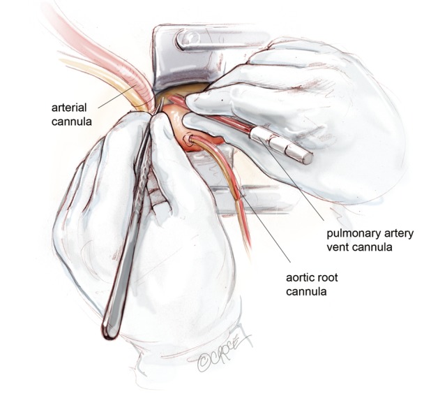
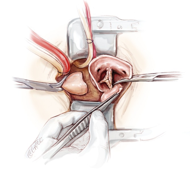
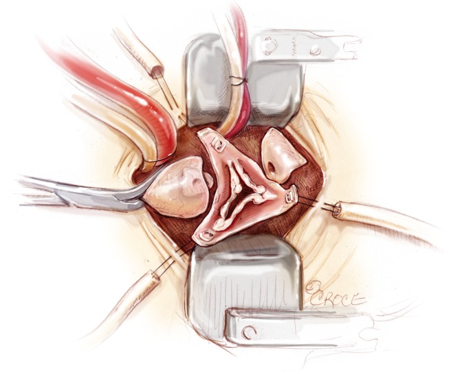
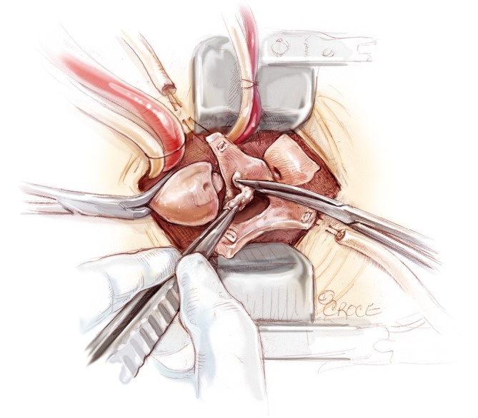
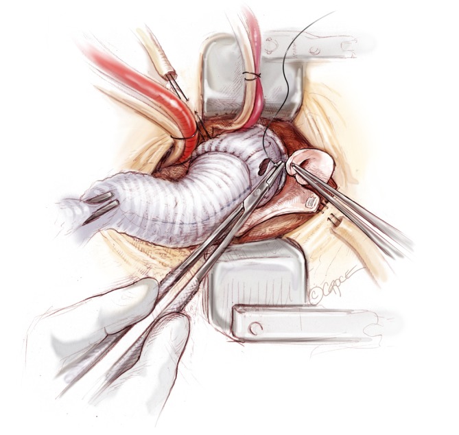
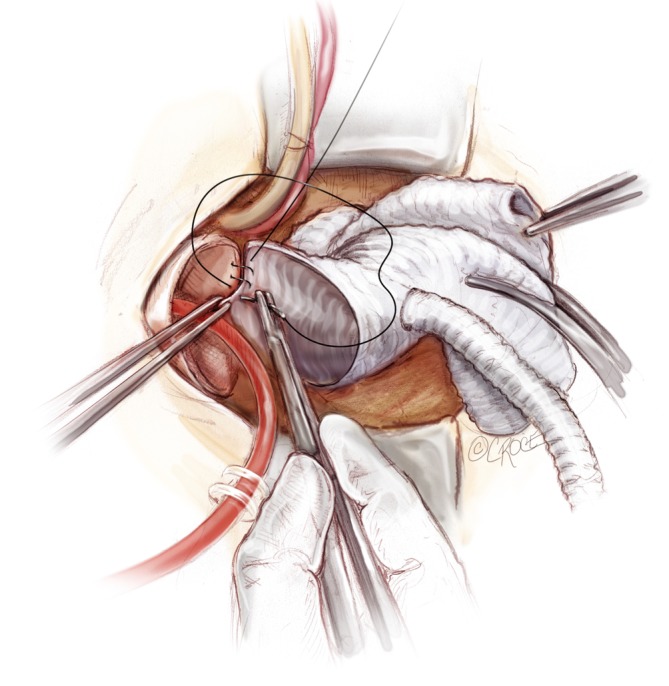
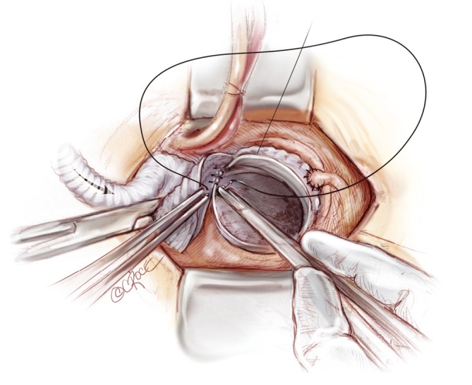
An important goal in cardiovascular and thoracic surgery is reducing surgical trauma to achieve faster recovery for our patients. Mini-Bentall procedure encompasses aortic root and ascending aortic replacement with re-implantation of coronary buttons, performed via a mini-sternotomy. The skin incision extends from the angle of Louis to the third intercostal space, usually measuring 5-7 cm in length. Through this incision, it is possible to perform isolated aortic root surgery and/or hemi-arch replacement. The present illustrated article describes the technical details on how I perform a Mini-Bentall procedure with hemi-arch replacement.

Keywords: Aortic root surgery; Bentall procedure; minimally invasive techniques.


Figures

Figure 1
Figure 1
A midline skin incision is performed from the manubrio-sternal junction to the level of the third intercostal space. A reversed “J” upper hemi-sternotomy is terminated to the left fourth intercostal space.
Figure 2
Figure 2
The mid-ascending aorta is slung with a Nylon tape. The access should adequately expose the aorta from the level of sinotubular junction to that of the distal ascending aorta.
Figure 3
Figure 3
Cardiopulmonary bypass is established via central aortic cannulation and femoral venous cannulation. A 16 Fr DLP pulmonary artery vent cannula (Medtronic Inc, Minneapolis, MN, USA) is inserted in the main pulmonary trunk.
Figure 4
Figure 4
The ascending aortic aneurysm is resected, leaving 1 cm cuff of aortic tissue proximal to the cross-clamp and 1 cm above the sinotubular junction.
Figure 5
Figure 5
The aortic root is carefully mobilized circumferentially and resected, leaving a rim of aortic wall just above the aortic annulus. The coronary buttons are prepared.
Figure 6
Figure 6
To provide an excellent exposure, the aortic root is brought in the cephalad direction by pulling on three commissural traction sutures.
Figure 7
Figure 7
The aortic leaflets are resected and the aortic annulus is decalcified.
Figure 8
Figure 8
Horizontal mattress sutures are used, with pledgets placed neatly below the aortic annulus.
Figure 9
Figure 9
The valve conduit is parachuted down by gently pulling the sutures vertically upwards with one hand and firmly pushing the valve conduit down onto the annulus with the other.
Figure 10
Figure 10
The left coronary button is trimmed, leaving a 3 mm circumferential cuff and re-implanted using a 5-0 running polypropylene suture.
Figure 11
Figure 11
The right coronary button is prepared and re-implanted using a 5-0 running polypropylene suture.
Figure 12
Figure 12
The graft is trimmed and anastomosed to the distal ascending aortic cuff by using a 3-0 running polypropylene suture.
Figure 13
Figure 13
The mid to distal ascending aorta is resected completely, together with the cannulation site. Selective antegrade cerebral perfusion is achieved by cannulating the innominate artery with or without left common carotid artery.
Figure 14
Figure 14
The open distal anastomosis is performed with a separate Ante-Flo graft with a single side arm (Vascutek Ltd, Renfrewshire, Scotland), using a continuous 3-0 running polypropylene suture.
Figure 15
Figure 15
A graft-to-graft anastomosis is performed using a continuous 3-0 running polypropylene suture and reinforced using pledgeted 4-0 polypropylene sutures where necessary.

References

    1. Bentall H, De Bono A.A technique for complete replacement of the ascending aorta. Thorax 1968;23:338-9. - PMC - PubMed
    1. Navia JL, Cosgrove DM, 3rd. Minimally invasive mitral valve operations. Ann Thorac Surg 1996;62:1542-4. - PubMed
    1. Cohn LH, Adams DH, Couper GS, et al. Minimally invasive cardiac valve surgery improves patient satisfaction while reducing costs of cardiac valve replacement and repair. Ann Surg 1997;226:421-6; discussion 427-8. - PMC - PubMed
    1. Phan K, Xie A, Di Eusanio M, et al. A meta-analysis of minimally invasive versus conventional sternotomy for aortic valve replacement. Ann Thorac Surg 2014;98:1499-511. - PubMed
    1. Phan K, Xie A, Tsai YC, et al. Ministernotomy or minithoracotomy for minimally invasive aortic valve replacement: a Bayesian network meta-analysis. Ann Cardiothorac Surg 2015;4:3-14. - PMC - PubMed