. 2015 Apr 13;13(4):2063-84.
doi: 10.3390/md13042063.

Structural analysis and anticoagulant activities of the novel sulfated fucan possessing a regular well-defined repeating unit from sea cucumber

Affiliations

Structural analysis and anticoagulant activities of the novel sulfated fucan possessing a regular well-defined repeating unit from sea cucumber

Mingyi Wu et al. Mar Drugs. .

Abstract

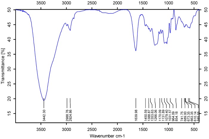
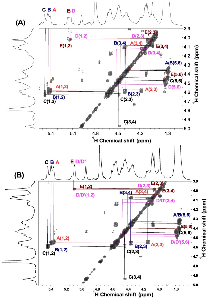
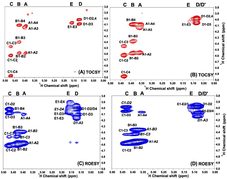
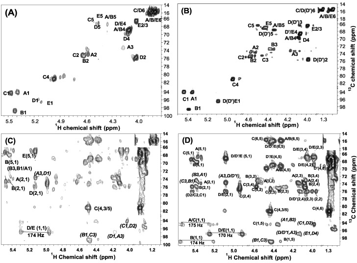
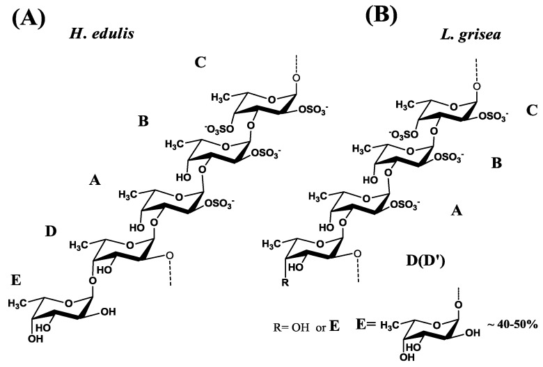
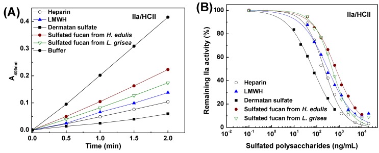
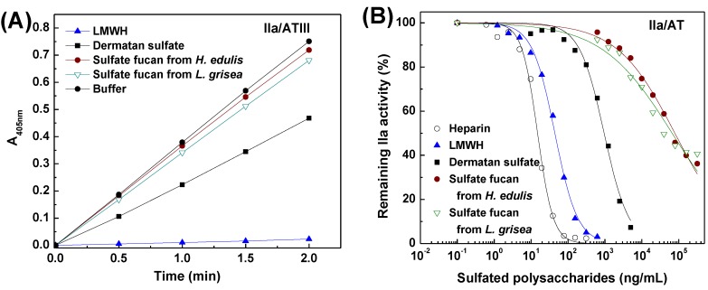
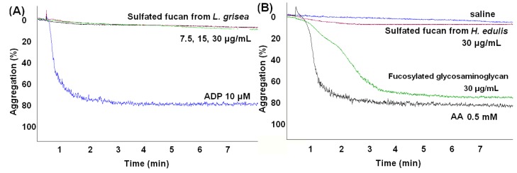
Sulfated fucans, the complex polysaccharides, exhibit various biological activities. Herein, we purified two fucans from the sea cucumbers Holothuria edulis and Ludwigothurea grisea. Their structures were verified by means of HPGPC, FT-IR, GC-MS and NMR. As a result, a novel structural motif for this type of polymers is reported. The fucans have a unique structure composed of a central core of regular (1→2) and (1→3)-linked tetrasaccharide repeating units. Approximately 50% of the units from L. grisea (100% for H. edulis fucan) contain sides of oligosaccharides formed by nonsulfated fucose units linked to the O-4 position of the central core. Anticoagulant activity assays indicate that the sea cucumber fucans strongly inhibit human blood clotting through the intrinsic pathways of the coagulation cascade. Moreover, the mechanism of anticoagulant action of the fucans is selective inhibition of thrombin activity by heparin cofactor II. The distinctive tetrasaccharide repeating units contribute to the anticoagulant action. Additionally, unlike the fucans from marine alga, although the sea cucumber fucans have great molecular weights and affluent sulfates, they do not induce platelet aggregation. Overall, our results may be helpful in understanding the structure-function relationships of the well-defined polysaccharides from invertebrate as new types of safer anticoagulants.


Figures

Figure 1
Figure 1
FT-IR spectrum of the sulfated fucan from sea cucumber.
Figure 2
Figure 2
1H (A,B) and 13C (C) one-dimensional NMR spectra at 500 MHz of the sulfated fucan from H. edulis. The spectra were recorded at 300 K for samples in D2O solution. Chemical shifts are relative to external trimethylsilylpropionic acid at 0 ppm. The residual water has been suppressed by pre-saturation. The anomeric signals assigned by 1H/13C HSQC (see Figure 5) are labeled A–E in the sulfated fucan. Expansion of the 4.9–5.6 ppm region of the 1H spectrum is shown in the inset in (A). The integrals were listed under the anomeric signals (B).
Figure 3
Figure 3
1H/1H COSY spectra of the sulfated fucans from H. edulis (A) and L. grisea (B). The spectra were recorded at 300 K for samples in D2O solution. Chemical shifts are relative to external trimethylsilylpropionic acid at 0 ppm. The residual water has been suppressed by pre-saturation. The anomeric signals assigned by 1H/13C HSQC (see Figure 5) are labeled AE in the sulfated fucans.
Figure 4
Figure 4
Expansions of the TOCSY (A,B) and ROESY (C,D) spectra of two sulfated fucans from H. edulis and L. grisea (B,D).The TOCSY spectra (A,B)show some cross-peaks used in the assignment of the fucose residue, especially positions bearing sulfate esters. The ROESY spectra (B,D) show ROEs, the sequence-defining A1–B3, B1–C3, C1–D2, D1–A3 and E1–D4. The five fucose residues in the repeating unit are marked A–E as described in the legend of Figure 6.
Figure 5
Figure 5
1H/13C HSQC (A,B) and HMBC (C,D) spectra of two sulfated fucans from two sea cucumbers H. edulis (A,C) and L. grisea (B,D). The assignments were based on TOCSY and COSY spectra. The anomeric signals were identified by the characteristic carbon chemical shifts and are marked A–E. The HMBC spectra (C,D) also show the sequence-defining A1–B3, B1–C3, C1–D2, D/D'1–A3 and E1–D4. The five fucose residues in the repeating unit are marked A–E as described in the legends of Figure 6.
Figure 6
Figure 6
Proposed regular repeating units of sulfated fucan isolated from two sea cucumbers H. edulis (A) and L. grisea (B). The five fucose residues in the repeating unit are marked A–E as described in the legends.
Figure 7
Figure 7
Inhibitory effects of the sulfated fucans, heparin, low molecular weight heparin (LMWH) and dermatan sulfate (DS) on thrombin mediated by heparin cofactor II. (A) Shows the time course of thrombin inhibition. HCII (~1 μM) was incubated with thrombin (20 NIH/mL) in the presence of 30 μL (625 ng/mL) samples at 37 °C. After 2 min, 30 μL of 4.5 mM CS-01 (38) was added, the residual thrombin activity was recorded by absorbance at 405 nm; (B) Shows the dependence on the sulfated polysaccharide concentration for thrombin inactivation in the presence of HCII. The reaction mixtures were as described in (A), except that different concentrations of sulfated polysaccharides were used. Results are shown as means of duplicates. See Table 4 for IC50 values.
Figure 8
Figure 8
Inhibitory effects of the sulfated fucans, heparin, LMWH and DS on thrombin in the presence of antithrombin. (A) Shows the time course of thrombin inhibition. Mixed samples of 30 μL of polysaccharides (625 ng/mL) and 30 μL of 0.25 IU/mL AT were incubated at 37 °C for 2 min, and 30 μL of 24 NIH/mL IIa was then added. After incubation for 2 min, 30 μL of 1.25 mM CS-01 (38) was added, the residual factor IIa activity was recorded by absorbance at 405 nm; (B) Shows the dependence on the sulfated polysaccharide concentration for thrombin inactivation mediated by AT. The reaction mixtures were as described in (A), except that different concentrations of sulfated polysaccharides were used. Results are shown as means of duplicates. See Table 4 for IC50 values.
Figure 9
Figure 9
Inhibitory effects of the sulfated fucans, heparin, LMWH and DS on factor Xa in the presence of antithrombin. (A) Shows the time course of Xa inhibition. Mixed samples of 30 μL of polysaccharides (625 ng/mL) and 30 μL of 1 IU/mL AT were incubated at 37 °C for 2 min, and 30 μL of 8 μg/mL bovine Xa was then added. After incubation for 1 min, 30 μL of 1.20 mM CS-11(65) was added, the residual Xa activity was recorded by absorbance at 405 nm; (B) Shows the dependence on the sulfated polysaccharide concentration for Xa inactivation in the presence of AT. The reaction mixtures were as described in (A), except that different concentrations of sulfated polysaccharides were used. Results are shown as means of duplicates. See Table 4 for IC50 values.
Figure 10
Figure 10
Profile of the platelet aggregation induced by the sea cucumber polysaccharides: the sulfated fucan from L. grisea (A); the sulfated fucan and fucosylated glycosaminoglycan from H. edulis (B). The profile showed that the sulfated fucans from sea cucumber do not cause platelets to aggregate at several concentrations.

References

    1. Streiff M.B., Bockenstedt P.L., Cataland S.R., Chesney C., Eby C., Fanikos J., Fogarty P.F., Gao S.W., Grcia-Aguilara J., Goldhaber S.Z., et al. Venous thromboembolic disease. J. Natl. Compr. Cancer Netw. 2011;9:714–777. - PMC - PubMed
    1. Warkentin T.E., Levine M.N., Hirsh J., Horsewood P., Roberts R.S., Gent M., Kelton J.G. Heparin-induced thrombocytopenia in patients treated with low-molecular weight heparin or unfractionated heparin. N. Engl. J. Med. 1995;332:1330–1335. doi: 10.1056/NEJM199505183322003. - DOI - PubMed
    1. Mourão P.A.S., Pereira M. Searching for alternatives to heparin: Sulfated fucans from marine invertebrates. Trends Cardiovasc. Med. 1999;9:225–232. doi: 10.1016/S1050-1738(00)00032-3. - DOI - PubMed
    1. Rocha H.A.O., Moraes F.A., Trindade E.S., Franco C.R.C., Torquato R.J.S., Veiga S.S., Valente A.P., Mourão P.A.S., Leite E.L., Nader H.B., et al. Structural and hemostatic activities of a sulfated galactofucan from the brown alga Spatoglossum schroederi: An ideal antihrombotic agent? J. Biol. Chem. 2005;280:41278–41288. - PubMed
    1. Bordbar S., Anwar F., Saari N. High-value components and bioactives from sea cucumbers for functional foods—A review. Mar. Drugs. 2011;9:1761–1805. doi: 10.3390/md9101761. - DOI - PMC - PubMed

Publication types

MeSH terms