. 2015 Apr 30;6(12):10532-47.
doi: 10.18632/oncotarget.3300.

Multiple myeloma induces Mcl-1 expression and survival of myeloid-derived suppressor cells

Affiliations

Multiple myeloma induces Mcl-1 expression and survival of myeloid-derived suppressor cells

Kim De Veirman et al. Oncotarget. .

Abstract

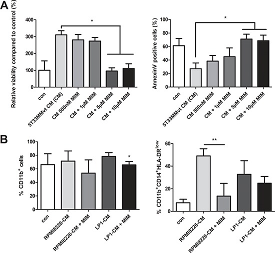
Myeloid-derived suppressor cells (MDSC) are contributing to an immunosuppressive environment by their ability to inhibit T cell activity and thereby promoting cancer progression. An important feature of the incurable plasma cell malignancy Multiple Myeloma (MM) is immune dysfunction. MDSC were previously identified to be present and active in MM patients, however little is known about the MDSC-inducing and -activating capacity of MM cells. In this study we investigated the effects of the tumor microenvironment on MDSC survival. During MM progression in the 5TMM mouse model, accumulation of MDSC in the bone marrow was observed in early stages of disease development, while circulating myeloid cells were increased at later stages of disease. Interestingly, in vivo MDSC targeting by anti-GR1 antibodies and 5-Fluorouracil resulted in a significant reduced tumor load in 5TMM-diseased mice. In vitro generation of MDSC was demonstrated by increased T cell immunosuppressive capacity and MDSC survival was observed in the presence of MM-conditioned medium. Finally, increased Mcl-1 expression was identified as underlying mechanism for MDSC survival. In conclusion, our data demonstrate that soluble factors from MM cells are able to generate MDSC through Mcl-1 upregulation and this cell population can be considered as a possible target in MM disease.

Keywords: generation; mechanism; multiple myeloma; myeloid-derived suppressor cells; targeting.


Conflict of interest statement

DISCLOSURE OF POTENTIAL CONFLICTS OF INTEREST

The authors disclose no potential conflicts of interest.

Figures

Figure 1
Figure 1. MDSC distribution in the bone marrow, blood and spleen of 5T33MM mice during disease progression
C57BL/KaLwRij mice were inoculated with 5T33MMvv cells and sacrificed at one, two and three weeks after inoculation and at the end-stage of the disease (n = 3/group). Blood, BM and spleen were investigated. A. % Plasmacytosis determined by microscopic examination of cytospins stained by the May-Grünwald-Giemsa method. B. % Idiotype+ cells detected by anti-idiotype (3H2) FACS staining to detect tumor load. C. The percentage of CD11b+ cells (gated on 3H2 cells) was determined by flow cytometry. D. Ly6G expression in the CD11b+ population was analyzed by flow cytometry. E. In the CD11b+ Ly6Glow population, Ly6C expression was analyzed by flow cytometry to distinguish inflammatory monocytes (MO) (Ly6Chi), eosinophils (Ly6Cint), and immature myeloid cells (IMC) (Ly6Clow). Error bars represent the SD. * indicates p < 0.05 and represents the significant increase compared to week 1 (Figure 1A and 1B) or day 0 (Figure 1C and 1E).
Figure 2
Figure 2. In vivo MDSC targeting by anti-GR1
A. Naive mice were treated with 200 μg/mL anti-GR1 antibody and sacrificed two days later. The percentage CD11b+ and Ly6G+ cells were analyzed by flow cytometry (n = 2). B–E. Mice were inoculated with 5TGM1-GFP+ cells and treated with vehicle (n = 5) or anti-GR1 antibodies (n = 7) (200 μg/mL, every two days) for 4 weeks. The effect on tumor load in the BM and spleen and IFNγ secreting CD8+ T cells in the BM was assessed by flow cytometry. M-spike was measured by means of serum electrophoresis. * indicate p < 0.05, ** indicate p < 0.01 (Mann–Whitney U-test). Error bars represent the SD.
Figure 3
Figure 3. In vivo MDSC targeting by 5-Fluorouracil
A. 5T33MMvv cells and CD11b+ cells were treated with increasing concentrations of 5FU for 48 h and analyzed for viability by CellTiter-Glo assay (n = 3). B–C. 5T33MM mice were treated with 50 mg/kg of 5-fluorouracil (5FU). Mice were sacrificed four days after drug treatment. The percentage CD11b+ cells in the BM and spleen was determined by flow cytometry (n = 4). D. Ly6G expression in the BM analyzed by flow cytometry (n = 4). E. In the CD11b+Ly6Glow population three MDSC subtypes were distinguished based on Ly6C (Ly6Chi inflammatory monocytes (MO), Ly6Cint eosinophils, and Ly6Clow immature myeloid cells (IMC)) (n = 4). F–G. 5T33MM mice were treated with 50 mg/kg of 5-fluorouracil (5FU) (one dose, i.p., 4 days after 5T33MMvv cell inoculation) and sacrificed 17 days after MM cell inoculation (n = 3/group). Tumor load was assessed by M-spike detection and 3H2 (anti-idiotype) FACS staining. * indicate p < 0.05 (Mann–Whitney U-test). Error bars represent the SD.
Figure 4
Figure 4. Generation of murine MDSC in myeloma cell conditioned medium
A. Mouse CD11b+ cells were cultured in 5T33MMvt-CM for 3 days at different MDSC/spleen cell ratios. CFSE labeled cells were activated with anti-CD3/CD28 Dynabeads and T cell proliferation was determined by FACS staining (n = 3). B. CD11b+ cells were isolated from the BM of naive C57BL/KaLwRij mice and cultured in 5T33MMvt-CM for 48 h. Viability was measured by CellTiter-Glo assay (n = 5). C and D. Apoptosis was determined by AnnexinV FITC (n = 4) and active Caspase 3 FITC (n = 3) with flow cytometry. In some conditions, 10 μg/ml anti GM-CSF or control rat IgG1 was added. E. Cytospins of murine CD11b+ cells, cultured in 5T33MMvt-CM (2 days) and stained by the May-Grünwald-Giemsa method, are shown. * indicate p < 0.05, ** indicate p < 0.01 (Mann–Whitney U-test). Error bars represent the SD.
Figure 5
Figure 5. Generation of human MDSC in myeloma cell conditioned medium
A. Human PBMC were cultured in control medium or HMCL-CM (RPMI8226, OPM2 and LP1) for 6 days and T cell proliferation was determined by FACS staining (n = 4). B. Peripheral blood mononuclear cells derived from healthy donor blood samples were cultured for 24 h in MM-CM (RPMI8226, OPM2, LP1) and viability was analyzed by CellTiter-Glo assay (n = 3). C. After 72 h in HMCL-CM, PBMC were analyzed by flow cytometry for MDSC markers CD11b, CD33, CD14 and HLA-DRlow (n = 4). Gating strategy is shown. D. Human CD11b+ cells, cultured in RPMI8226-CM (3 days) and stained by the May-Grünwald-Giemsa method, are shown. Bright-field pictures were taken with a Nikon Eclipse 90i microscope at 400x original magnification. * indicate p < 0.05 (Mann–Whitney U-test). Error bars represent the SD.
Figure 6
Figure 6. Underlying mechanisms for MDSC survival
Western blot analysis for pSTAT3, STAT3, Mcl-1, Bcl-xL and Bcl-2 on mouse CD11b+ cells and human CD11b+ cells. A. Mouse CD11b+ cells were incubated for 5 min, 30 min, 1 h, 6 h and 18 h in 5T33MMvt-CM. B. Mouse CD11b+ cells were incubated with 5T33MMvt-CM in the presence of 10–20 μg/mL anti-GM-CSF for 24 h. C. CD11b+ cells derived from naive and 5T33MM mice were compared after in vivo isolation. D. Human CD11b+ cells were incubated in RPMI8226-CM and LP1-CM for 18 h. One experiment representing three is shown. The pixel densities of proteins were normalized to B-actin and quantified by ImageJ.
Figure 7
Figure 7. Effect of Mcl-1 targeting by MIM1 on MDSC survival
A. Murine CD11b+ cells were cultured in 5T33MMvt-CM for 48 h with increasing concentrations of the Mcl-1inhibitor MIM1 (MIM). Viability was measured by CellTiter-Glo and apoptosis by AnnexinV staining (n = 3). B. Human CD11b+ cells were incubated with MIM1 in RPMI8226-CM and LP1-CM for 72 h and analyzed for MDSC markers by FACS (n = 5). * indicates p < 0.05, ** indicate p < 0.01 (Mann–Whitney U-test). Error bars represent the SD.

References

    1. Kyle R a, Rajkumar SV. Multiple myeloma. N Engl J Med. 2004;351:1860–73. - PubMed
    1. Nur H, Fostier K, Aspeslagh S, Renmans W, Bertrand E, Leleu X, Favreau M, Breckpot K, Schots R, De Waele M, Van Valckenborgh E, De Bruyne E, Facon T, et al. Preclinical evaluation of invariant natural killer T cells in the 5T33 multiple myeloma model. PLoS One. 2013;8:e65075. - PMC - PubMed
    1. Nur H, Van Valckenborgh E, De Bruyne E, Vanderkerken K, Menu E. The Role of Invariant Natural Killer T Cells in Cancer. Int Blood Res Rev. 2014;1:44–71.
    1. Rutella S, Locatelli F. Targeting multiple-myeloma-induced immune dysfunction to improve immunotherapy outcomes. Clin Dev Immunol. 2012;2012:196063. - PMC - PubMed
    1. Pan B, Lentzsch S. The application and biology of immunomodulatory drugs (IMiDs) in cancer. Pharmacol Ther. 2012;136:56–68. - PubMed

Publication types

Substances