. 2015 May 8;589(11):1173-8.
doi: 10.1016/j.febslet.2015.03.035. Epub 2015 Apr 11.

Fluorescence quenching studies of structure and dynamics in calmodulin-eNOS complexes

Affiliations

Fluorescence quenching studies of structure and dynamics in calmodulin-eNOS complexes

David C Arnett et al. FEBS Lett. .

Abstract

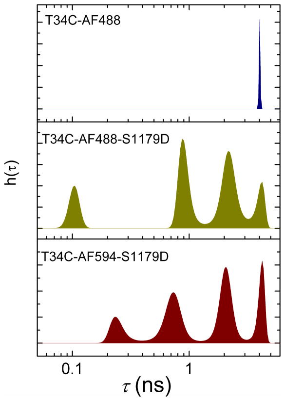
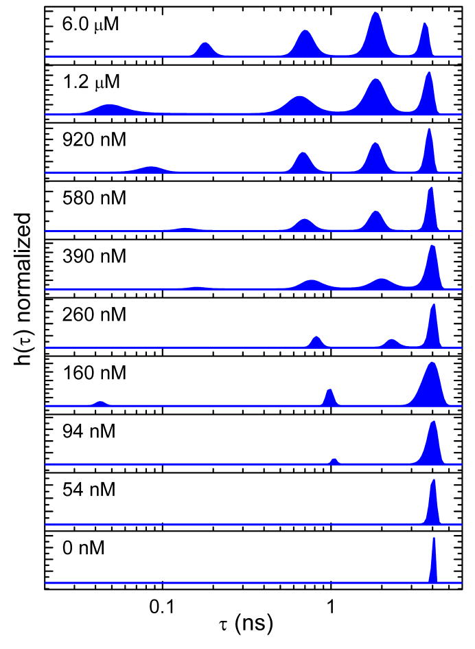
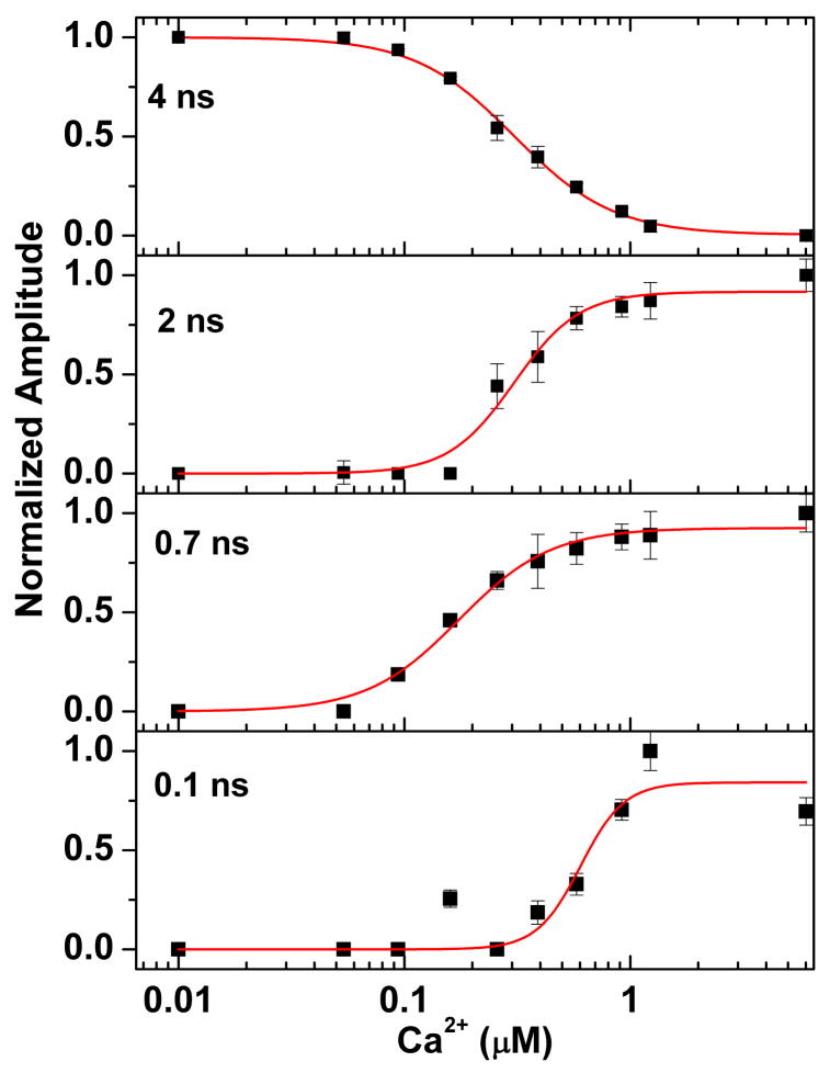
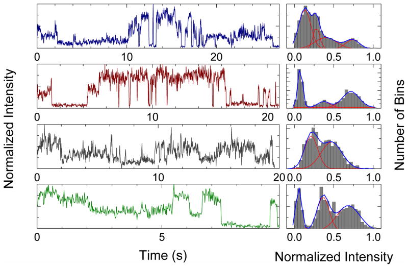
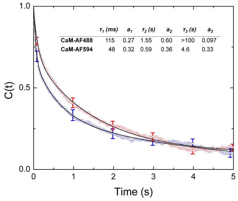
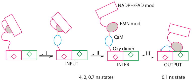
Activation of endothelial nitric oxide synthase (eNOS) by calmodulin (CaM) facilitates formation of a sequence of conformational states that is not well understood. Fluorescence decays of fluorescently labeled CaM bound to eNOS reveal four distinct conformational states and single-molecule fluorescence trajectories show multiple fluorescence states with transitions between states occurring on time scales of milliseconds to seconds. A model is proposed relating fluorescence quenching states to enzyme conformations. Specifically, we propose that the most highly quenched state corresponds to CaM docked to an oxygenase domain of the enzyme. In single-molecule trajectories, this state occurs with time lags consistent with the oxygenase activity of the enzyme.

Keywords: Calmodulin; Fluorescence decay; Förster resonance energy transfer; Nitric oxide synthase; Single-molecule fluorescence.


Figures

Figure 1
Figure 1
Distributions of fluorescence lifetimes recovered by maximum entropy analysis. The plots show the normalized amplitudes h(τ) obtained from maximum-entropy fits as a function of fluorescence lifetime τ. Top: CaM-AF488. Center: CaM-AF488 bound to S1179D-eNOS. Bottom: CaM-AF594 bound to S1179D-eNOS. Distributions were generated by maximum-entropy fits to fluorescence decays recorded by TCSPC. Samples were in high Ca2+ buffer with [Ca2+] = 2 mM, [CaM] • 200 nM, and [eNOS] • • 800 nM. Maximum entropy analysis gave the following lifetimes (and amplitudes): for CaM-AF488: 4.1 ns (100%); for CaM-AF488 with S1179D-eNOS: 104 ps (15%), 917 ps (33%), 2.22 ns (39%), 4.05 ns (13%); for CaM-AF594: 154 ps (8%), 698 ps (21%), 2.37 ns (44%), 4.00 ns (27%). Lifetime values are the average over each peak in the maximum entropy distribution. Uncertainties in lifetime and amplitude are 3 to 10 %. AF488 was excited at 488 nm and AF594 at 594 nm. Emission was collected at 517 nm for AF488 and 617 nm for AF594.
Figure 2
Figure 2
Dependence of fluorescence quenching on Ca2+ concentration for CaM-AF488 (200 nM) in the presence of S1179D-eNOS (800 nM) at Ca2+ concentrations from near zero to 6 μM. The Ca2+ concentration noted in each panel was generated by adding various amounts of a high-Ca2+ buffer containing 10 mM Ca2+ to a low-Ca2+ buffer containing 2 mM EGTA. Ca2+ concentrations were calibrated as described in Methods.
Figure 3
Figure 3
The Ca2+ dependence of fluorescence quenching for CaM-AF488 binding to S1179D-eNOS complexes. The fractional amplitude for each peak in the lifetime distribution was obtained from the sum of the amplitudes for each peak. Lines show fits to a Hill equation. Amplitudes and errors were obtained from maximum entropy fits.
Figure 4
Figure 4
Single-molecule fluorescence trajectories and histograms. Left panels: Examples of single-molecule trajectories and intensity histograms for S1179D-eNOS immobilized on a Cu2+ surface; top two panels, with CaM-AF594; bottom two panels, with CaM-AF488. Fluorescence intensities are normalized to the maximum signal level in each trajectory. Histograms of fluorescence counts per bin are shown in the right panels. The blue lines show a fit to a sum of two to four Gaussian distributions with the individual Gaussian functions shown in red.
Figure 5
Figure 5
Time correlation functions, C(t), for CaM-AF594 and CaM-AF488 complexes with S1179D-eNOS. Correlation functions are averages of over 200 or more correlation functions from single-molecule trajectories. The solid lines represent three-exponential fits to C(t). The obtained time constants and amplitudes are given in the table inset.
Figure 6
Figure 6
Model for the activation of eNOS by CaM. The module depicts the FAD module (rectangle) and FMN module (grey oval) of one protomer, and oxygenase domains of both protoomers, with hemes represented by diamonds. Process I represents binding of CaM (blue oval) to eNOS and is not observed in our studies. Process II represents exchange among input and intermediate conformations that we observed as states with lifetimes of approximately 0.7 ns, 2 ns, and 4 ns. The intermediate conformation may represent multiple conformations with the FMN module undocked. Process III is docking of CaM to the heme domain to generate the output state, which we assigned as the conformation with fluorescence lifetime of approximately 0.1 ns.

References

    1. Masters BS, McMillan K, Sheta EA, Nishimura JS, Roman LJ, Martasek P. Neuronal nitric oxide synthase, a modular enzyme formed by convergent evolution: structure studies of a cysteine thiolate-liganded heme protein that hydroxylates L-arginine to produce NO. as a cellular signal. FASEB J. 1996;10:552–558. - PubMed
    1. Stuehr DJ, Tejero J, Haque MM. Structural and mechanistic aspects of flavoproteins: electron transfer through the nitric oxide synthase flavoprotein domain. FEBS J. 2009;276:3959–3974. - PMC - PubMed
    1. Leferink NG, Hay S, Rigby SE, Scrutton NS. Towards the free energy landscape for catalysis in mammalian nitric oxide synthases. FEBS J 2014 - PubMed
    1. Spratt DE, Taiakina V, Palmer M, Guillemette JG. Differential binding of calmodulin domains to constitutive and inducible nitric oxide synthase enzymes. Biochemistry. 2007;46:8288–8300. - PubMed
    1. Persechini A, Tran QK, Black DJ, Gogol EP. Calmodulin-induced structural changes in endothelial nitric oxide synthase. FEBS Lett. 2013;587:297–301. - PMC - PubMed

Publication types