. 2015 Jul;156(7):2608-18.
doi: 10.1210/en.2014-1762. Epub 2015 Apr 14.

Circadian Control of the Female Reproductive Axis Through Gated Responsiveness of the RFRP-3 System to VIP Signaling

Affiliations

Circadian Control of the Female Reproductive Axis Through Gated Responsiveness of the RFRP-3 System to VIP Signaling

Kimberly A Russo et al. Endocrinology. 2015 Jul.

Abstract

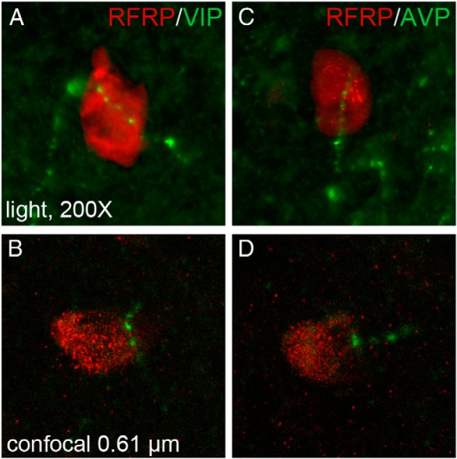
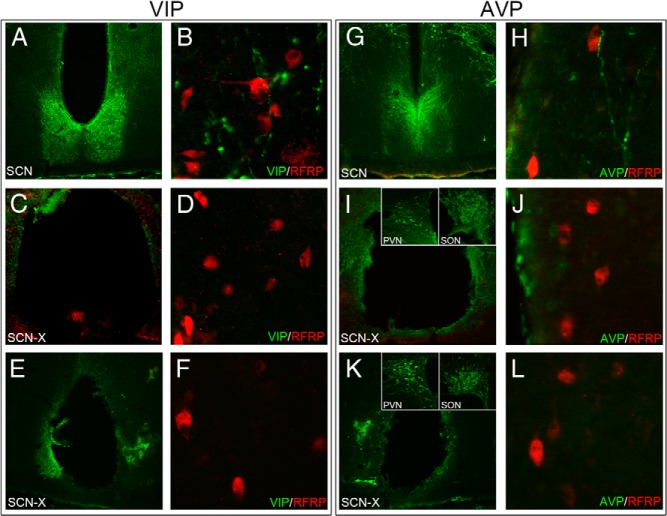
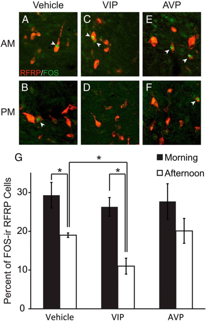
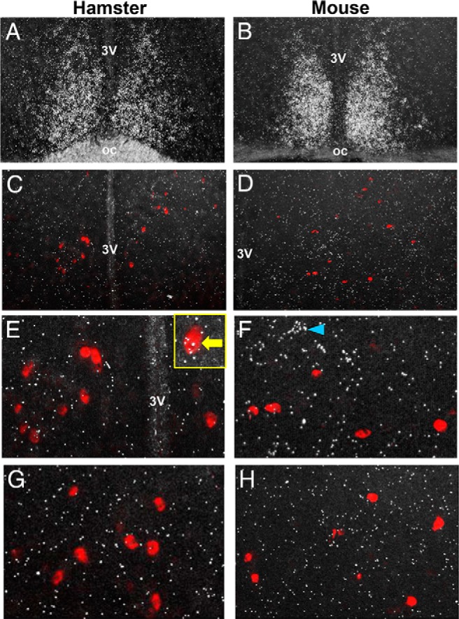
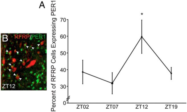
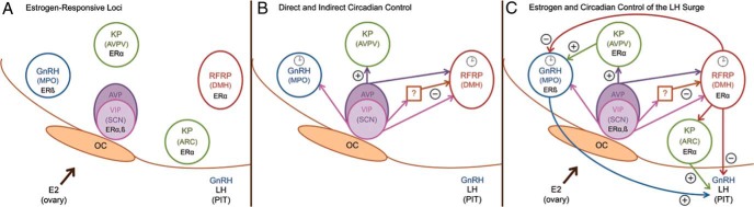
Throughout most of the ovulatory cycle, estrogen negative feedback restrains the GnRH neuronal system. Just before ovulation, however, estrogen negative feedback is removed to permit stimulation of the preovulatory GnRH/LH surge (positive feedback) by the circadian clock in the suprachiasmatic nucleus (SCN). The mammalian ortholog of avian gonadotropin-inhibitory hormone, RFamide-related peptide 3 (RFRP-3), participates in the circadian-timed removal of estrogen negative feedback to permit the LH surge. The present study examined the specific neurochemical means by which the SCN controls RFRP-3 activity and explored whether the RFRP-3 system exhibits time-dependent responsiveness to SCN signaling to precisely time the LH surge. We found that RFRP-3 cells in female Syrian hamsters (Mesocricetus auratus) receive close appositions from SCN-derived vasopressin-ergic and vasoactive intestinal peptide (VIP)-ergic terminal fibers. Central VIP administration markedly suppressed RFRP-3 cellular activity in the evening, but not the morning, relative to saline controls, whereas vasopressin was without effect at either time point. Double-label in situ hybridization for Rfrp-3 and the VIP receptors VPAC1 and VPAC2 revealed that the majority of RFRP-3 cells do not coexpress either receptor in Syrian hamsters or mice, suggesting that SCN VIP-ergic signaling inhibits RFRP-3 cells indirectly. The timing of this VIP-mediated disinhibition is further coordinated via temporally gated responsiveness of RFRP-3 cells to circadian signaling. Together, these findings reveal a novel circadian hierarchy of control coordinating the preovulatory LH surge and ovulation.


Figures

Figure 1.
Figure 1.
SCN fiber projections form close appositions with RFRP-ir cells in the DMH. A, Low-power conventional light photomicrograph (×200) depicting VIP fiber projections in close apposition to RFRP-3-ir cells in the DMH. B, Confocal microscopic image of VIP fiber within the same 0.5-μm plane as an RFRP-3-ir cell. C, Low-power conventional light photomicrograph of AVP fiber projections in close apposition to RFRP-3-ir cells in the DMH. D, Confocal microscopic image of AVP fiber within the same 0.5-μm plane as an RFRP-3-ir neuron.
Figure 2.
Figure 2.
SCN lesions abolish AVP and VIP fiber projections to RFRP-3 cells. A, Low-power photomicrograph of VIP-ir labeling in an intact SCN. B, VIP fibers (green) and RFRP-3 cells (red) (×400) in the DMH. C, SCN lesion with elimination of VIP labeling. D, SCN lesions abolishes VIP fiber expression in the DMH. E, Small SCN lesion eliminating VIP in the SCN. F, Elimination of VIP labeling in DMH. G, Low-power photomicrograph of AVP-ir labeling in an intact SCN. H, AVP fibers (green) and RFRP-3 cells (red) (×400) in the DMH. I, SCN lesion with AVP labeling (green) and unperturbed AVP populations in the supraoptic nucleus (SON) and PVN (insets). J, SCN lesions abolishes VIP fiber expression in the DMH. K, Small SCN lesion eliminating virtually all AVP labeling (green) and unperturbed AVP populations in the SON and PVN (insets). L, Elimination of AVP labeling in DMH.
Figure 3.
Figure 3.
Representative photomicrographs depicting RFRP-3-ir (red) and Fos-ir (green) neurons 1 hour after intracerebroventricular injection of (A) saline (vehicle) at ZT2 (morning) and (B) ZT12 (late afternoon), (C) AVP (morning) and (D) AVP (late afternoon), and (E) VIP (morning) and (F) VIP (late afternoon). G, Mean (±SEM) percentage of RFRP-3 cells expressing Fos after saline (vehicle), AVP, or VIP injections in the morning or late afternoon. *, significantly different (P < .05).
Figure 4.
Figure 4.
Representative photomicrographs of VPAC1 and VPAC2 mRNA expression in the brains of female hamsters and mice at the time of the LH surge. A and B, Abundant VPAC2 expression (silver grain clusters) in the SCN of both hamster and mouse, respectively. C and D, Rfrp-3 neurons (red fluorescence) in the DMH of hamster and mouse, respectively, with minimal observed VPAC2 expression in this region. E and F, Higher magnification of VPAC2 and Rfrp-3 expression in hamster and mouse, denoting virtually no coexpression of the 2 in either species. Yellow box inset in E denotes an example of 1 of the few Rfrp-3 cells weakly coexpressing VPAC2. Blue arrowhead in F denotes a VPAC2-expressing cell, which is not an Rfrp-3 cell. G and H, High magnification of VPAC1 and Rfrp-3 expression in hamster and mouse, denoting virtually no coexpression in either species.
Figure 5.
Figure 5.
RFRP-3 cells rhythmically express PER1 protein. A, Representative low-power photomicrograph of RFRP-3-ir (red) and PER1 (green) immunofluorescence at ZT12. B, Mean (±SEM) percentage of RFRP-3-ir cells expressing PER1-ir at ZT2, ZT7, ZT12, and ZT19 in ovariectomized estradiol-treated female hamsters. *, significantly greater (P < .05) than ZT7, comparison with ZT2 and ZT19 nearing significance, P = .052 and P = .057, respectively.
Figure 6.
Figure 6.
A proposed model depicting circadian control of the preovulatory LH surge and ovulation. A, Estrogen responsive circuitry. These cell populations integrate estrogenic, SCN-derived, and local endogenous circadian cues to appropriately time the preovulatory LH surge. B, Loci receiving direct and indirect input from the master clock in the SCN. In this model, the SCN projects directly to GnRH, kisspeptin, and RFRP-3 cells. Additionally, the SCN projects to a putative interneuron to drive the removal of RFRP-3 inhibition by SCN-derived VIP at the time of the surge. Clock icons indicate neural populations exhibiting circadian oscillations in clock genes/proteins and time-dependent sensitivity to SCN signaling, including the GnRH (35, 42, 43) and RFRP-3 systems (46). C, Estrogen- and clock-controlled hierarchy. RFRP-3 neurons project to GnRH cells and to the median eminence to regulate gonadotropin secretion. It is proposed that RFRP-3 neurons respond to estradiol by inhibiting the reproductive axis throughout most of the estrous cycle. At the time of the LH surge, the SCN inhibits the RFRP-3 system through indirect VIP-ergic signaling, removing negative feedback to allow concomitant, SCN-controlled surge generation. oc, optic chiasm; MPO, medial preoptic area; ARC, arcuate nucleus; PIT, pituitary; SON, supraoptic nucleus.

Similar articles

Cited by

References

    1. Ahlborg G, Jr, Axelsson G, Bodin L. Shift work, nitrous oxide exposure and subfertility among Swedish midwives. Int J Epidemiol. 1996;25(4):783–790. - PubMed
    1. Mahoney MM. Shift work, jet lag, and female reproduction. Int J Endocrinol. 2010;2010:813764. - PMC - PubMed
    1. Bisanti L, Olsen J, Basso O, Thonneau P, Karmaus W. Shift work and subfecundity: a European multicenter study. European Study Group on Infertility and Subfecundity. J Occup Environ Med. 1996;38(4):352–358. - PubMed
    1. Nurminen T. Shift work and reproductive health. Scand J Work Environ Health. 1998;24(suppl 3):28–34. - PubMed
    1. Endo A, Watanabe T. Effects of non-24-hour days on reproductive efficacy and embryonic development in mice. Gamete Res. 1989;22(4):435–441. - PubMed

Publication types

MeSH terms

Substances