. 2015 Apr 13;16(4):8168-85.
doi: 10.3390/ijms16048168.

Quercetin improves postischemic recovery of heart function in doxorubicin-treated rats and prevents doxorubicin-induced matrix metalloproteinase-2 activation and apoptosis induction

Affiliations

Quercetin improves postischemic recovery of heart function in doxorubicin-treated rats and prevents doxorubicin-induced matrix metalloproteinase-2 activation and apoptosis induction

Monika Barteková et al. Int J Mol Sci. .

Abstract

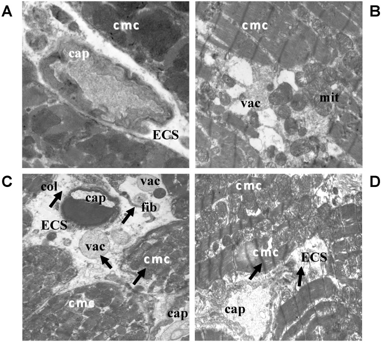
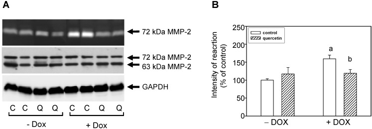
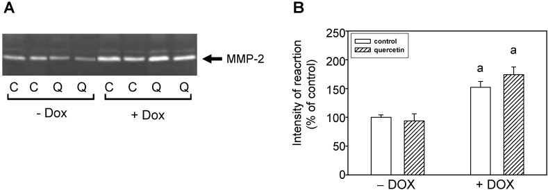
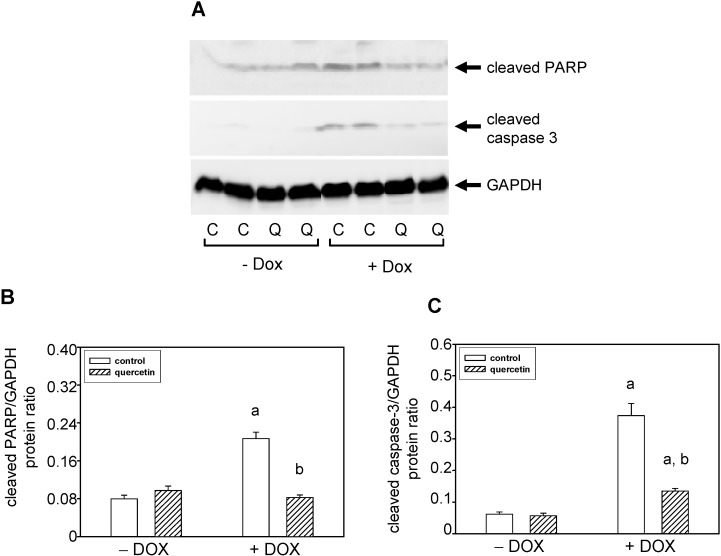
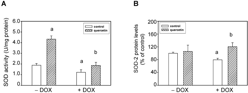
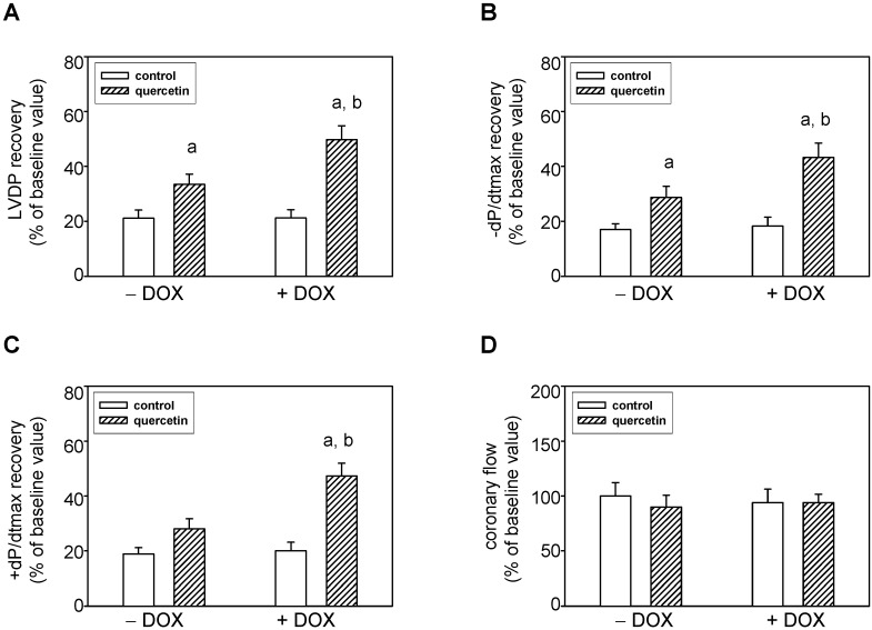
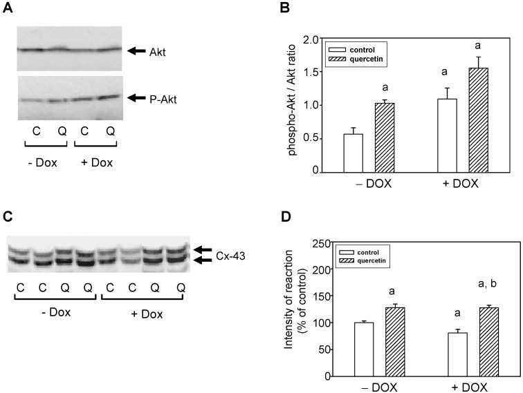
Quercetin (QCT) is flavonoid that possesses various biological functions including anti-oxidative and radical-scavenging activities. Moreover, QCT exerts some preventive actions in treatment of cardiovascular diseases. The aim of present study was to explore effects of prolonged administration of QCT on changes induced by repeated application of doxorubicin (DOX) in rat hearts. We focused on the ultrastructure of myocardium, matrix metalloproteinases (MMPs), biometric parameters, and apoptosis induction. Our aim was also to examine effects of QCT on ischemic tolerance in hearts exposed to chronic effects of DOX, and to determine possible mechanisms underlying effects of QCT. Our results showed that QCT prevented several negative chronic effects of DOX: (I) reversed DOX-induced blood pressure increase; (II) mediated improvement of deleterious effects of DOX on ultrastructure of left ventricle; (III) prevented DOX-induced effects on tissue MMP-2 activation; and (iv) reversed effects of DOX on apoptosis induction and superoxide dismutase inhibition. Moreover, we showed that rat hearts exposed to effects of QCT were more resistant to ischemia/reperfusion injury. Effects of QCT on modulation of ischemic tolerance were linked to Akt kinase activation and connexin-43 up-regulation. Taken together, these results demonstrate that prolonged treatment with QCT prevented negative chronic effects of DOX on blood pressure, cellular damage, MMP-2 activation, and apoptosis induction. Moreover, QCT influenced myocardial responses to acute ischemic stress. These facts bring new insights into mechanisms of QCT action on rat hearts exposed to the chronic effects of DOX.


Figures

Figure 1
Figure 1
Electron microscopic images showing qualitative changes in ultrastructure of the left ventricle of rat hearts. (A) Electron micrograph of control rat heart showing normal architecture of cardiomyocytes and without changes in ECS; (B) Electron micrograph of the myocardium after treatment with QCT; (C) Electron micrograph of the myocardium affected with DOX demonstrating subcellular alterations of cardiomyocytes and extracellular space (arrows); (D) Ultrastructure of the myocardium of rats treated with both QCT and DOX. Arrows indicate improvement of some deleterious subcellular alterations induced by DOX. ECS: extracellular space; cmc: cardiomyocytes; cap: capillary; col: collagen; mit: mitochondria; vac: vacuole; fib: fibroblast. (A) original magnification ×8000; (B) original magnification ×8000; (C) original magnification ×6000; (D) original magnification ×6000.
Figure 2
Figure 2
Effect of QCT and DOX treatment on tissue matrix metalloproteinase-2. (A) At the top is a record showing the activities of MMP-2 analyzed using gelatin zymography; in the middle, a western blot record showing MMP-2 protein levels analyzed using a specific antibody that reacts with both the 72 and 63 kDa forms of MMP-2, and at the bottom is documented the protein loading using GAPDH; (B) Quantitative analysis of the tissue 72-kDa MMP-2 activities. Data are expressed as a percentage of value for corresponding control. Each bar represents mean ± S.E.M. of seven independent tissue samples per group. Statistical significance is revealed by Student’s unpaired t-test, a p < 0.05 vs. control saline-treated (−DOX) rats; b p < 0.05 vs. DOX-treated (+DOX) rats.
Figure 3
Figure 3
Effects of QCT and DOX treatment on plasma MMP-2 activities. (A) Zymogram showing the activities of circulating plasma MMP-2 analyzed using gelatin zymography; (B) Quantitative analysis of plasma MMP-2 activities. Data are expressed as a percentage of value for corresponding control. Each bar represents mean ± S.E.M. of seven independent plasma samples per group. Statistical significance is revealed by Student’s unpaired t-test, a p < 0.05 vs. control saline-treated (−DOX) rats.
Figure 4
Figure 4
Effect of QCT and DOX on markers of apoptosis induction in the left ventricle. (A) The protein levels of cleaved caspase-3 and cleaved PARP were determined by western blot analysis using specific antibodies. The protein loading is documented using GAPDH; (B) Quantification of cleaved caspase-3 content normalized to the GAPDH protein levels; (C) Quantification of cleaved PARP content normalized to the GAPDH protein levels. Data were obtained from western blot records and each bar represents mean ± S.E.M. of seven tissue samples per group. Statistical significance is revealed by Student’s unpaired t-test, a p < 0.05 vs. control saline-treated (−DOX) rats; b p < 0.05 vs. DOX-treated (+DOX) rats.
Figure 5
Figure 5
Effects of QCT and DOX treatment on superoxide dismutase (SOD) total activities and protein levels. (A) The SOD activities were analyzed using the SOD Assay kit in tissue samples of the left ventricle. The specific activities are expressed in units per mg of proteins and are presented as mean ± S.E.M.; (B) Quantitative analysis of SOD-2 protein levels. Data are expressed as a percentage of value for corresponding control. Each bar represents mean ± S.E.M. of seven independent tissue samples per group. Statistical significance is revealed by Student’s unpaired t-test, a p < 0.05 vs. control saline-treated (−DOX) rats; b p < 0.05 vs. DOX-treated (+DOX) rats.
Figure 6
Figure 6
Effects of quercetin on ischemic tolerance of hearts isolated from normal and doxorubicin-treated rats. (A) Postischemic recovery of the left ventricular developed pressure LVDP; (B) Postischemic recovery of the maximal rate of pressure development +(dP/dt)max; (C) Postischemic recovery of the maximal rates of pressure fall −(dP/dt)max; (D) Postischemic recovery of the coronary flow. The recovery of parameters was determined after 30 min lasting global ischemia followed by a 40-min reperfusion. The recovery of parameters was expressed as a percentage of pre-ischemic baseline values. Each bar represents mean ± S.E.M. of eight independent measurements. a p < 0.05 vs. control saline-treated (−DOX) rats; b p < 0.05 vs. DOX-treated (+DOX) rats.
Figure 7
Figure 7
Effect of QCT and DOX on Akt kinase and connexin-43. (A) Western blot records showing the changes in protein levels and specific phosphorylation of Akt kinase. The changes in activation of Akt kinase were determined using an antibody which reacts with Akt kinase phosphorylated specifically on Ser473; (B) The quantification of Akt kinase phosphorylation (activation). Data were obtained from western blot records and are expressed as a ratio of content of phosphorylated Akt kinase to total Akt kinase. Each bar represents mean ± S.E.M. of seven tissue samples per group. Statistical significance is revealed by Student’s unpaired t-test, a p < 0.05 vs. control saline-treated (−DOX) rats; b p < 0.05 vs. DOX-treated (+DOX) rats; (C) Protein levels of Connexin-43 (Cx-43) were determined by western blot analysis using specific antibody. The upper Cx-43 band represents the expression of highly phosphorylated Cx-43, while the lower band corresponds to Cx-43 phosphorylated to a lower extent (degree); (D) Quantification of content of the lower phosphorylated form of Cx-43. Bars represent mean ± S.E.M. of measurements of seven independent tissue samples per group. Statistical significance is revealed by Student’s unpaired t-test; a p < 0.05 vs. control saline-treated (−DOX) rats; b p < 0.05 vs. DOX-treated (+DOX) rats.

References

    1. Hertog M.G., Bueno-de-Mesquita H.B., Fehily A.M., Sweetnam P.M., Elwood P.C., Kromhout D. Fruit and vegetable consumption and cancer mortality in the Caerphilly Study. Cancer Epidemiol. Biomark. Prev. 1996;5:673–677. - PubMed
    1. Erden Inal M., Kahraman A. The protective effect of flavonol quercetin against ultraviolet a induced oxidative stress in rats. Toxicology. 2000;154:21–29. - PubMed
    1. Wu J., Xu X., Li Y., Kou J., Huang F., Liu B., Liu K. Quercetin, luteolin and epigallocatechin gallate alleviate TXNIP and NLRP3-mediated inflammation and apoptosis with regulation of AMPK in endothelial cells. Eur. J. Pharmacol. 2014;745:59–68. - PubMed
    1. Yu P.X., Zhou Q.J., Zhu W.W., Wu Y.H., Wu L.C., Lin X., Chen M.H., Qiu B.T. Effects of quercetin on LPS-induced disseminated intravascular coagulation (DIC) in rabbits. Thromb. Res. 2013;131:270–273. - PubMed
    1. Duarte J., Perez-Palencia R., Vargas F., Ocete M.A., Perez-Vizcaino F., Zarzuelo A., Tamargo J. Antihypertensive effects of the flavonoid quercetin in spontaneously hypertensive rats. Br. J. Pharmacol. 2001;133:117–124. doi: 10.1038/sj.bjp.0704064. - DOI - PMC - PubMed

Publication types

MeSH terms

LinkOut - more resources