. 2015 Apr 14;8(372):ra35.
doi: 10.1126/scisignal.aaa0441.

Casein kinase 2 (CK2) phosphorylates the deubiquitylase OTUB1 at Ser16 to trigger its nuclear localization

Affiliations

Casein kinase 2 (CK2) phosphorylates the deubiquitylase OTUB1 at Ser16 to trigger its nuclear localization

Lina Herhaus et al. Sci Signal. .

Abstract

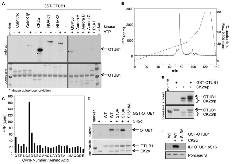
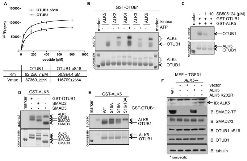
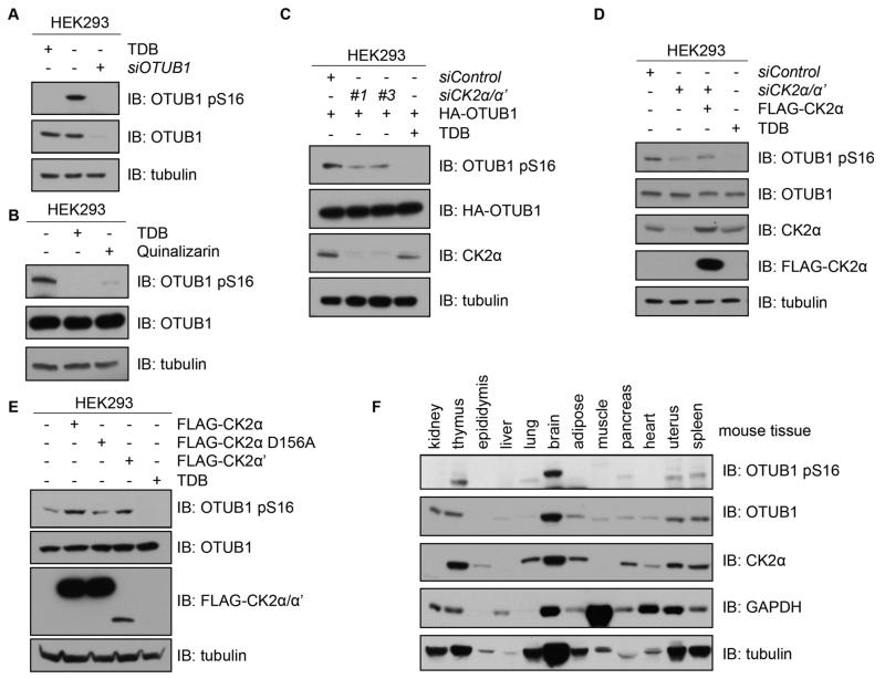
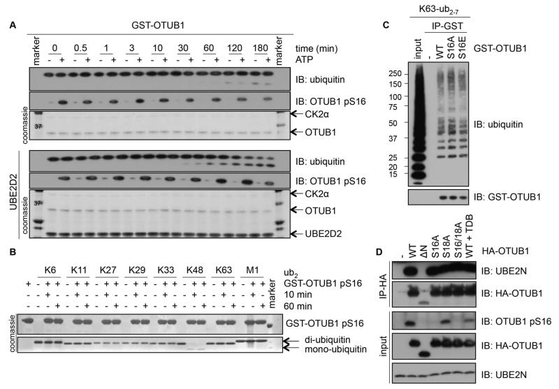
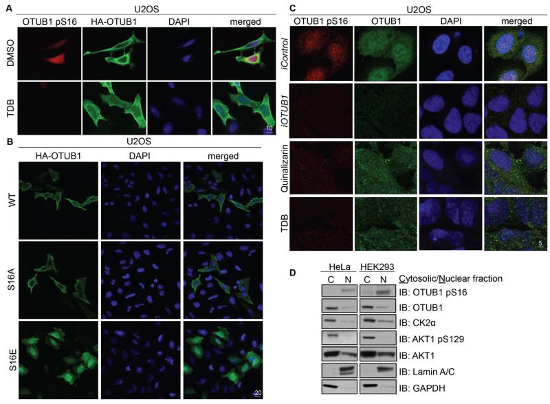
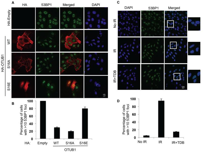
The deubiquitylating enzyme OTUB1 is present in all tissues and targets many substrates, in both the cytosol and nucleus. We found that casein kinase 2 (CK2) phosphorylated OTUB1 at Ser(16) to promote its nuclear accumulation in cells. Pharmacological inhibition or genetic ablation of CK2 blocked the phosphorylation of OTUB1 at Ser(16), causing its nuclear exclusion in various cell types. Whereas we detected unphosphorylated OTUB1 mainly in the cytosol, we detected Ser(16)-phosphorylated OTUB1 only in the nucleus. In vitro, Ser(16)-phosphorylated OTUB1 and nonphosphorylated OTUB1 exhibited similar catalytic activity, bound K63-linked ubiquitin chains, and interacted with the E2 enzyme UBE2N. CK2-mediated phosphorylation and subsequent nuclear localization of OTUB1 promoted the formation of 53BP1 (p53-binding protein 1) DNA repair foci in the nucleus of osteosarcoma cells exposed to ionizing radiation. Our findings indicate that the activity of CK2 is necessary for the nuclear translocation and subsequent function of OTUB1 in DNA damage repair.


Figures

Figure 1
Figure 1. OTUB1 is phosphorylated by CK2 in vitro
(A) Coomassie stain and autoradiography of SDS-PAGE after an in vitro kinase assay with various kinases and GST-OTUB1 as the substrate. (B) γ32P-release chromatograph of CK2-phosphorylated GST-OTUB1, which was digested with trypsin and resolved by HPLC on a C18 column on an increasing acetonitrile gradient as shown. (C) γ32P radioactivity release of the peak after each cycle of Edman degradation. (D) Coomassie stain and autoradiography of SDS-PAGE after an in vitro kinase assay with CK2α and wild-type (WT) or various mutants of GST-OTUB1. (E) As in (D), with the holoenzyme CK2α/β used as the kinase and GST-OTUB1 as the substrate. (F) Western blot (IB) of the phosphorylation of OTUB1 at Ser16 (pS16) after the in vitro kinase assay as in (D). Total GST-OTUB1 was detected with Ponceau S. All blots are representative of 3 independent experiments.
Figure 2
Figure 2. ALKs can phosphorylate OTUB1, but phosphorylation of OTUB1 at Ser16 is specific to CK2
(A) Kinetics of an in vitro kinase assay assessing CK2α-mediated phosphorylation of increasing amounts of OTUB1 and pSer18-OTUB1 peptides. The Km and Vmax values are indicated. Data are mean ± S.D. from 3 experiments. (B) Coomassie stain and autoradiography of an in vitro kinase assay with different ALKs and GST-OTUB1. (C) As in (B), with GST-ALK5 and GST-OTUB1 in the presence of increasing amounts of the ALK5 inhibitor SB505124. (D) As in (B), with GST-ALK5 and SMAD2, SMAD3 and GST-OTUB1. (E) As in (B), with GST-ALK5 and wild-type or mutant GST-OTUB1. (F) Western blotting (IB) for the indicated proteins in lysates from MEF cells (wild-type, ALK5−/− and ALK5−/− transfected with wild-type or kinase-deficient mutant ALK5) treated with TGFβ (50 pM, 1 hour). (SMAD2-TP: SMAD2 tail phosphorylated at residues Ser465 and Ser467). All blots are representative of 3 independent experiments.
Figure 3
Figure 3. CK2 phosphorylates OTUB1 in vivo
(A) Western blotting (IB) of lysates from HEK293 cells that were untreated, treated with TDB (10 μM, 4 hours) or transfected with OTUB1 siRNA and lysed 48 hours later. (B) Western blotting (IB) of lysates from HEK293 cells that were untreated or, treated with TDB (10 μM, 4 hours) or quinalizarin (10 μM, 4 hours). (C) Western blotting (IB) of lysates from HEK293 cells transfected with HA-OTUB1 and FOXO4 siRNA (siControl) or one of two siRNAs against both CK2α and CK2α’ splice variants. A separate culture of cells was treated with TDB (10 μM, 4 hours). (D) Western blotting (IB) of lysates from HEK293 cells transfected with FOXO4 siRNA (control) or CK2α/α’ siRNA alone or reconstituted with N-terminal FLAG-tagged CK2α. A separate culture of cells was treated with TDB (10 μM, 4 hours) prior to lysis. (E) Western blotting (IB) of lysates from HEK293 cells transfected with vectors encoding N-terminal FLAG-tagged CK2α, FLAG-CK2α[D156A] or FLAG-CK2α’, or treated with TDB (10 μM, 4 hours). (F) Western blotting of homogenized lysates from the indicated mouse tissues. All blots are representative of 3 independent experiments.
Figure 4
Figure 4. The catalytic activity, ubiquitin- or E2-binding ability of OTUB1 are not altered by OTUB1 Ser16 phosphorylation
(A) A time course of K48-diubiquitin cleavage assay using unphosphorylated or in vitro CK2-phosphorylated OTUB1 (0.5 μM), in the absence (top panel) or presence of UBE2D2 (bottom panel). Coomassie stains of OTUB1, CK2α and UBE2D2 or Western blotting (IB) of ubiquitin and the phosphorylation of OTUB1 at Ser16 (pS16) are indicated. (B) As in (A), with phosphorylated GST-OTUB1 (5 μM) incubated with K6-, K11-, K27-, K29-, K33-, K48-, K63-, or M1-di-ubiquitin chains for 10 and 60 min in the absence of UBE2D2 and Coomassie stains indicated. (C) Western blotting (IB) of GST (-), GST-OTUB1 (WT), GST-OTUB1-S16A (S16A) or GST-OTUB1-S16E (S16E) incubated in vitro with K63-linked polyubiquitin chains in an interaction assay. (D) Western blotting (IB) of lysates or HA-immunoprecipitates from HEK293 cells transfected with HA-OTUB1 or HA-OTUB1 mutants treated with or without TDB (10 μM, 4 h). All blots are representative of 3 independent experiments.
Figure 5
Figure 5. OTUB1 phosphorylation at Ser16 determines its subcellular localization
(A) Fixed cell immunofluorescence in U2OS cells transfected with HAOTUB1 and untreated or treated with TDB (10 μM, 4 hours). Individual and merged images show phosphorylated OTUB1 pSer16 (red), OTUB1 (HA, green) and DAPI (blue). Scale bar, 10 μm. (B) As in (A) in U2OS cells transfected with HA-OTUB1, HA-OTUB1[S16A] or HA-OTUB1[S16E]. Scale bar, 20 μm. (C) As in (A) in U2OS cells 48 hours after RNA interference [iOTUB1 or iControl (FOXO4)] or after treatment with CK2 inhibitors quinalizarin (10 μM, 4 hours) or TDB (10 μM, 4 hours). Scale bar, 5 μm. (D) Western blotting (IB) for the indicated proteins in cytosolic and nuclear fractions from HeLa or HEK293 cells. All data are representative of 3 independent experiments.
Figure 6
Figure 6. The phosphorylation and nuclear localization of OTUB1 promotes IR-induced 53BP1 foci formation
(A) Fixed cell immunofluorescence for OTUB1 localization and 53BP1 foci in U2OS cells transfected with HA-tag (HA-Empty), wild-type (WT) or mutant (S16E or S16A) HA-tagged OTUB1, then exposed to 5 Gy IR and fixed 3.5 hours later. Scale bar, 15 μm. (B) Quantification of cells displaying more than 10 53BP1 foci from each condition in (A). (C) Fixed cell immunofluorescence in U2OS cells exposed to 5 Gy IR alone or treated with TDB (10 μM, 4 hours) prior to IR exposure. Individual and merged pictures are shown displaying 53BP1 foci formation in green and DAPI in blue. Scale bar, 20 μm. (D) Quantification as in (B). Images are representative, and data are mean ± S.D. from 3 experiments, at least 33 cells quantified in each.

References

    1. Mevissen Tycho E. T., Hospenthal Manuela K., Geurink Paul P., Elliott Paul R., Akutsu M, Arnaudo N, Ekkebus R, Kulathu Y, Wauer T, El Oualid F, Freund Stefan M. V., Ovaa H, Komander D. OTU Deubiquitinases Reveal Mechanisms of Linkage Specificity and Enable Ubiquitin Chain Restriction Analysis. Cell. 2013;154:169–184. - PMC - PubMed
    1. Komander D, Clague MJ, Urbe S. Breaking the chains: structure and function of the deubiquitinases. Nature reviews. Molecular cell biology. 2009;10:550–563. - PubMed
    1. Balakirev MY, Tcherniuk SO, Jaquinod M, Chroboczek J. Otubains: a new family of cysteine proteases in the ubiquitin pathway. EMBO Rep. 2003;4:517–522. - PMC - PubMed
    1. Edelmann MJ, Iphöfer A, Akutsu M, Altun M, di Gleria K, Kramer HB, Fiebiger E, Dhe-Paganon S, Kessler BM. Structural basis and specificity of human otubain 1-mediated deubiquitination. Biochemical Journal. 2009;418:379. - PubMed
    1. Messick TE, Russell NS, Iwata AJ, Sarachan KL, Shiekhattar R, Shanks JR, Reyes-Turcu FE, Wilkinson KD, Marmorstein R. Structural basis for ubiquitin recognition by the Otu1 ovarian tumor domain protein. J Biol Chem. 2008;283:11038–11049. - PMC - PubMed

Publication types

MeSH terms