. 2015 Apr 15:5:9578.
doi: 10.1038/srep09578.

The effects of interfacial potential on antimicrobial propensity of ZnO nanoparticle

Affiliations

The effects of interfacial potential on antimicrobial propensity of ZnO nanoparticle

Manoranjan Arakha et al. Sci Rep. .

Abstract

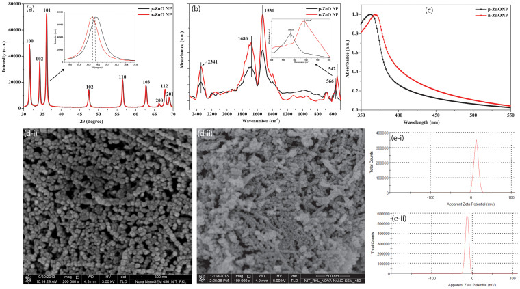
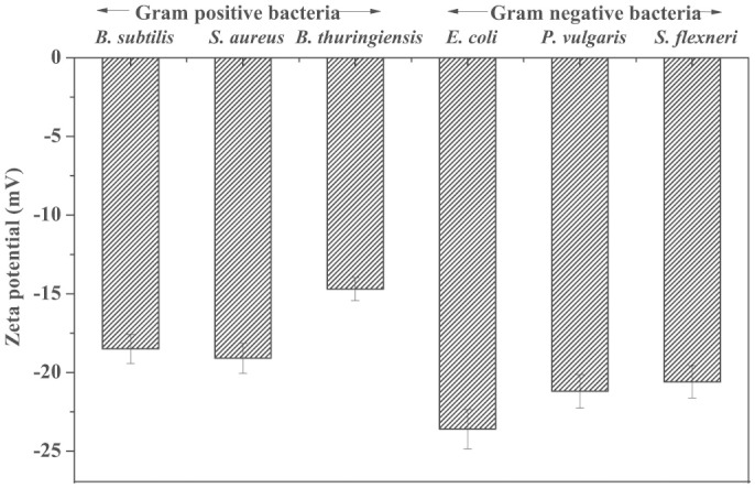
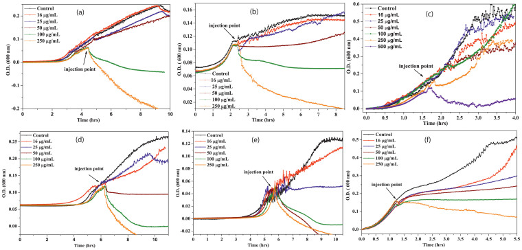
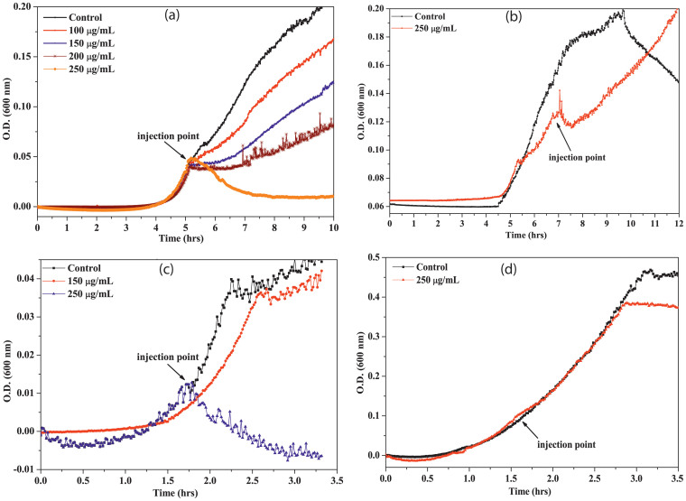
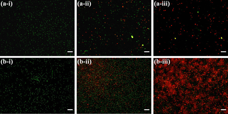
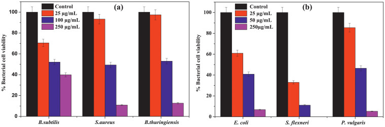
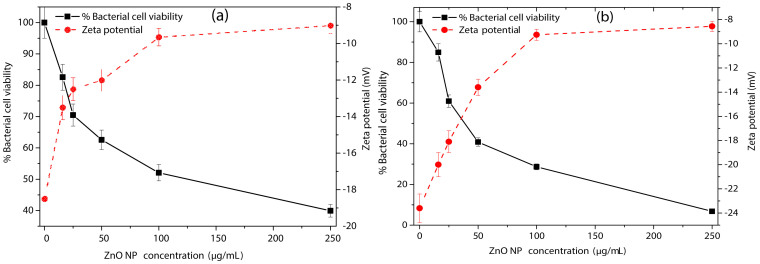
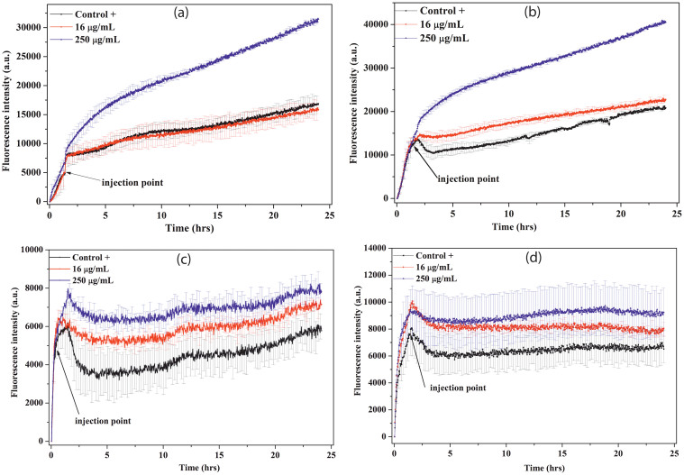
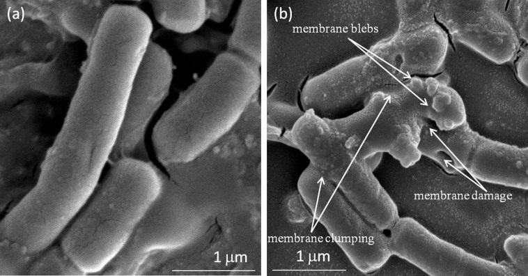
The work investigates the role of interfacial potential in defining antimicrobial propensity of ZnO nanoparticle (ZnONP) against different Gram positive and Gram negative bacteria. ZnONPs with positive and negative surface potential are tested against different bacteria with varying surface potentials, ranging -14.7 to -23.6 mV. Chemically synthesized ZnONPs with positive surface potential show very high antimicrobial propensity with minimum inhibitory concentration of 50 and 100 μg/mL for Gram negative and positive bacterium, respectively. On other hand, ZnONPs of the same size but with negative surface potential show insignificant antimicrobial propensity against the studied bacteria. Unlike the positively charged nanoparticles, neither Zn(2+) ion nor negatively charged ZnONP shows any significant inhibition in growth or morphology of the bacterium. Potential neutralization and colony forming unit studies together proved adverse effect of the resultant nano-bacterial interfacial potential on bacterial viability. Thus, ZnONP with positive surface potential upon interaction with negative surface potential of bacterial membrane enhances production of the reactive oxygen species and exerts mechanical stress on the membrane, resulting in the membrane depolarization. Our results show that the antimicrobial propensity of metal oxide nanoparticle mainly depends upon the interfacial potential, the potential resulting upon interaction of nanoparticle surface with bacterial membrane.


Figures

Figure 1
Figure 1. Characterization of ZnONPs.
(a) XRD, (b) ATR-FTIR absorption spectra, (c) UV-Vis absorption spectra of p-ZnONP and n-ZnONP, (d) FE-SEM image of p-ZnONP (d-i) and n-ZnONP (d-ii), (e) Zeta potential analysis of p-ZnONP and n-ZnONP showing value of +12.9 mV (e-i) & -12.9 mV (e-ii), respectively.
Figure 2
Figure 2. Zeta potentials of Gram positive and Gram negative bacteria.
Figure 3
Figure 3. Growth kinetics of bacteria (a. B. subtilis, b. S. aureus, c. B. thuringiensis, d. E. coli, e. S. flexneri, and f. P. vulgaris) in presence of different concentrations of p-ZnONPs.
In each case, black line shows the growth kinetic curve of untreated cells. Different concentrations of p-ZnONP taken were 16, 25, 50, 100, 250, and 500 (only for B. thuringiensis) μg/mL, and injected at the mid log phase of growth kinetics, as shown by arrow.
Figure 4
Figure 4. Growth kinetics of bacteria in the presence of different concentrations of n-ZnONP.
In each case, black line shows the growth kinetic curve of untreated cells. Both Gram positive (a. B. subtilis) and Gram negative (b. E. coli, c. S. flexneri, d. P. vulgaris) bacteria were treated up to 250 μg/mL of n-ZnONP (injected at the mid log phase of growth kinetics, as shown by arrow).
Figure 5
Figure 5. Fluorescence microscopic images of the green and red fluorescence stained B. subtilis and E. coli in absence and presence of p-ZnONP; B. subtilis (a-i), B. subtilis in presence of 100 μg/mL of p-ZnONP (a-ii), and 250 μg/mL of p-ZnONP (a-iii), E. coli (b-i), E. coli in presence of 50 μg/mL of p-ZnONP (b-ii), and 250 μg/mL of p-ZnONP (b-iii).
The scale bars represent for 20 μm.
Figure 6
Figure 6. Quantification of bacterial cell viability at different concentrations of p-ZnONP.
Colony forming units (CFU) were quantified for both Gram positive and Gram negative bacteria, and expressed as percentage of viable cells.
Figure 7
Figure 7. Effect of p-ZnONP on bacterial cell viability and surface zeta potential of B. subtilis and E. coli cells.
B. subtilis (a) and E. coli (b) cells were treated with increasing concentrations of p-ZnONP like 16, 25, 50, 100, 250 μg/mL. Solid black lines represent the relative percentage of viable bacterial cells, whereas dashed red lines correspond to zeta potential values at different concentrations of p-ZnONP. Triplicate experiments were done for each reactions, and error bar represents the standard error of mean.
Figure 8
Figure 8. ZnONPs induced ROS detection.
B. subtilis cells (figure a and c) and E. coli cells (figure b and d) were treated with 16 μg/mL (red curve) and 250 μg/mL (blue curve) of positively charged (panel a and b) and negatively charged (panel c and d) ZnONPs, and ROS were detected by measuring fluorescence emission intensity at 523 nm. In each case, except control, NPs were added in the log phase of bacterial growth. The fluorescence emission intensity are compared with positive control (without injection of NPs, black curve) in each case. Each curve represents the average of three independent measurements with corresponding standard error of mean.
Figure 9
Figure 9. Visualization of ZnONP treated E. coli cell surface by FE-SEM, (a) control (without ZnONP treated cells), (b) showing membrane blebbings, membrane damage, and membrane clumping in ZnONP treated cells.

Similar articles

Cited by

References

    1. Nel A. E. et al. Understanding biophysicochemical interactions at the nano-bio interface. Nat. Mater. 8, 543–557 (2009). - PubMed
    1. Moyano D. F. & Rotello V. M. Nano meets biology: structure and function at the nanoparticle interface. Langmuir 27, 10376–10385 (2011). - PMC - PubMed
    1. Dewan S. et al. Structure of water at charged interfaces: a molecular dynamics study. Langmuir 30, 8056–8065 (2014). - PubMed
    1. Monopoli M. P., berg C., Salvati A. & Dawson K. A. Biomolecular coronas provide the biological identity of nanosized materials. Nat. Nanotechnol. 7, 779–786 (2012). - PubMed
    1. Elsaesser A. & Howard C. V. Toxicology of nanoparticles. Adv. Drug Del. Rev. 64, 129–137 (2012). - PubMed

Publication types

MeSH terms