. 2015 Apr 13;7(2):1759091415569908.
doi: 10.1177/1759091415569908. Print 2015 Mar-Apr.

AAV-mediated gene delivery in a feline model of Sandhoff disease corrects lysosomal storage in the central nervous system

Affiliations

AAV-mediated gene delivery in a feline model of Sandhoff disease corrects lysosomal storage in the central nervous system

Hannah E Rockwell et al. ASN Neuro. .

Abstract

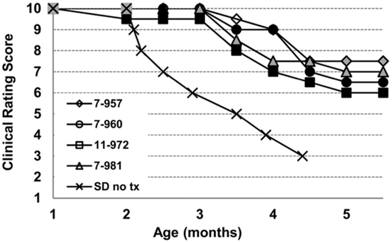
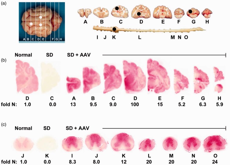
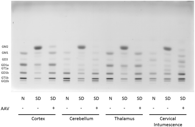
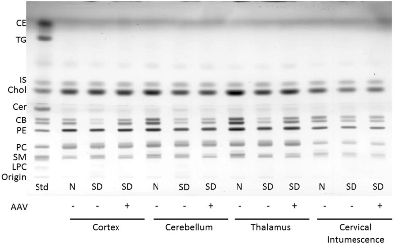
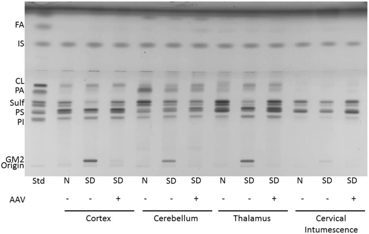
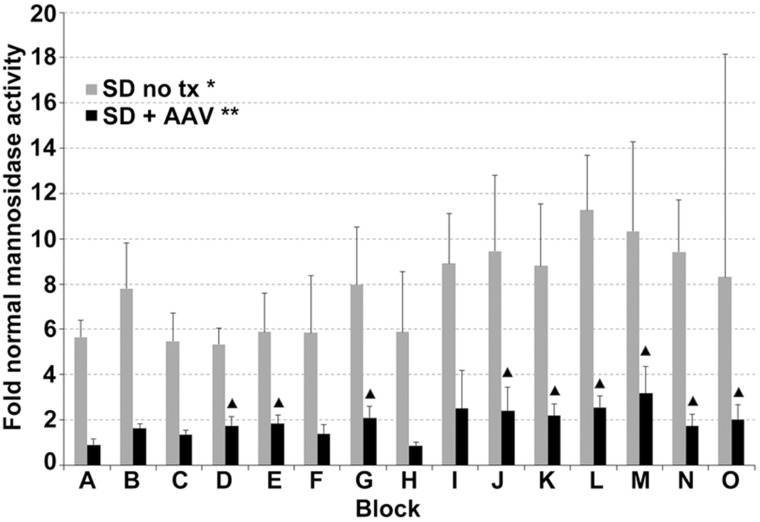
Sandhoff disease (SD) is an autosomal recessive neurodegenerative disease caused by a mutation in the gene for the β-subunit of β-N-acetylhexosaminidase (Hex), resulting in the inability to catabolize ganglioside GM2 within the lysosomes. SD presents with an accumulation of GM2 and its asialo derivative GA2, primarily in the central nervous system. Myelin-enriched glycolipids, cerebrosides and sulfatides, are also decreased in SD corresponding with dysmyelination. At present, no treatment exists for SD. Previous studies have shown the therapeutic benefit of adeno-associated virus (AAV) vector-mediated gene therapy in the treatment of SD in murine and feline models. In this study, we treated presymptomatic SD cats with AAVrh8 vectors expressing feline Hex in the thalamus combined with intracerebroventricular (Thal/ICV) injections. Treated animals showed clearly improved neurologic function and quality of life, manifested in part by prevention or attenuation of whole-body tremors characteristic of untreated animals. Hex activity was significantly elevated, whereas storage of GM2 and GA2 was significantly decreased in tissue samples taken from the cortex, cerebellum, thalamus, and cervical spinal cord. Treatment also increased levels of myelin-enriched cerebrosides and sulfatides in the cortex and thalamus. This study demonstrates the therapeutic potential of AAV for feline SD and suggests a similar potential for human SD patients.

Keywords: Sandhoff disease; adeno-associated virus; ganglioside; gene therapy; β-hexosaminidase.


Figures

Figure 1.
Figure 1.
Clinical progression of AAV-treated Sandhoff cats. SD cats from 1 to approximately 5.5 months of age were assigned numerical scores corresponding to discrete stages of clinical disease progression, as defined in the “Materials and Methods” section. Shown are clinical rating scores of four AAV-treated SD cats (7-957, 7-960, 11-972, 7-981) and mean scores for 14 untreated SD cats (SD no tx). The humane endpoint is triggered by a score of 3, which occurs in untreated SD cats at 4.4 ± 0.6 months of age. Treated cats survived in good condition to the predetermined experimental endpoint of approximately 5.5 months of age. Shading of individual symbols represents tremor severity as follows: no shading, no tremor; gray shading, fine tremors; black shading, mild whole-body tremors. Untreated SD cats developed fine tremors at 1.3 (±0.2) months of age and whole-body tremors at 2.4 (± 0.1) months of age. The mean clinical rating scores and standard deviations for treated cats at each age are as follows: 1 mo, 10 ± 0.0; 2 mo, 9.9 ± 0.3; 2.5 mo, 9.9 ± 0.3; 3 mo, 9.9 ± 0.3; 3.5 mo, 8.8 ± 0.6; 4 mo, 8.1 ± 1.0; 4.5 mo, 7.1 ± 0.5; 5 mo, 6.8 ± 0.6; 5.5 mo, 6.8 ± 0.6. Supplemental videos 1 and 2 depict representative untreated and AAV-treated SD cats, respectively. AAV = adeno-associated virus; SD = Sandhoff disease.
Figure 2.
Figure 2.
Therapeutic enzyme distribution in the CNS of Sandhoff cats after AAV treatment. SD cats were injected bilaterally in the thalamus and the left lateral ventricle with AAVrh8-fHEXA and AAVrh8-fHEXB (1.1 × 1012 vector genomes total), and tissues were collected 16 weeks later. (a) Shown are injection sites (white circles) and 0.6  cm coronal blocks of the brain (A–H) and spinal cord (I–O) collected at necropsy. Blocks were halved and analyzed for enzyme activity (right) or for storage material (left; black circles show biopsy sites for HPTLC). Lysosomal Hex activity (red) detected with naphthol substrate at acidic pH was visualized throughout the brain (b) and spinal cord (c) of a representative, treated SD cat (SD+AAV; 7-960). Corresponding Hex activity against MUGS substrate is shown below each block as fold normal level (fold N). Representative control sections are shown from untreated normal cats along with untreated SD cats, which express ≤0.02 fold normal Hex activity in the brain and spinal cord. The range of specific activities for normal control blocks were brain, 28.1 (G) – 57.4 (D); spinal cord, 8.3 (M) – 17.3 (K) nmol 4MU/mg/hr. AAV = adeno-associated virus; SD = Sandhoff disease.
Figure 3.
Figure 3.
HPTLC of gangliosides from Sandhoff cat cortex, cerebellum, thalamus, and spinal cord (cervical intumescence). Gangliosides from normal cats (N), Sandhoff disease cats (SD), and Sandhoff disease cats treated with AAV vectors were separated in a single ascending run with CHCl3:CH3OH;dH20, 55:45:10 (v/v/v) with 0.02% CaCl2. 1.5µg of sialic acid was spotted for each sample. The bands were visualized with the resorcinol-HCl spray. Ganglioside positions are shown on the left-hand side of the plate. AAV = adeno-associated virus; SD = Sandhoff disease; HPTLC = high-performance thin layer chromatography.
Figure 4.
Figure 4.
HPTLC of neutral lipids from Sandhoff cat cortex, cerebellum, thalamus, and cervical intumescence. Sample abbreviations are as listed in Figure 3. Std, standard. The plate was developed to a height of 4.5  cm with CHCl3:CH3OH: CH3COOH:CHOOH:dH2O 35:15:6:2:1 (v/v/v/v/v) and then developed to the top with C6H14:C6H14O:CH3COOH 65:35:2 (v/v/v). The amount of neutral lipid spotted per lane was equivalent to 70 µg tissue dry weight. The bands were visualized by charring with 3% cupric acetate in 8% phosphoric acid solution. CE = cholesterol esters; TG = triglycerides; IS = internal standard (oleyl alcohol); Chol = cholesterol; Cer = ceramide; CB = cerebrosides (doublet); PE = phosphatidylethanolamine; PC = phosphatidylcholine; SM = sphingomyelin; LPC = lysophosphatidylcholine; AAV = adeno-associated virus; SD = Sandhoff disease; HPTLC = high-performance thin layer chromatography.
Figure 5.
Figure 5.
HPTLC of the acidic lipids from Sandhoff cat cortex, cerebellum, thalamus, and cervical intumescence. Sample abbreviations are as listed in Figure 3. Std, standard. The plate was developed to a height of 6  cm with CHCl3:CH3OH: CH3COOH:CHOOH:dH2O 35:15:6:2:1 (v/v/v/v/v) and then developed to the top with C6H14:C6H14O:CH3COOH 65:35:2 (v/v/v). The amount of acidic lipid spotted per lane was equivalent to 200 µg tissue dry weight. The bands were visualized by charring with 3% cupric acetate in 8% phosphoric acid solution. FA = fatty acids; IS = internal standard (oleyl alcohol); CL = cardiolipin; PA = phosphatidic acid; Sulf = sulfatides (doublet); PS = phosphatidylserine; PI = phosphatidylinositol.
Figure 6.
Figure 6.
Normalization of lysosomal α-mannosidase activity in the CNS of SD cats 16 weeks posttreatment. Lettering of brain and spinal cord blocks correspond to Figure 2. Error bars represent standard deviation. *, all samples from untreated SD cats were significantly higher than normal (p = .010 for each block); **, all samples from treated SD cats were significantly lower than untreated (p ≤ .019 for each brain block; p ≤ .037 for each spinal cord block); ▴, samples from treated cats that were significantly higher than normal (p ≤ .030). AAV = adeno-associated virus; SD = Sandhoff disease; CNS = central nervous system.

Similar articles

Cited by

References

    1. Ando S., Chang N. C., Yu R. K. (1978) High-performance thin-layer chromatography and densitometric determination of brain ganglioside compositions of several species. Analytical Biochemistry 89: 437–450. - PubMed
    1. Arthur J. R., Wilson M. W., Larsen S. D., Rockwell H. E., Shayman J. A., Seyfried T. N. (2013) Ethylenedioxy-PIP2 oxalate reduces ganglioside storage in juvenile Sandhoff disease mice. Neurochemical Research 38: 866–875. - PMC - PubMed
    1. Baek R. C., Broekman M. L., Leroy S. G., Tierney L. A., Sandberg M. A., d'Azzo A., Sena-Esteves M. (2010) AAV-mediated gene delivery in adult GM1-gangliosidosis mice corrects lysosomal storage in CNS and improves survival. PLoS One 5: e13468. - PMC - PubMed
    1. Baek R. C., Kasperzyk J. L., Platt F. M., Seyfried T. N. (2008) N-butyldeoxygalactonojirimycin reduces brain ganglioside and GM2 content in neonatal Sandhoff disease mice. Neurochemistry International 52: 1125–1133. - PubMed
    1. Baek R. C., Martin D. R., Cox N. R., Seyfried T. N. (2009) Comparative analysis of brain lipids in mice, cats, and humans with Sandhoff disease. Lipids 44: 197–205. - PMC - PubMed

Publication types

MeSH terms