. 2015 Apr 14;6(2):e00441-15.
doi: 10.1128/mBio.00441-15.

Changes in expression induced by Epstein-Barr Virus LMP1-CTAR1: potential role of bcl3

Affiliations

Changes in expression induced by Epstein-Barr Virus LMP1-CTAR1: potential role of bcl3

Rachel Hood Edwards et al. mBio. .

Abstract

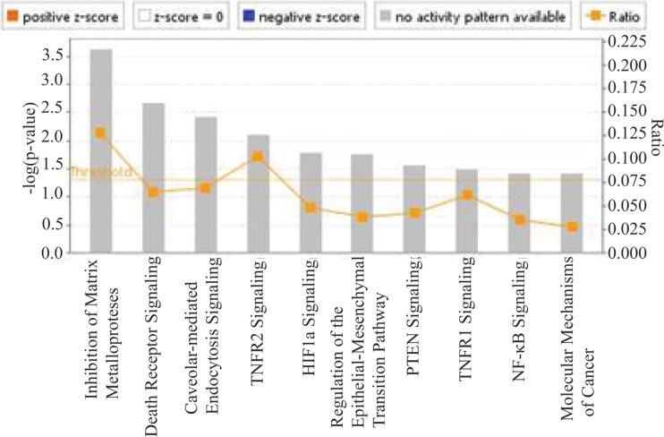
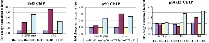
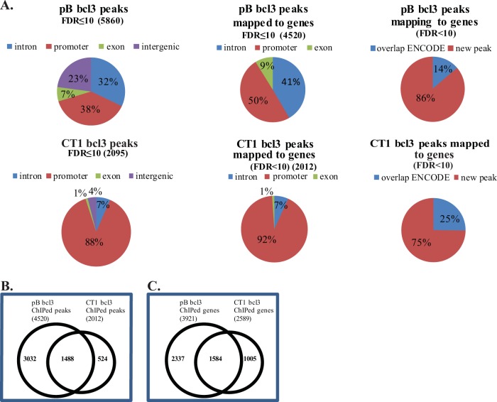
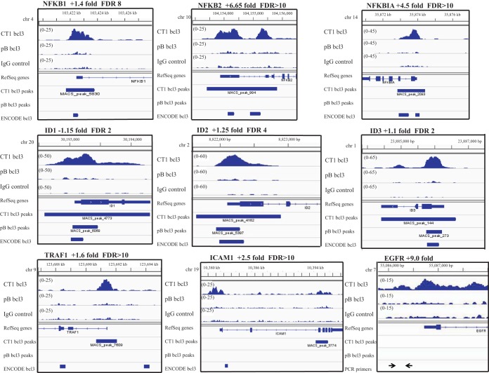
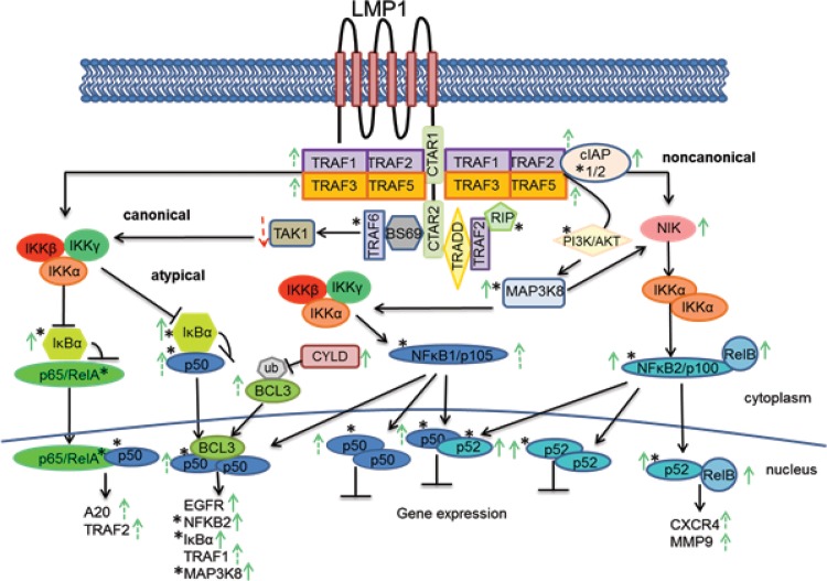
The Epstein-Barr virus protein latent membrane protein 1 (LMP1) has two NF-κB activating domains within its intracellular carboxy terminus (carboxy-terminal activating region 1 [CTAR1] and CTAR2). LMP1-CTAR1 is required for B-lymphocyte transformation, is capable of transforming rodent fibroblasts, and uniquely activates phosphoinositol (PI3) kinase, the noncanonical NF-κB pathway, and expression of the epidermal growth factor receptor (EGFR). In this study, the effects of LMP1-CTAR1 on cellular gene expression were determined by high-throughput sequencing. Additionally, the binding of bcl3 was determined using chromatin immunoprecipitation (ChIP) and sequencing. LMP1-CTAR1 induced few changes in transcription with more genes showing decreased expression. Ingenuity pathway analysis indicated significant enrichment for genes involved in cancer and cellular movement, survival, growth, and proliferation pathways. ChIP in combination with high-throughput sequencing (ChIP-Seq) identified bcl3 binding for more than 2,000 genes in LMP1-CTAR1-expressing cells with more than 90% of the peaks at genes detected within the probable promoter region. Only a small subset of the genes with significant changes in expression had corresponding peaks in the bcl3 ChIP. However, both NFKB2 and PI3 kinase were identified in the bcl3 ChIP. Additionally, many of the predicted upstream regulators for the changes in expression were identified in the bcl3 ChIP. Analysis of the proteins in the NF-κB pathway revealed many changes identified by the high-throughput RNA sequencing (RNA-Seq) and bcl3 ChIP that would likely activate noncanonical NF-κB signaling and possibly inhibit canonical NF-κB signaling. These findings suggest that the two LMP1 signaling domains modulate their combined activity and that the bcl3 transcription factor is likely responsible for some of the unique effects of CTAR1 on cellular expression.

Importance: The Epstein-Barr virus protein latent membrane protein 1 (LMP1) has potent effects on cell growth. LMP1 has two regions, carboxy-terminal activating region 1 (CTAR1) and CTAR2, that distinctly activate NF-κB, a transcription factor complex involved in activation of important host genes. In this study, analysis of the effects on cellular gene expression revealed that CTAR1 significantly affected cellular expression in part through effects on a specific form of NF-κB. The data suggest that LMP1 can activate a distinct subset of host gene expression through its CTAR1 domain which in combination with other signaling effects induced by the CTAR2 domain likely affects cell movement, survival, and growth.


Figures

FIG 1
FIG 1
Canonical pathways associated with genes with 2-fold expression change by RNA-Seq in CTAR1-expressing C33a cells. Significant canonical pathways determined by IPA analysis of the genes with 2-fold change in expression by RNA-Seq in the CTAR1-expressing cells are shown. The height of the bars reflects the P value, and the orange boxes reflect the ratio of the number of genes in the data set that are represented in the pathway.
FIG 2
FIG 2
Quantitative PCR of known binding sites of the transcription factors on chromatin. To confirm the success of the ChIP, the sample chromatin from the pBabe (pB)- and CTAR1 (CT1)-expressing cells was subjected to quantitative PCR for known target/binding sites of the transcription factors. For the bcl3 and p50 ChIPs, primer pairs were used to amplify known binding sites within the EGFR promoter, EGFR-pro and KB2. For the pStat3 ChIP, primer pairs were used to amplify known binding sites within the bcl3 gene, the bcl3-promoter and intronic sites HS3 and HS4.
FIG 3
FIG 3
Distribution of bcl3 peaks. (A) Distribution of peaks with FDRs of <10 across the genome, those mapped to genes (within 2 kb of start site or within intron or exon) and those that overlap ENCODE bcl3 peaks. (B) Overlap of bcl3 peaks between the pB-containing and CT1-expressing cells. (C) Overlap of genes ChIPed by bcl3 in the pB-containing and CT1-expressing cells.
FIG 4
FIG 4
Visualization of bcl3 peaks called by MACS. A representation of peaks called for bcl3 in the CT1-expressing and pB-containing cells, showing the height of the peak, location on the genome, gene bound, and bcl3 peaks in the ENCODE database. The gene is listed with the fold change by RNA-Seq and the FDR of the peak called by MACS for the CT1-expressing cells. For the EGFR gene, the locations of primers used to check chromatin for known NF-κB sites are shown.
FIG 5
FIG 5
LMP1 signaling through CTAR1. LMP1-CTAR1 binds TRAF1, -2, -3, and -5. TRAF2 and -3 recruit cIAP1 (Birc2) which targets TRAF3 for degradation, thereby releasing NIK from TRAF3 negative regulation. NIK is ubiquitinated and stabilized and now available to activate IKKα, which in turn phosphorylates p100 leading to p100 processing to p52, and its subsequent translocation with RelB into the nucleus where the complex will bind DNA and increase transcription of the bound genes. Through activation of PI3K by CTAR1, the increased levels of Map3K8 (Cot) are activated leading to phosphorylation and activation of NIK and its downstream effects. Activated Map3K8 also leads to the processing of p105 to p50 and its subsequent translocation to the nucleus where it can bind with bcl3 to turn on transcription or when bound as homodimers or heterodimers with p52, suppress transcription. The increased CYLD expression would lead to deubiquitination of bcl3 and prevent bcl3 nuclear accumulation. A decrease in canonical NF-κB signaling would result from the decreased expression of TAK1 and the increased expression of IκBα. Genes ChIPed by bcl3 with a FDR of ≤10 and/or match ENCODE peak are indicated by an asterisk. Solid green or red arrows reflect changes in expression with P < 0.05 in RNA-Seq data. Dashed green or red arrows reflect changes in expression with P > 0.05 in RNA-Seq data.

References

    1. Raab-Traub N. 2007. EBV-induced oncogenesis, p 986–1006. In Arvin A, Campadelli-Fiume G, Mocarski E, Moore PS, Roizman B, Whitley R, Yamanishi K (ed), Human herpesviruses: biology, therapy, and immunoprophylaxis. Cambridge University Press, Cambridge, United Kingdom. - PubMed
    1. Raab-Traub N. 2002. Epstein-Barr virus in the pathogenesis of NPC. Semin Cancer Biol 12:431–441. doi:10.1016/S1044579X0200086X. - DOI - PubMed
    1. Dawson CW, Port RJ, Young LS. 2012. The role of the EBV-encoded latent membrane proteins LMP1 and LMP2 in the pathogenesis of nasopharyngeal carcinoma (NPC). Semin Cancer Biol 22:144–153. doi:10.1016/j.semcancer.2012.01.004. - DOI - PubMed
    1. Mosialos G, Birkenbach M, Yalamanchili R, VanArsdale T, Ware C, Kieff E. 1995. The Epstein-Barr virus transforming protein LMP1 engages signaling proteins for the tumor necrosis factor receptor family. Cell 80:389–399. doi:10.1016/0092-8674(95)90489-1. - DOI - PubMed
    1. Mainou BA, Everly DN Jr, Raab-Traub N. 2005. Epstein-Barr virus latent membrane protein 1 CTAR1 mediates rodent and human fibroblast transformation through activation of PI3K. Oncogene 24:6917–6924. doi:10.1038/sj.onc.1208846. - DOI - PubMed

Publication types

MeSH terms

Substances