. 2015 May 22;290(21):13556-66.
doi: 10.1074/jbc.M114.587923. Epub 2015 Apr 14.

Cyclic AMP Response Element-binding Protein H (CREBH) Mediates the Inhibitory Actions of Tumor Necrosis Factor α in Osteoblast Differentiation by Stimulating Smad1 Degradation

Affiliations

Cyclic AMP Response Element-binding Protein H (CREBH) Mediates the Inhibitory Actions of Tumor Necrosis Factor α in Osteoblast Differentiation by Stimulating Smad1 Degradation

Won-Gu Jang et al. J Biol Chem. .

Abstract

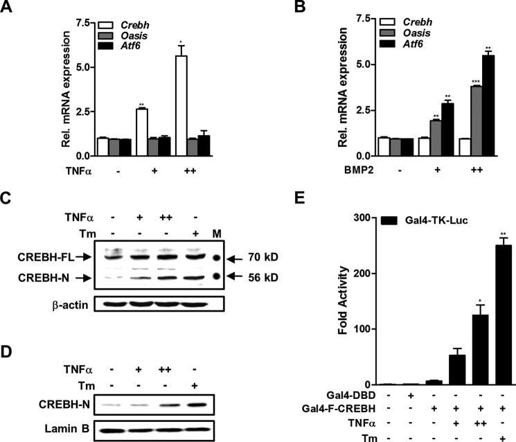
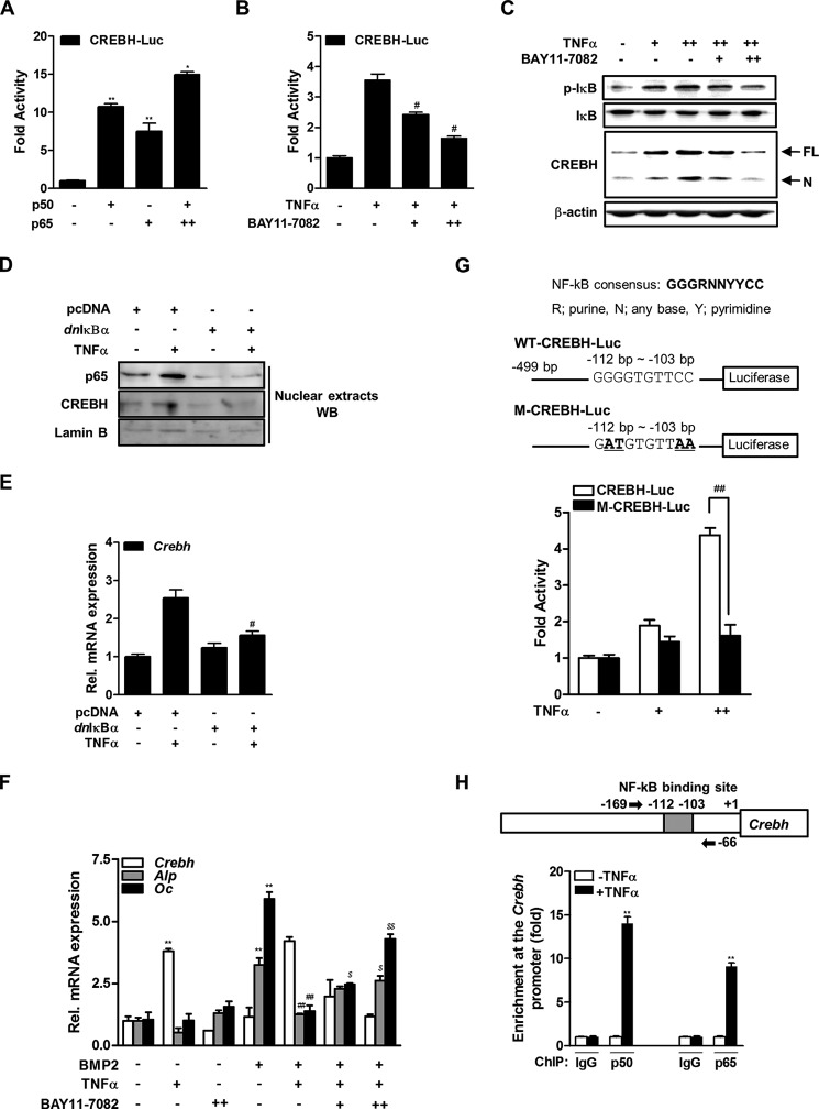
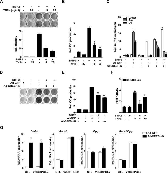
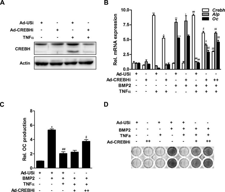
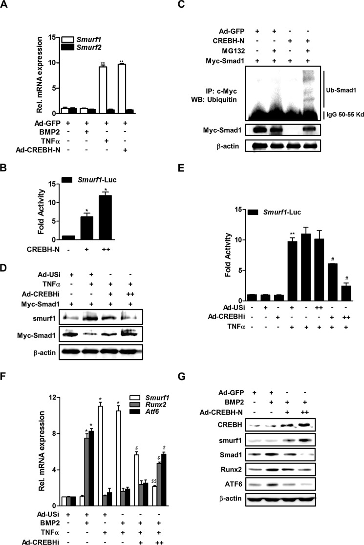
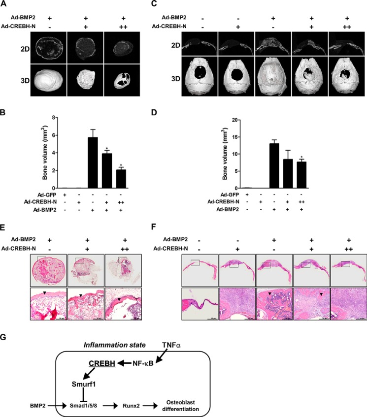
Endoplasmic reticulum (ER) stress transducers, such as old astrocyte specifically induced substance (OASIS) and activating transcription factor 6 (ATF6), which are induced by bone morphogenetic protein 2 (BMP2), regulate bone formation and osteoblast differentiation. Here, we examined the role of cAMP response element-binding protein H (CREBH), a member of the same family of ER membrane-bound basic leucine zipper (bZIP) transcription factors as OASIS and ATF6, in osteoblast differentiation and bone formation. Proinflammatory cytokine TNFα increased CREBH expression by up-regulating the nuclear factor-κB (NF-κB) signaling pathway in osteoblasts, increased the level of N-terminal fragment of CREBH in the nucleus, and inhibited BMP2 induction of osteoblast specific gene expression. Overexpression of CREBH suppressed BMP2-induced up-regulation of the osteogenic markers runt-related transcription factor 2 (Runx2), alkaline phosphatase (ALP), and osteocalcin (OC) in MC3T3-E1 cells and primary osteoblasts, as well as BMP2-induced ALP activity and OC protein production. In contrast, knockdown of CREBH attenuated the inhibitory effect of TNFα on BMP2-induced osteoblast differentiation. Mechanistic studies revealed that CREBH increased the expression of Smad ubiquitination regulatory factor 1 (Smurf1), leading to ubiquitin-dependent degradation of Smad1, whereas knockdown of CREBH inhibited TNFα-mediated degradation of Smad1 by Smurf1. Consistent with these in vitro findings, administration of Ad-CREBH inhibited BMP2-induced ectopic and orthotopic bone formation in vivo. Taken together, these results suggest that CREBH is a novel negative regulator of osteoblast differentiation and bone formation.

Keywords: SMAD transcription factor; cAMP response element-binding protein (CREB); endoplasmic reticulum stress (ER stress); osteoblast; tumor necrosis factor (TNF).


Figures

FIGURE 1.
FIGURE 1.
TNFα induces CREBH expression and cleavage of the N-terminal fragment in osteoblasts. A and B, real-time qRT-PCR measurements of the expression levels of the Crebh, Oasis, and Atf6 mRNAs. MC3T3-E1 cells were treated with or without TNFα (+, 5 ng/ml; ++, 20 ng/ml) for 6 h (A) or BMP2 (+, 200 ng/ml; ++, 500 ng/ml) for 24 h (B). *, p < 0.05; **, p < 0.01; and ***, p < 0.001 versus the untreated group. C and D, Western blot analyses of cellular (C) and nuclear (D) levels of CREBH. MC3T3-E1 cells were treated with or without TNFα (+, 5 ng/ml; ++, 20 ng/ml) and with or without tunicamycin (Tm; 1 μg/ml) as a positive control for 12 h. FL, full-length; N, nuclear; M, molecular size marker. E, the effects of TNFα and tunicamycin on the luciferase activity of Gal4-fused full-length CREBH. MC3T3-E1 cells were co-transfected with Gal4-TK-Luc (200 ng) and Gal4-FL-CREBH (100 ng) or Gal4-DBD (100 ng) as a negative control. At 12 h post-transfection, cells were exposed to TNFα for 24 h. The results are expressed as the luciferase activity relative to that of the control. *, p < 0.05 and **, p < 0.01 versus the Gal4-FL-CREBH transfected group.
FIGURE 2.
FIGURE 2.
NF-κB signaling pathway is involved in TNFα induction of CREBH expression. A, the effects of p50 and p65 overexpression on CREBH-Luc activity in MC3T3-E1 cells. Luciferase activity was measured 48 h after transfection. β-Galactosidase plasmid was used as an internal control. The results are expressed as the luciferase activity relative to that of the control. *, p < 0.05 and **, p < 0.01 versus the untransfected control. B, the effects of TNFα and BAY11-7082 on CREBH-Luc activity. At 24 h post-transfection of CREBH-Luc, MC3T3-E1 cells were treated with TNFα (20 ng/ml) and/or BAY11-7082 (+, 0.1 μm; ++, 0.5 μm) for 6 h, and then luciferase activity was measured as in A. #, p < 0.05 versus the TNFα-treated group. C, the cells were treated with TNFα (20 ng/ml) and BAY11-7082 (0.1 or 0.5 μm) for 6 h. After total protein was extracted, and Western blot analysis was performed with the indicated antibodies. p-IκB, phosphorylated IκB; FL, full-length; N, nuclear CREBH. D and E, effects of dnIκBα overexpression on TNFα-induced CREBH expression. The cells were transiently transfected with pcDNA or dnIκBα expression construct and incubated in the presence of TNFα (20 ng/ml) for 24 h. D, nuclear fractions were prepared and Western blotting was performed with the designated antibodies. E, expression of CREBH mRNA was determined by qRT-PCR analysis. The expressions were normalized to those of β-actin. #, p < 0.05 versus the TNFα-treated group. F, effects of BMP2, TNFα, and BAY11-7082 on Crebh, Alp, and Oc mRNA levels. The cells were cultured with BMP2 (200 ng/ml) for 2 days and then treated with TNFα and/or BAY11-7082 for 6 h. The expression levels were normalized to those of β-actin. **, p < 0.01 versus the untreated control; ##, p < 0.01 versus the BMP2-treated group; $, p < 0.05 and $$, p < 0.01 versus the BMP2 with TNFα-treated group. G, effects of TNFα on activities of WT-CREBH-Luc or M-CREBH-Luc plasmid. The upper panel shows the consensus NF-κB binding site identified in the Crebh promoter. The lower panel shows the effects of TNFα on the luciferase activities of WT-CREBH-Luc and M-CREBH-Luc, the latter of which contained two point mutations of the NF-κB binding site (underlined in the upper panel). The cells were transfected with the plasmids for 24 h, and then treated with TNFα (20 ng/ml) for 6 h prior to measuring luciferase activity. H, ChIP-qPCR analysis for p50 or p65 binding to Crebh gene. The upper panel shows a schematic representation of the Crebh promoter region depicting the NF-κB binding motif and the qPCR primer binding sites (arrow). The lower panel shows relative binding of p50 or p65 to Crebh promoter gene. The cells were cultured with TNFα (20 ng/ml) for 12 h and then immunoprecipitated with anti-p50 or anti-p65 antibody or IgG as a negative control. Precipitated DNA was subjected to qPCR analysis with the primer pairs described in the upper panel. Fold-differences were calculated by the ΔCT method. All data represent the mean ± S.E. from three independent experiments. **, p < 0.01 versus the TNFα-untreated control.
FIGURE 3.
FIGURE 3.
TNFα inhibits BMP2-induced osteoblast differentiation by stimulating CREBH expression. A and B, the effects of TNFα on BMP2-induced ALP activity in primary calvarial osteoblasts (A) and OC protein production in osteoblasts (B). Cells were cultured with BMP2 (200 ng/ml) for 3 days and then treated with TNFα for 24 h. Cells were stained with a BCIP®/nitro blue tetrazolium solution to determine ALP activity (A), and the level of OC protein in the culture medium was measured using an OC-specific ELISA kit (B). C–E, the effects of BMP2 and/or infection with an Ad-CREBH-N on the expression levels of the Crebh, Alp, and Oc mRNAs (C), ALP activity (D), and OC production (E) in MC3T3-E1 cells (C) or primary osteoblasts (D and E). Cells were infected with Ad-CREBH-N (+, 50 m.o.i.; ++, 100 m.o.i.) for 24 h and then treated with BMP2 (200 ng/ml) for 3 days. The expression levels of the target mRNAs were measured by qRT-PCR and normalized to those of β-actin. *, p < 0.05; **, p < 0.01; #, p < 0.05; and ##, p < 0.01 versus the BMP2-treated group. F, the effects of BMP2 and TNFα on CREBH-Luc activity. Twelve hours after transfection, cells were treated with BMP2 (200 ng/ml) for 48 h and/or TNFα (+, 5 ng/ml; ++, 20 ng/ml) for 6 h, and then luciferase activity was measured. **, p < 0.01 versus the untreated control; #, p < 0.01 versus the BMP2-treated group. G, the effects of a 3-day exposure to vitamin D3 (VitD3, 10 nm) and prostaglandin E2 (PGE2, 1 μm) on the expression levels of the Crebh, Rankl, and Opg mRNAs in primary osteoblasts infected with Ad-CREBH-N. The expression levels of the mRNAs were evaluated by qRT-PCR and normalized to those of β-actin. CTL, control.
FIGURE 4.
FIGURE 4.
Inhibition of CREBH attenuates TNFα-mediated suppression of BMP2-induced osteoblast differentiation. A, Western blot analysis for efficiency of Ad-CREBHi. MC3T3-E1 cells were infected with adenoviruses encoding an unspecific siRNA (Ad-USi) as a control virus or CREBH-specific shRNA (Ad-CREBHi) for 24 h, and then treated with TNFα (20 ng/ml) for 12 h. Western blotting was performed with CREBH and β-actin antibodies. B, the effect of knockdown of CREBH using adenoviruses encoding a CREBH-specific shRNA (Ad-CREBHi) on the expression levels of the Crebh, Alp, and Oc mRNAs in BMP2- and/or TNFα-treated MC3T3-E1 cells. The cells were infected with Ad-CREBHi (+, 50 m.o.i.; ++, 100 m.o.i.) or Ad-USi (100 m.o.i.). Ad-USi for unspecific shRNA was used as a control. Twenty-four hours after infection, the cells were treated with BMP2 (200 ng/ml) for 3 days and then treated with TNFα (20 ng/ml). qRT-PCR analyses were used to examine the expression levels of the Alp, Oc, and Crebh mRNAs. The mRNA expression levels were normalized to those of β-actin. *, p < 0.05; **, p < 0.01; ***, p < 0.001 versus the untreated control; ##, p < 0.01 versus the BMP2-treated group; and $, p < 0.05 and $$, p < 0.01 versus the BMP2 with TNFα-treated group. C and D, the effect of Ad-CREBHi on TNFα-mediated suppression of osteoblast differentiation. MC3T3-E1 cells (C) or primary osteoblasts (D) were cultured as described in B. The level of OC protein (C) and ALP activity (D) were measured as described in the legend to Fig. 3. *, p < 0.05 versus the BMP2-untreated control; ##, p < 0.01 versus the BMP2-treated group; and $, p < 0.05 versus the BMP2 with TNFα treated group.
FIGURE 5.
FIGURE 5.
CREBH promotes Smad1 degradation by inducing Smurf1 expression. A, the effects of TNFα and Ad-CREBH-N on Smurf1 and Smurf2 mRNA levels. MC3T3-E1 cells were infected with Ad-CREBH-N or Ad-GFP as a control virus (100 m.o.i.). Twenty-four hours after infection, the cells were treated with BMP2 (200 ng/ml) for 3 days and then with TNFα (20 ng/ml) for 6 h. The mRNA levels were determined by qRT-PCR and normalized to those of β-actin. **, p < 0.01 versus the untreated control. B, the effect of CREBH overexpression on Smurf1 promoter activity. Cells were transfected with the Smurf1-Luc and CREBH-N expression plasmids (+, 50 ng; ++, 200 ng) and luciferase activity was measured 24 h later. *, p < 0.05 versus the untransfected control. C, the effects of CREBH-N overexpression and proteasome inhibition on Smad1 ubiquitination. Cells were transfected with a Myc-Smad1 expression plasmid with or without a CREBH-N expression plasmid for 24 h, and then exposed to 10 μm MG132 (lanes 2 and 4) for 12 h. Immunoprecipitation (IP) and Western blotting (WB) analyses were performed with an anti-Myc or anti-ubiquitin antibody, respectively. D, the effects of inhibition of CREBH on TNFα-mediated changes in Smad1 and Smurf1 protein levels. Cells were transfected with a Myc-Smad1 with or without an Ad-USi or a CREBH-specific shRNA (+, 50 m.o.i.; ++, 100 m.o.i.). Twenty-four hours after transfection, the cells were treated with or without TNFα (20 ng/ml) for 6 h. Western blotting was performed with the designated antibodies. E, the effects of inhibition of CREBH on TNFα-mediated activation of the Smurf1 promoter. Cells were transfected with Smurf1-Luc and Ad-CREBHi or Ad-USi, and then exposed to TNFα (20 ng/ml) for 12 h. Luciferase activity was measured 24 h later. **, p < 0.01 versus the untreated control and #, p < 0.05 versus the TNFα treated group. F, the effects of inhibition of CREBH on TNFα-mediated changes in Smurf1, Runx2, and Atf6 mRNA levels. Cells were transfected with Ad-USi or Ad-CREBHi for 12 h and then exposed to TNFα (20 ng/ml) for 6 h prior to treatment with BMP2 (200 ng/ml) for 2 days. Total viral titer was held constant at 100 m.o.i. by addition of the appropriate amount of Ad-USi, a control Ad-shRNA. The levels of the mRNAs were determined by qRT-PCR and normalized to those of β-actin. *, p < 0.05 versus the untreated control, and $, p < 0.05 and $$, p < 0.01 versus the BMP2/TNFα-treated group. G, Western blot analyses of the effects of overexpression of CREBH on BMP2-mediated changes in Smad1, Runx2, ATF6, and CREBH protein levels. Cells were treated with BMP2 (200 ng/ml) for 2 days and infected with Ad-CREBH-N (+, 50 m.o.i.; ++, 100 m.o.i.) or Ad-GFP (50 m.o.i.) for an additional 24 h.
FIGURE 6.
FIGURE 6.
CREBH suppresses BMP2-induced ectopic and orthotopic bone formation. Ad-GFP (5 × 10¹⁰ particle number, PN), Ad-BMP2 (5 × 10¹⁰ PN), and/or Ad-CREBH-N (5 × 10¹⁰ or 10 × 1010 PN) with absorbable collagen sponges were subcutaneously implanted into the backs or critical sized cranial defects of mice. The total amount of implanted virus was adjusted by adding Ad-GFP virus. Three weeks after implantation, the implants were harvested and ectopic or orthotopic bone formation was evaluated. A and B, two- and three-dimensional μCT reconstructions (A) and volume (B) of subcutaneously formed ectopic bones. *, p < 0.05 versus the Ad–MP2treated group. C and D, μCT reconstructions (C) and volume (D) of newly formed orthotopic bones in critical-sized calvarial defects. *, p < 0.05 versus the Ad-BMP2-treated group. E and F, histology of ectopic (E) or orthotopic (F) regenerated bones. Lower panels are magnified images of the squared areas in upper panel, respectively. Black arrowheads indicate newly formed mineralized tissues. Representative data are shown (n = 4). G, an overview of the role of CREBH in BMP2-induced osteoblast differentiation. Under normal conditions, BMP2 stimulates osteoblast differentiation via Smad1/5/8-dependent intracellular signaling. In the presence of severe inflammation, TNFα induces the expression of CREBH and Smurf1 by NF-κB pathway, leading to the suppression of BMP2-induced osteoblast differentiation through Smurf1-dependent degradation of Smad1.

Similar articles

Cited by

References

    1. Urist M. R. (1965) Bone: formation by autoinduction. Science 150, 893–899 - PubMed
    1. Wozney J. M., Rosen V., Celeste A. J., Mitsock L. M., Whitters M. J., Kriz R. W., Hewick R. M., Wang E. A. (1988) Novel regulators of bone formation: molecular clones and activities. Science 242, 1528–1534 - PubMed
    1. Ron D., Walter P. (2007) Signal integration in the endoplasmic reticulum unfolded protein response. Nat. Rev. Mol. Cell Biol. 8, 519–529 - PubMed
    1. Malhotra J. D., Kaufman R. J. (2007) The endoplasmic reticulum and the unfolded protein response. Semin. Cell Dev. Biol. 18, 716–731 - PMC - PubMed
    1. Harding H. P., Zhang Y., Ron D. (1999) Protein translation and folding are coupled by an endoplasmic-reticulum-resident kinase. Nature 397, 271–274 - PubMed

Publication types

MeSH terms

LinkOut - more resources