. 2015 May 12;54(18):2919-30.
doi: 10.1021/acs.biochem.5b00199. Epub 2015 Apr 28.

Function discovery and structural characterization of a methylphosphonate esterase

Affiliations

Function discovery and structural characterization of a methylphosphonate esterase

Dao Feng Xiang et al. Biochemistry. .

Abstract

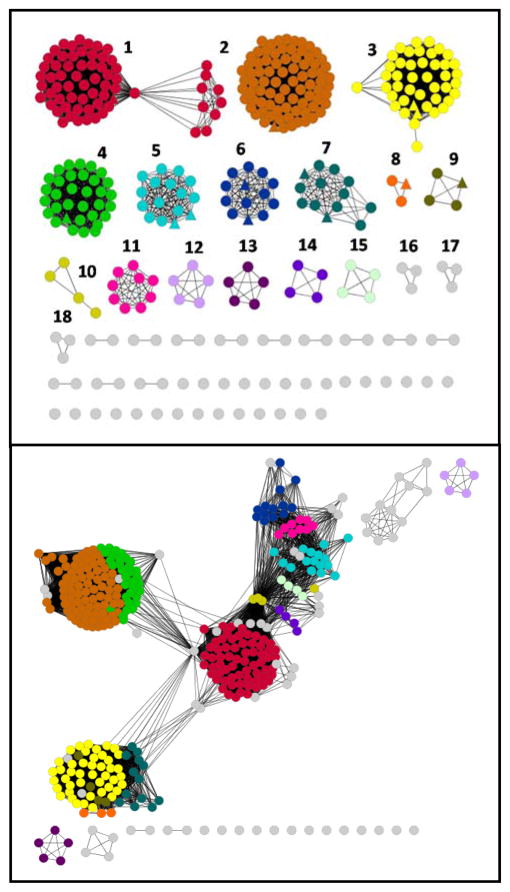
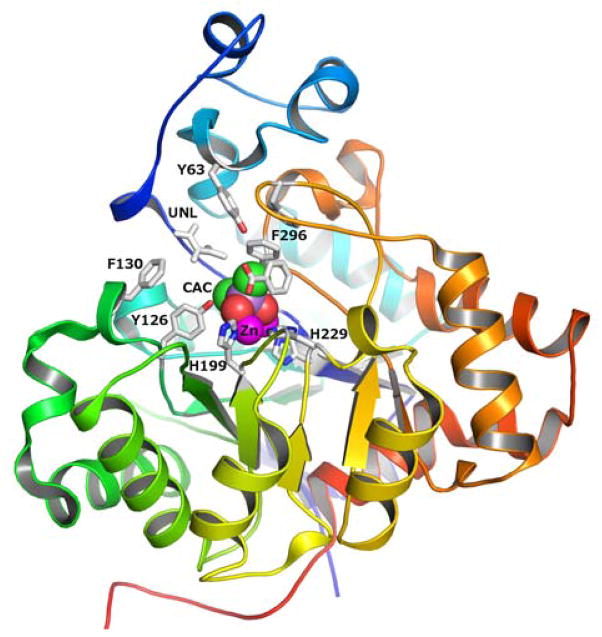
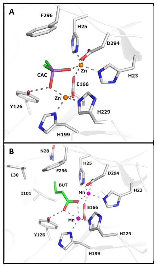
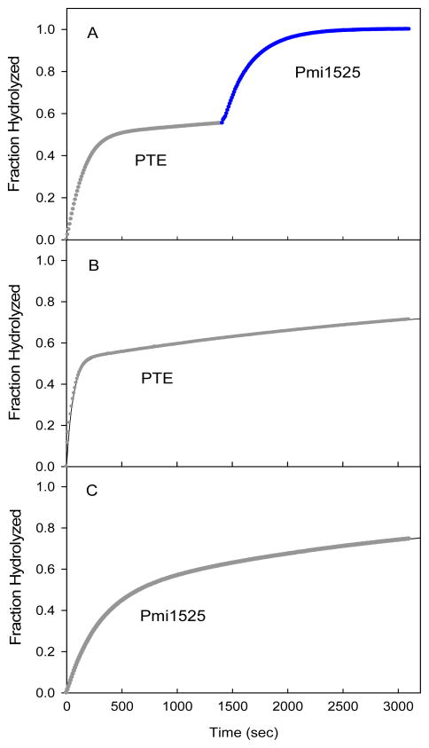
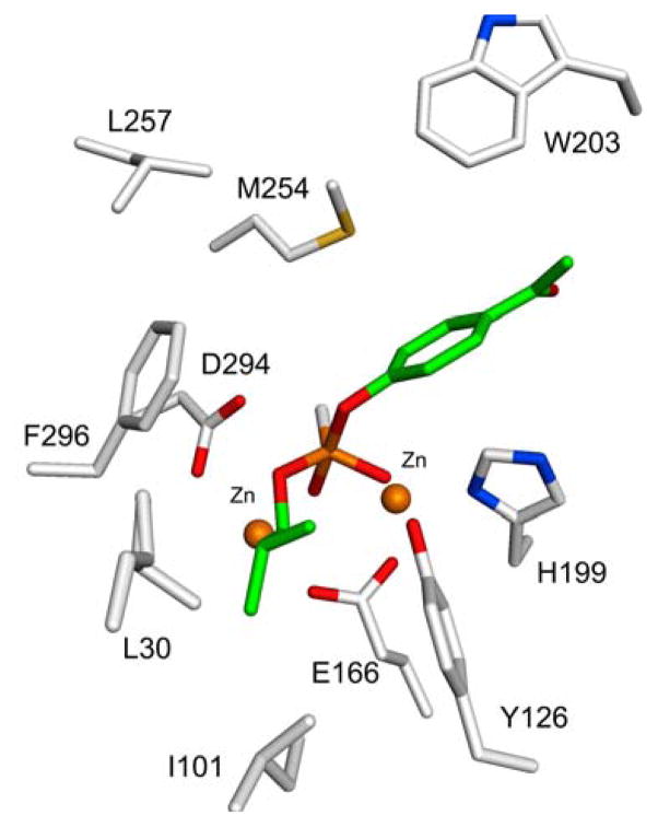
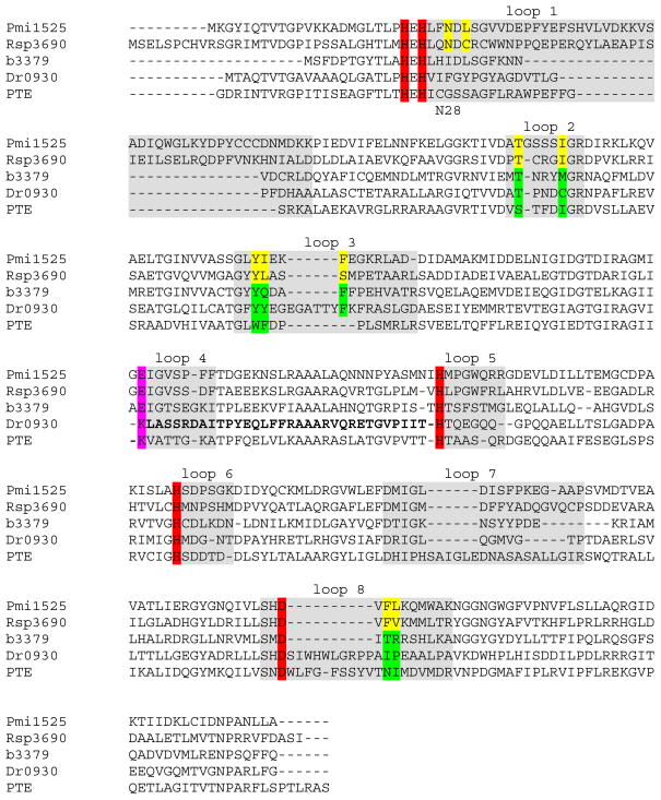
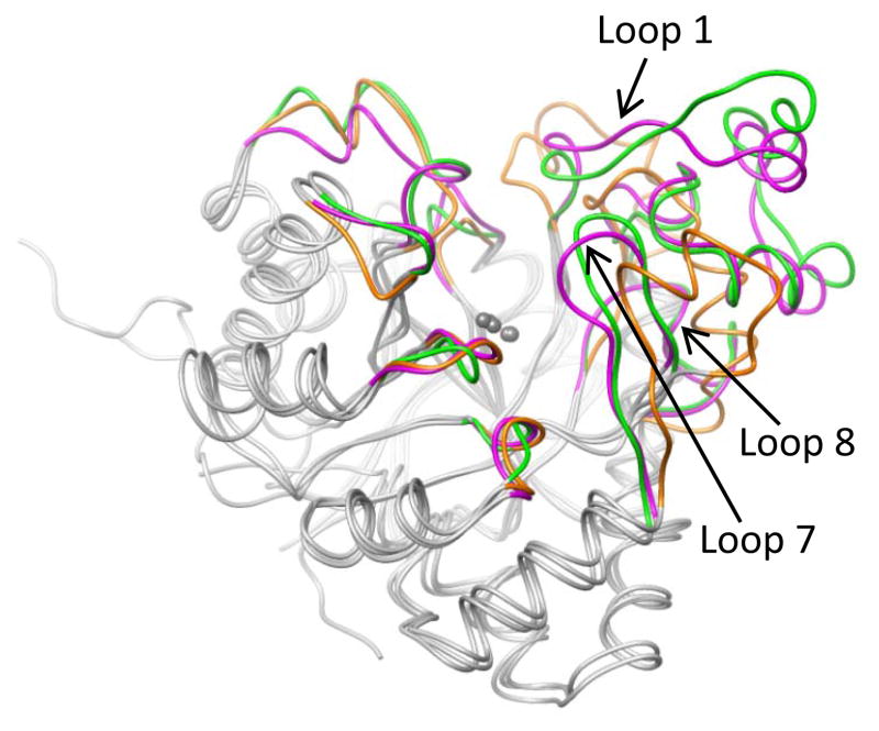
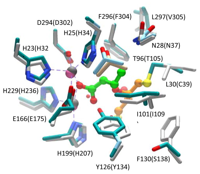
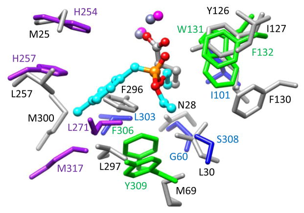
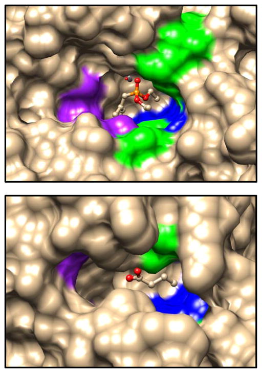
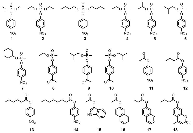
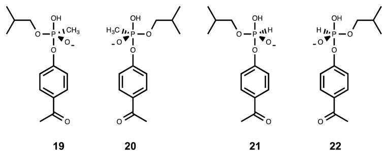
Pmi1525, an enzyme of unknown function from Proteus mirabilis HI4320 and the amidohydrolase superfamily, was cloned, purified to homogeneity, and functionally characterized. The three-dimensional structure of Pmi1525 was determined with zinc and cacodylate bound in the active site (PDB id: 3RHG ). The structure was also determined with manganese and butyrate in the active site (PDB id: 4QSF ). Pmi1525 folds as a distorted (β/α)8-barrel that is typical for members of the amidohydrolase superfamily and cog1735. The substrate profile for Pmi1525 was determined via a strategy that marshaled the utilization of bioinformatics, structural characterization, and focused library screening. The protein was found to efficiently catalyze the hydrolysis of organophosphonate and carboxylate esters. The best substrates identified for Pmi1525 are ethyl 4-nitrophenylmethyl phosphonate (kcat and kcat/Km values of 580 s(-1) and 1.2 × 10(5) M(-1) s(-1), respectively) and 4-nitrophenyl butyrate (kcat and kcat/Km values of 140 s(-1) and 1.4 × 10(5) M(-1) s(-1), respectively). Pmi1525 is stereoselective for the hydrolysis of chiral methylphosphonate esters. The enzyme hydrolyzes the (SP)-enantiomer of isobutyl 4-nitrophenyl methylphosphonate 14 times faster than the corresponding (RP)-enantiomer. The catalytic properties of this enzyme make it an attractive template for the evolution of novel enzymes for the detection, destruction, and detoxification of organophosphonate nerve agents.


Figures

Figure 1
Figure 1
Sequence similarity network for proteins of cog1735 from the amidohydrolase superfamily at BLAST E-value cutoffs of 10−80 (top) and 10−40 (bottom) created using Cytoscape (http://www.cytoscape.org). For these proteins an E-value of 10−80 corresponds to a sequence identity of approximately 42%, whereas an E-value of 10−40 corresponds to a sequence identity of approximately 30%. All protein sequences available through NCBI that are designated as belonging to cog1735 were used to create the network. Each node in the network represents a single sequence, and each edge (depicted as lines) represents the pairwise connection between two sequences at the given BLAST E-value (32). Lengths of edges are not significant except that sequences in tightly clustered groups are relatively more similar to each other than sequences with few connections. The triangular nodes represent proteins that have been functionally and/or structurally characterized. Additional information is available in the text.
Figure 2
Figure 2
Cartoon representation of the Pmi1525 (PDB id: 3RHG) with bound zinc (colored in magenta) and cacodylate ion (CAC, carbon atoms are green, oxygen is red). Zinc and cacodylate are shown as vdw spheres. The protein subunit is rainbow colored with the N-terminus as dark blue and the C-terminus as red. The active site residues, an unknown ligand (UNL) and benzoic acid are drawn as stick models with carbon atoms gray colored, oxygen is red and nitrogen is blue.
Figure 3
Figure 3
Metal and ligand coordination in the active site of Pmi1525. All residues are drawn as stick models, metals are shown as non-bonded spheres. (A) The structure of the zinc bound enzyme with cacodylate (CAC) (PDB id: 3RHG). (B) The structure the manganese bound enzyme with bound butyrate (BUT) (PDB id: 4QSF). Carbon atoms are gray colored, oxygen is red, nitrogen is blue. The coordination and selected hydrogen bonds are presented as dashed gray lines.
Figure 4
Figure 4
Time courses for the hydrolysis of 90 μM racemic substrate 6 using wild-type PTE and Pmi1525. (A) The reaction was initiated with the addition of 0.5 nM wild-type PTE. After 23 minutes, 50 nM of Pmi1525 was added to hydrolyze the remaining material. (B) Hydrolysis of 90 μM racemic 6 using 1.0 nM wild-type PTE. (C) Hydrolysis of 90 μM racemic 6 using 50 nM Pmi1525.
Figure 5
Figure 5
Active site of Pmi1525 computationally docked with compound 22. The axial OH group attached to the pentacoordinated phosphorus occupies the position of the nucleophilic hydroxide that is bridged by the two metal ions. The isobutyl side chain makes hydrophobic contacts with the residues (Leu-30, Ile-101, and Tyr-126) in the larger pocket of Pmi1525
Figure 6
Figure 6
Amino acid sequence alignment of Pmi1525 and four other proteins from cog1735, including the non-specific carboxylesterase Rsp3690 from subgroup 4, phosphotriesterase homology protein (PHP, b3379) from subgroup 1, γ,δ-lactonase Dr0930 from subgroup 7, and phosphotriesterase PTE from subgroup 9. The four histidine and the aspartate residues coordinated to the two metal ions are highlighted in red. The residue bridging the two metal ions is colored in pink. The residues forming the hydrophobic pocket in the active site in Pmi1525 and Rsp3690 are colored yellow and the corresponding residues in the other four proteins are colored green.
Figure 7
Figure 7
Superposition of coordinates for structures Pmi1525 (PDB id: 3RHG), Rsp3690 (PDB id: 3k2g), and PTE (PDB id: 1dpm). The eight β/α loops from Pmi1525, Rsp23690, and PTE are depicted in magenta, green, and orange, respectively. The central β-barrel, the surrounding α-helices, and the two metal atoms are depicted in grey. The RMSD between grey colored parts of Pmi1525/Rsp3690, Pmi1525/PTE, and Rsp3690/PTE are 0.9, 1.0, and 1.1 Å, respectively.
Figure 8
Figure 8
Superimposition of active site residues coordinated to the two metal ions and residues around the butyrate ion in Pmi1525 and homologous residues near the DTT bound in the active site of Rsp3690. The residues of Pmi1525 are colored in light gray; the residues of Rsp3690 are colored in cyan and the residue labels are enclosed in brackets. The carbon chain of butyrate bound in the active site of Pmi1525 is shown in green and the carbon chain of DTT bound in Rsp3690 is shown in orange. Manganese atoms are colored pink and the zinc atoms are colored in dark grey. Oxygen is shown in red and sulfur in yellow.
Figure 9
Figure 9
Superimposition of the residues of the three binding pockets in the active site of PTE (PDB id: 1dpm) and the corresponding residues in Mn-containing structure Pmi1525. For PTE, the large pocket residues are colored in purple, the small pocket residues are colored in blue, and leaving group pocket residues are colored in green. The corresponding residues of Pmi1525 are colored in light grey. The labels of the residues of PTE are colored the same as the residues themselves. The residue labels of Pmi1525 are colored in black.
Figure 10
Figure 10
Substrate binding pocket comparison of wild-type PTE with the bound substrate analog, diethyl 4-methylbenzylphosphonate (top) and Pmi1525 with the bound product butyrate (bottom). The color schemes are the same as shown in Figure 9 (purple, blue and green represents the large, small, and leaving group residues, respectively.
Scheme 1
Scheme 1
Scheme 2
Scheme 2

Similar articles

Cited by

References

    1. Benson DA, Karsch-Mizrachi I, Lipman DJ, Sayers EW. GenBank. Nucleic Acids Res. 2009;37:D26–31. - PMC - PubMed
    1. Schnoes AM, Brown SD, Dodevski I, Babbitt PC. Annotation error in public database: Misannotation of molecular function in enzyme superfamilies. PLoS Comput Biol. 2009;5:e1000605. - PMC - PubMed
    1. Seffernick JL, de Souza ML, Sadowsky MJ, Wackett LP. Melamine deaminase and atrazine chlorohydrolase: 98 percent identical but functionally different. J Bacterial. 2001;183:2405–2410. - PMC - PubMed
    1. Odokonyero D, Ragumani S, Lopez MS, Bonanno JB, Ozerova ND, Woodard DR, Machala BW, Swaminathan S, Burley SK, Almo SC, Glasner ME. Divergent evolution of ligand binding in the o-succinylbenzoate synthase family. Biochemistry. 2013;52:7512–7521. - PMC - PubMed
    1. Hall RS, Fedorov AA, Marti-Arbona R, Fedorov EV, Kolb P, Sander JM, Burley SK, Shoichet BK, Almo SC, Raushel FM. The hunt for 8-oxoguanine deaminase. J Am Chem Soc. 2010;113:1762–1763. - PMC - PubMed

Publication types

Substances

LinkOut - more resources