

Extensive survey of STAT6 expression in a large series of mesenchymal tumors
- PMID: 25873501
- PMCID: PMC4505928
- DOI: 10.1309/AJCPN25NJTOUNPNF
Extensive survey of STAT6 expression in a large series of mesenchymal tumors
Abstract
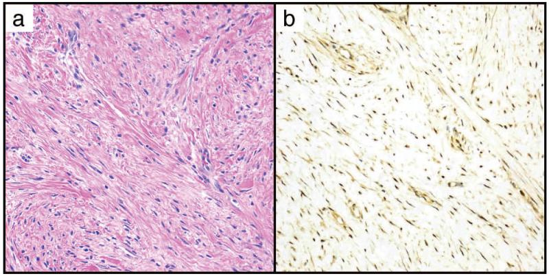
Objectives: Expression of strong nuclear STAT6 is thought to be a specific marker for solitary fibrous tumors (SFTs). Little is known about subtle expression patterns in other mesenchymal lesions.
Methods: We performed immunohistochemical studies against the C-terminus of STAT6 in tissue microarrays and whole sections, comprising 2366 mesenchymal lesions.
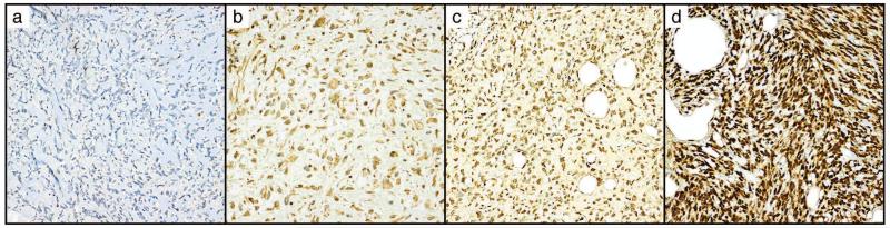
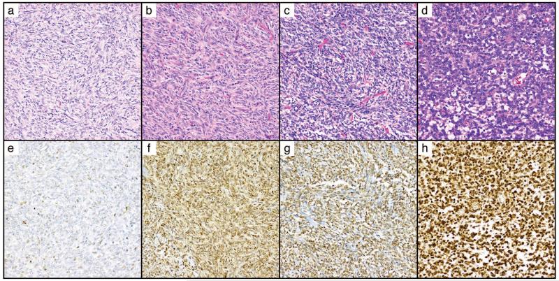
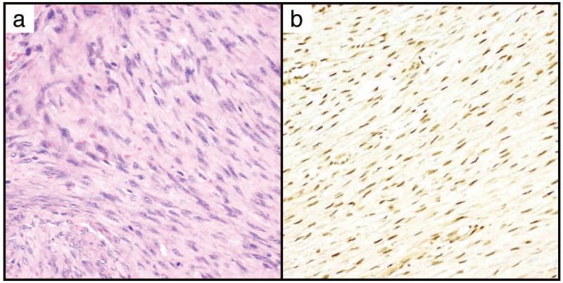
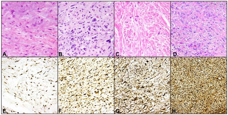
Results: Strong nuclear STAT6 was expressed in 285 of 2,021 tumors, including 206 of 240 SFTs, 49 of 408 well-differentiated/dedifferentiated liposarcomas, eight of 65 unclassified sarcomas, and 14 of 184 desmoid tumors, among others. Expression in SFTs was predominately limited to the nucleus. Other positive tumors typically expressed both nuclear and cytoplasmic STAT6. Complete absence of STAT6 was most common in pleomorphic liposarcoma and alveolar soft part sarcoma (60% and 72% cases negative, respectively).
Conclusions: Strong nuclear STAT6 is largely specific for SFTs. Physiologic low-level cytoplasmic/nuclear expression is common in mesenchymal neoplasia and is of uncertain significance.
Keywords: Immunohistochemistry; STAT6; Sarcoma; Solitary fibrous tumor.
Copyright© by the American Society for Clinical Pathology.
Figures
References
-
- Hou J, Schindler U, Henzel WJ, Ho TC, Brasseur M, McKnight SL. An interleukin-4-induced transcription factor: IL-4 Stat. Science. 1994;265:1701–1706. - PubMed
-
- Izuhara K, Heike T, Otsuka T, Yamaoka K, Mayumi M, Imamura T, Niho Y, Harada N. Signal transduction pathway of interleukin-4 and interleukin-13 in human B cells derived from X-linked severe combined immunodeficiency patients. J Biol Chem. 1996;271:619–622. - PubMed
Publication types
MeSH terms
Substances
Grants and funding
LinkOut - more resources
Full Text Sources
Medical
Research Materials
Miscellaneous
