

Comparison of high-risk histopathological features in eyes with primary or secondary enucleation for retinoblastoma
- PMID: 25873648
- PMCID: PMC5148135
- DOI: 10.1136/bjophthalmol-2014-306364
Comparison of high-risk histopathological features in eyes with primary or secondary enucleation for retinoblastoma
Abstract
Aims: To compare high-risk histopathology of eyes with primary versus secondary enucleation from patients with retinoblastoma.
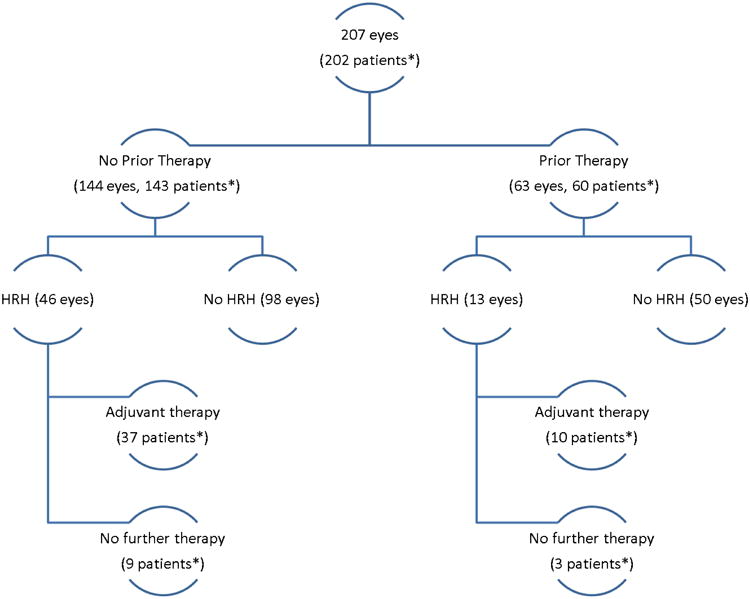
Patients and methods: A retrospective histopathology review identified 207 eyes enucleated from 202 patients between March 1997 and August 2013. Our review considered high-risk histopathological features to include extraocular disease or invasion of the anterior chamber, iris, ciliary body, choroid (massive), postlaminar optic nerve or sclera.
Results: Most eyes (144, 70%) were primarily enucleated; 63 (30%) were secondarily enucleated after neoadjuvant therapy. The primary enucleation group had more advanced disease (Reese-Ellsworth group V: 95% vs 59%; International Classification Group D/E: 97% vs 59%; p<0.001). The incidence of high-risk histopathology features was similar between groups (32% vs 21%, n=59; p=0.132). The type of prior therapy was not associated with high-risk histopathology features. Time to enucleation was longer for secondarily enucleated eyes with high-risk features. Choroid and postlaminar optic nerve invasion were more frequent in eyes primarily enucleated (p<0.001). Forty-six of the 59 (78%) patients with high-risk features received adjuvant chemotherapy and/or external beam radiation therapy. Three patients who received primary enucleation and adjuvant therapy died of metastatic recurrence.
Conclusions: Despite the more favourable classification of eyes treated with neoadjuvant therapy, the risk of high-risk histopathology features at enucleation was comparable with eyes undergoing primary enucleation. Delayed enucleation was associated with these features, and the majority of patients required further adjuvant therapy. Caution must be exercised in treating recalcitrant intraocular retinoblastoma to promptly pursue definitive enucleation in an effort to minimise further treatment exposures and metastases.
Keywords: Eye (Globe); Pathology; Retina.
Published by the BMJ Publishing Group Limited. For permission to use (where not already granted under a licence) please go to http://group.bmj.com/group/rights-licensing/permissions.
Conflict of interest statement
No conflicting relationship exists for any author.
Figures
Similar articles
-
High-Risk Histopathology Features in Primary and Secondary Enucleated International Intraocular Retinoblastoma Classification Group D Eyes.Ophthalmology. 2017 Jun;124(6):851-858. doi: 10.1016/j.ophtha.2017.01.048. Epub 2017 Mar 13. Ophthalmology. 2017. PMID: 28302322
-
Optic Nerve Invasion in Retinoblastoma: Impact of Eye Salvage and Adjuvant Chemotherapy.Invest Ophthalmol Vis Sci. 2025 Jun 2;66(6):65. doi: 10.1167/iovs.66.6.65. Invest Ophthalmol Vis Sci. 2025. PMID: 40548635 Free PMC article.
-
A histopathologic analysis of 50 eyes primarily enucleated for retinoblastoma in a tertiary cancer center in Jordan.Turk Patoloji Derg. 2014;30(3):171-7. doi: 10.5146/tjpath.2014.01260. Turk Patoloji Derg. 2014. PMID: 24913302
-
Current treatment of retinoblastoma.Curr Opin Ophthalmol. 2002 Oct;13(5):331-6. doi: 10.1097/00055735-200210000-00007. Curr Opin Ophthalmol. 2002. PMID: 12218465 Review.
-
Management of retinoblastoma in children: current status.Paediatr Drugs. 2015 Jun;17(3):185-98. doi: 10.1007/s40272-015-0121-9. Paediatr Drugs. 2015. PMID: 25742925 Review.
Cited by
-
Current Indications of Secondary Enucleation in Retinoblastoma Management: A Position Paper on Behalf of the European Retinoblastoma Group (EURbG).Cancers (Basel). 2021 Jul 6;13(14):3392. doi: 10.3390/cancers13143392. Cancers (Basel). 2021. PMID: 34298608 Free PMC article.
-
Pathologic comparisons of enucleated eyes with retinoblastoma after superselective ophthalmic arterial chemotherapy with or without intravenous chemotherapy.Int J Ophthalmol. 2020 Nov 18;13(11):1794-1799. doi: 10.18240/ijo.2020.11.17. eCollection 2020. Int J Ophthalmol. 2020. PMID: 33215012 Free PMC article.
-
Tumor Environment of Retinoblastoma, Intraocular Cancer.Adv Exp Med Biol. 2020;1296:349-358. doi: 10.1007/978-3-030-59038-3_21. Adv Exp Med Biol. 2020. PMID: 34185303
-
Prior non-irradiative focal therapies do not compromise the efficacy of delayed episcleral plaque brachytherapy in retinoblastoma.Br J Ophthalmol. 2018 Jun 28:bjophthalmol-2018-311923. doi: 10.1136/bjophthalmol-2018-311923. Online ahead of print. Br J Ophthalmol. 2018. PMID: 29954786 Free PMC article.
-
Histopathological assessment of optic nerve invasion guided by radiological findings in enucleated globes with retinoblastoma.BMC Ophthalmol. 2020 Sep 29;20(1):386. doi: 10.1186/s12886-020-01654-z. BMC Ophthalmol. 2020. PMID: 32993566 Free PMC article.
References
-
- Kingston JE, Hungerford JL, Madreperla SA, et al. Results of combined chemotherapy and radiotherapy for advanced intraocular retinoblastoma. Arch Ophthalmol. 1996;114:1339–43. - PubMed
-
- Shields CL, Shields JA, Needle M, et al. Combined chemoreduction and adjuvant treatment for intraocular retinoblastoma. Ophthalmology. 1997;104:2101–11. - PubMed
-
- Abramson DH, Dunkel IJ, Brodie SE, et al. Superselective ophthalmic artery chemotherapy as primary treatment for retinoblastoma (chemosurgery) Ophthalmology. 2010;117:1623–9. - PubMed
-
- Shields CL, Honavar SG, Meadows AT, et al. Chemoreduction for unilateral retinoblastoma. Arch Ophthalmol. 2002;120:1653–8. - PubMed
-
- Chan HS, Gallie BL, Munier FL, et al. Chemotherapy for retinoblastoma. Ophthalmol Clin North Am. 2005;18:55–63. viii. - PubMed
Publication types
MeSH terms
Grants and funding
LinkOut - more resources
Full Text Sources
Other Literature Sources