Review
. 2015 Jul;361(1):95-114.
doi: 10.1007/s00441-014-2102-7. Epub 2015 Jan 22.

Relating structure and function of inner hair cell ribbon synapses

Affiliations
Review

Relating structure and function of inner hair cell ribbon synapses

C Wichmann et al. Cell Tissue Res. 2015 Jul.

Abstract

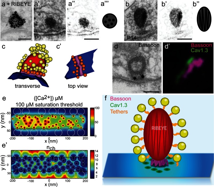
In the mammalian cochlea, sound is encoded at synapses between inner hair cells (IHCs) and type I spiral ganglion neurons (SGNs). Each SGN receives input from a single IHC ribbon-type active zone (AZ) and yet SGNs indefatigably spike up to hundreds of Hz to encode acoustic stimuli with submillisecond precision. Accumulating evidence indicates a highly specialized molecular composition and structure of the presynapse, adapted to suit these high functional demands. However, we are only beginning to understand key features such as stimulus-secretion coupling, exocytosis mechanisms, exo-endocytosis coupling, modes of endocytosis and vesicle reformation, as well as replenishment of the readily releasable pool. Relating structure and function has become an important avenue in addressing these points and has been applied to normal and genetically manipulated hair cell synapses. Here, we review some of the exciting new insights gained from recent studies of the molecular anatomy and physiology of IHC ribbon synapses.


Figures

Fig. 1
Fig. 1
Spatial distribution of IHC AZ proteins. a RIBEYE is the main component of the ribbon as shown by pre-embedding immunogold labeling of a P14 IHC synaptic ribbon using an anti-CtBP2 antibody (courtesy of Susann Michanski, InnerEarLab, University Medical Center, Göttingen, Germany); a’ Representative image of an electron micrograph of a round-shaped P9 immature ribbon exhibiting a dotted pattern possibly caused by RIBEYE arrangement (contrast enhanced image in a”), see also schematic representation (a”’). b A P14 mature ribbon with the typical multi-lamellar pattern (contrast enhanced image in b’), see also scheme in b”. Scale bars (a, a”, b’) 100 nm. c A serial 3D reconstruction of a mature ribbon with two distinct morphological vesicle pools (yellow: ribbon-associated vesicles; orange: membrane-proximal vesicles; red: ribbon; blue: AZ membrane; magenta: presynaptic density). c’ The membrane-proximal vesicles (orange) are arranged around the presynaptic density (magenta) that is containing the scaffolding protein bassoon as shown by the pre-embedding immunogold labeling in (d), Scale bar (d) 100 nm (courtesy of Susann Michanski, InnerEarLab, University Medical Center, Göttingen, Germany); d’ 2-color STED image of immunolabeled bassoon (magenta) and CaV1.3 channel clusters (green) in mature IHCs: stripe‐like morphology and closely aligned immunofluorescence of bassoon and CaV1.3 can be observed. Scale image:700 × 700 nm; e, e’ Mathematic model showing the total mean steady state [Ca2+] profile at the AZ membrane (e); e’ effective number of CaV1.3 channels contributing to total mean steady state [Ca2+] as shown in (e). (c, c’, d’, e, e’ modified from Wong et al. , EMBO J; reprinted with permission © 2014 Wong et al.). f Schematic summary of the protein arrangement at mature IHC ribbon synapses
Fig. 2
Fig. 2
Principle of functional heterogeneity in IHCs. a Schematic of an organ of Corti showing afferent and efferent innervations at IHCs. Modified from Meyer and Moser , Curr Opin Otolaryngol Head Neck Surg, reprinted with permission from © 2010 Wolters Kluwer Health. b, b’ Heterogeneous Ca2+ signaling in IHCs. b Mean and SD of ΔF (gray) as a function of depolarizing potential (V m), obtained from spot-detection experiments at the center of the Ca2+ microdomain; ΔF was averaged over the last 15 ms of a 20-ms stimulus. ΔF (mean gray) and I Ca (mean black) show a similar voltage dependence (thin lines corresponding SDs). b’ Heterogeneous voltage dependence and Ca2+ channel number of synaptic Ca2+ channel clusters in IHCs. Pronounced variability in the voltage dependence of activation, even within the same cell (dashed traces individual data curves from 3 Ca2+ microdomains in an IHC). Modified from Frank et al. (2009), PNAS USA, with permission from © Frank et al. c, c’ Colorized spatial distribution of vesicles and cisterns around the ribbon in low- and high-spontaneous rate (SR) fibers. Sections through a high-SR (c) and a low-SR (c’) synapse containing the synaptic ribbon are shown with cisternal (maroon) and vesicular (green) profiles. Scale bar (cm c’) 200 nm. d, d’ Distribution of docked vesicles and cisterns. d Mean density (± SE) of docked vesicles, i.e., within 20 nm of the presynaptic density along the presynaptic membrane. d’ Mean number (± SE) of cisterns within 20 nm of the presynaptic density versus distance along the presynaptic membrane is shown for all synapses. In addition, counts for low- and high-SR synapses are plotted separately. Rectangle the area of significant differences between low- and high-SR synapses. SE standard error; SR spontaneous rate. (c, c’, d, d’ modified from Kantardzhieva et al. , J Comp Neurol, reprinted with permission from © 2013 Wiley Periodicals)
Fig. 3
Fig. 3
Endocytosis in inner hair cells. a, a’ Representative recordings in response to 20 ms (a) or 200 ms (a’) depolarizations. After the C m increase upon 20 ms depolarization, the slope-corrected C m traces (middle) typically showed a linear decay (a). The 200-ms-long depolarization resulted in a combination of exponential and linear decay (a’). Modified from Neef et al. (2014) reprinted with permission from © 2014 Neef et al. bb”’ 3D reconstructions of resting (b), stimulated (b’) and recovered IHCs (b”, b”’). Endocytotic organelles are shown in purple. Note the presence of tubular organelles both before and after stimulation. Most organelles, including the tubular ones, are replaced by small vesicles during the recovery periods. Insets magnified regions from the four different cell regions (cuticular plate, top, nuclear and basal regions). Note the increased number of endosome-like organelles at the base of the cell after stimulation and during recovery. Modified from Kamin et al. (2014), reprinted with permission © 2014 Kamin et al. c mCLING-labeled organs of Corti were immunostained for Vglut3 and otoferlin (first row), for Vglut3 and syntaxin 6 (Sx 6, second row), for otoferlin and syntaxin 16 (Sx 16, third row) and finally for syntaxin 6 and syntaxin 16 (fourth row). The samples were cut into 20-nm sections and were imaged using an epifluorescence microscope. Dashed white lines the plasma membrane of the IHCs. White arrowheads organelles where the signals for mCLING and the two immunostained proteins colocalized. Scale bar 2 μm. d Graphic representation of Pearson’s correlation coefficients: otoferlin and syntaxin 6 (or syntaxin 16) correlate in the mCLING-labeled organelles at the top and nuclear levels. Vglut3 correlates best with otoferlin at the basal level. At least 100 organelles were analyzed for each condition. Error bars SEMs. e Model of membrane recycling in IHCs. Organelles with a different molecular composition recycle membrane in different regions, taking up mCLING. Apical endocytosis takes up the membrane into round organelles, a sizeable proportion of which is similar to late endosomes (light blue). Endocytosis in the top and nuclear regions reaches tubular organelles containing otoferlin and two endosome markers, syntaxin 16 and syntaxin 6. This suggests that these organelles participate in constitutive pathways, probably by maintaining membrane traffic between the plasma membrane and the trans-Golgi. At the base of the cell, stimulation induces the formation of membrane infoldings and cisterns that are characterized by the presence of Vglut3, Rab3 and also otoferlin. (ce modified from Revelo et al. , reprinted with permission from © 2014 Revelo et al.)

References

    1. Altrock WD, et al. Functional inactivation of a fraction of excitatory synapses in mice deficient for the active zone protein bassoon. Neuron. 2003;37:787–800. - PubMed
    1. Antonellis PJ, Pollock LM, Chou S-W, Hassan A, Geng R, Chen X, Fuchs E, Alagramam KN, Auer M, McDermott BM. ACF7 is a hair-bundle antecedent, positioned to integrate cuticular plate actin and somatic tubulin. J Neurosci. 2014;34:305–312. - PMC - PubMed
    1. Betz A, Ashery U, Rickmann M, Augustin I, Neher E, Südhof TC, Rettig J, Brose N. Munc13-1 is a presynaptic phorbol ester receptor that enhances neurotransmitter release. Neuron. 1998;21:123–136. - PubMed
    1. Betz A, Thakur P, Junge HJ, Ashery U, Rhee J-S, Scheuss V, Rosenmund C, Rettig J, Brose N. Functional interaction of the active zone proteins Munc13-1 and RIM1 in synaptic vesicle priming. Neuron. 2001;30:183–196. - PubMed
    1. Beurg M, Safieddine S, Roux I, Bouleau Y, Petit C, Dulon D. Calcium- and otoferlin-dependent exocytosis by immature outer hair cells. J Neurosci. 2008;28:1798–1803. - PMC - PubMed

Publication types

MeSH terms

LinkOut - more resources