

Fetal and umbilical Doppler ultrasound in normal pregnancy
- PMID: 25874722
- PMCID: PMC6464774
- DOI: 10.1002/14651858.CD001450.pub4
Fetal and umbilical Doppler ultrasound in normal pregnancy
Abstract
Background: One of the main aims of routine antenatal care is to identify the 'at risk' fetus in order to apply clinical interventions which could result in reduced perinatal morbidity and mortality. Doppler ultrasound study of umbilical artery waveforms helps to identify the compromised fetus in 'high-risk' pregnancies and, therefore, deserves assessment as a screening test in 'low-risk' pregnancies.
Objectives: To assess the effects on obstetric practice and pregnancy outcome of routine fetal and umbilical Doppler ultrasound in unselected and low-risk pregnancies.
Search methods: We searched the Cochrane Pregnancy and Childbirth Group Trials Register (28 February 2015) and reference lists of retrieved studies.
Selection criteria: Randomised and quasi-randomised controlled trials of Doppler ultrasound for the investigation of umbilical and fetal vessels waveforms in unselected pregnancies compared with no Doppler ultrasound. Studies where uterine vessels have been assessed together with fetal and umbilical vessels have been included.
Data collection and analysis: Two review authors independently assessed the studies for inclusion, assessed risk of bias and carried out data extraction. In addition to standard meta-analysis, the two primary outcomes and five of the secondary outcomes were assessed using GRADE software and methodology.
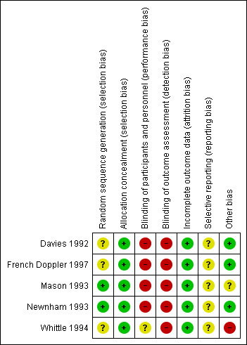
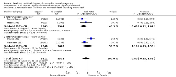
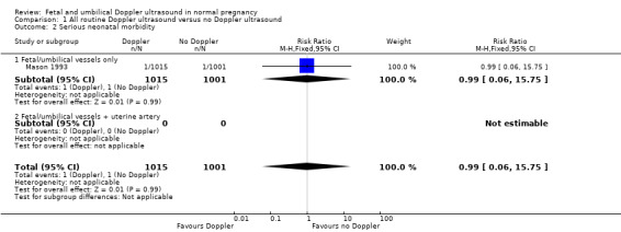
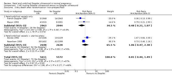
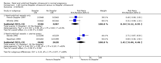
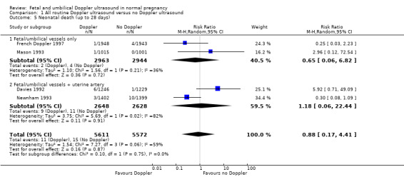
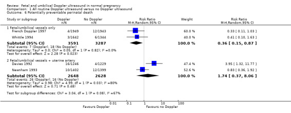
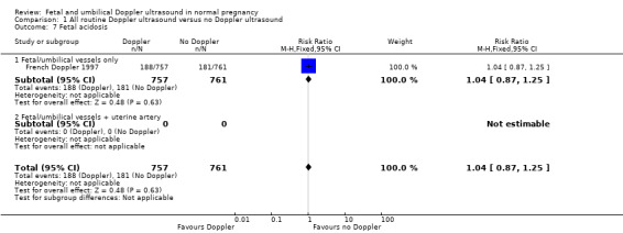
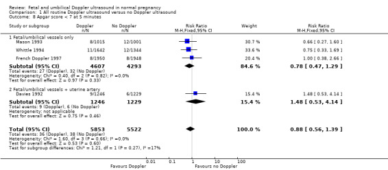
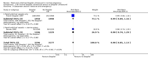
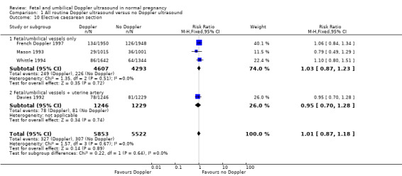
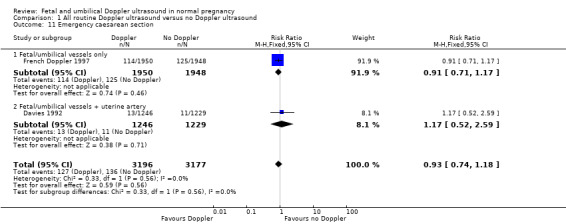
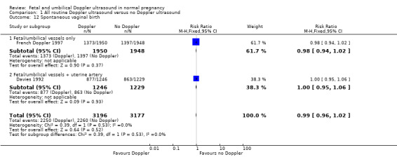
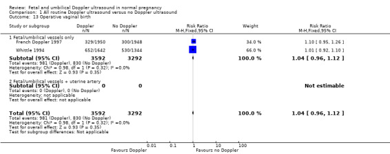
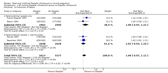
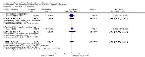
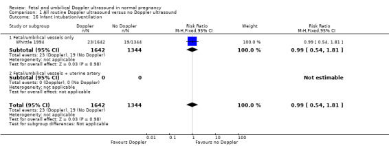
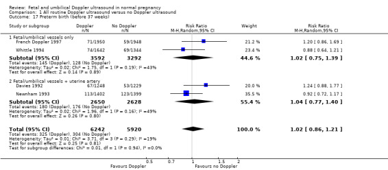
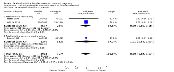
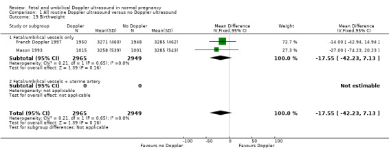
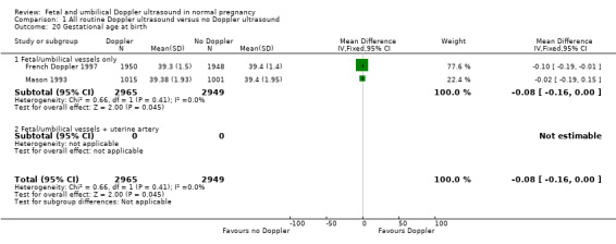
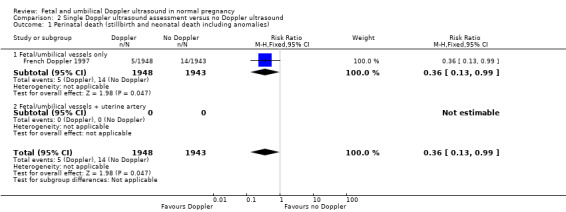
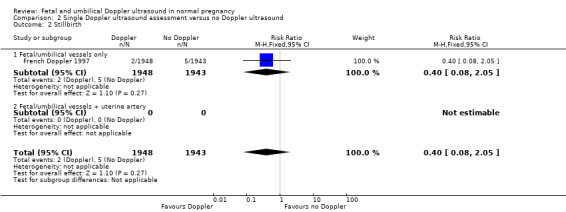
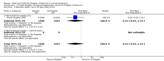
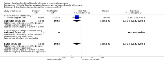
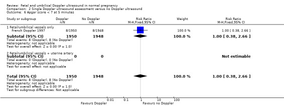
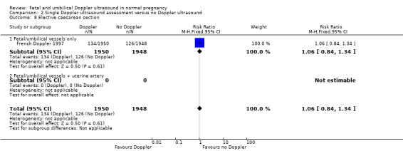
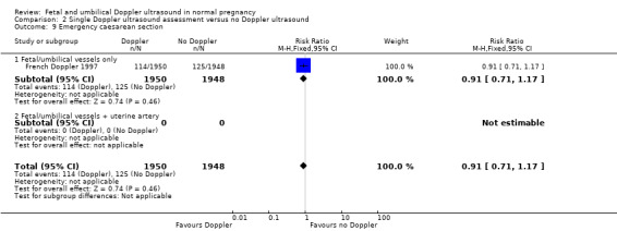
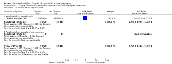
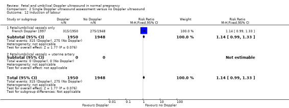
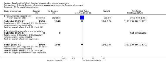
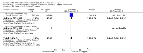
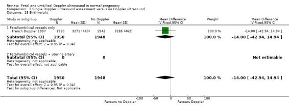
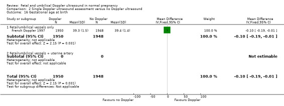
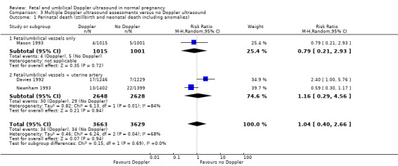
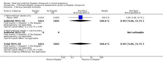
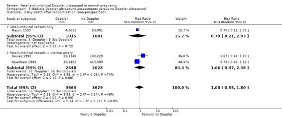
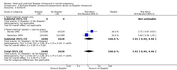
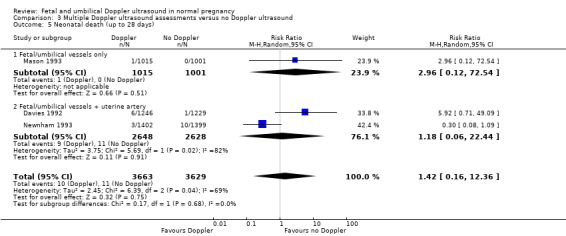
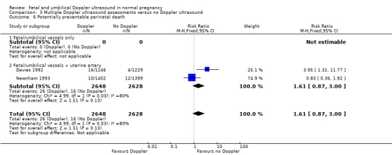
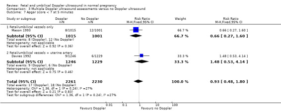
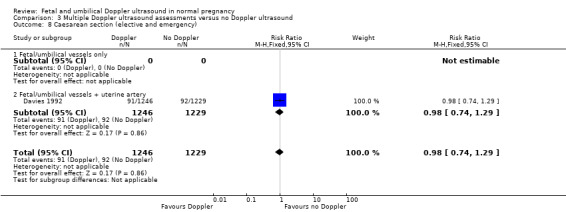
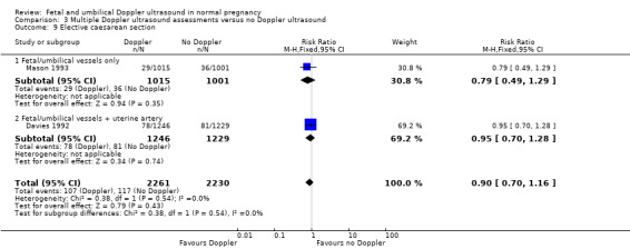
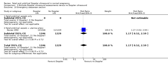
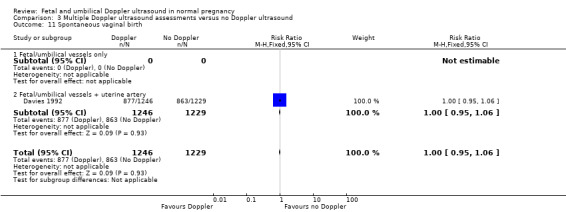
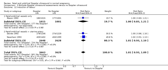
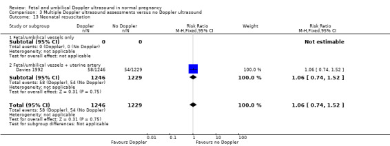
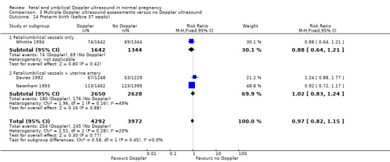
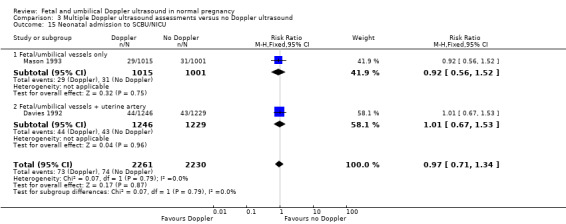
Main results: We included five trials that recruited 14,624 women, with data analysed for 14,185 women. All trials had adequate allocation concealment, but none had adequate blinding of participants, staff or outcome assessors. Overall and apart from lack of blinding, the risk of bias for the included trials was considered to be low.Overall, routine fetal and umbilical Doppler ultrasound examination in low-risk or unselected populations did not result in increased antenatal, obstetric and neonatal interventions. There were no group differences noted for the review's primary outcomes of perinatal death and neonatal morbidity. Results for perinatal death were as follows: (average risk ratio (RR) 0.80, 95% confidence interval (CI) 0.35 to 1.83; four studies, 11,183 participants). Only one included trial assessed serious neonatal morbidity and found no evidence of group differences (RR 0.99, 95% CI 0.06 to 15.75; one study, 2016 participants).For the comparison of a single Doppler assessment versus no Doppler, evidence for group differences in perinatal death was detected (RR 0.36, 95% CI 0.13 to 0.99; one study, 3891 participants). However, these results are based on a single trial, and we would recommend caution when interpreting this finding.There was no evidence of group differences for the outcomes of caesarean section, neonatal intensive care admissions or preterm birth less than 37 weeks.When the quality of the evidence for the main comparison of 'All Doppler versus no Doppler' was assessed with GRADE software, the outcomes of perinatal death and serious neonatal morbidity data were graded as of low quality. Evidence for the outcome of stillbirth was graded according to regimen subgroups - with a moderate quality rating for stillbirth (fetal/umbilical vessels only) and a low quality rating for stillbirth (fetal/umbilical vessels + uterine artery vessels). Evidence for admission to neonatal intensive care unit was assessed as of moderate quality, and evidence for the outcomes of caesarean section and preterm birth less than 37 weeks was graded as of high quality.There is no available evidence to assess the effect on substantive long-term outcomes such as childhood neurodevelopment and no data to assess maternal outcomes, particularly maternal satisfaction.
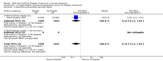
Authors' conclusions: Existing evidence does not provide conclusive evidence that the use of routine umbilical artery Doppler ultrasound, or combination of umbilical and uterine artery Doppler ultrasound in low-risk or unselected populations benefits either mother or baby. Future studies should be designed to address small changes in perinatal outcome, and should focus on potentially preventable deaths.
Conflict of interest statement
None known.
Figures


Update of
-
Fetal and umbilical Doppler ultrasound in normal pregnancy.Cochrane Database Syst Rev. 2010 Aug 4;(8):CD001450. doi: 10.1002/14651858.CD001450.pub3. Cochrane Database Syst Rev. 2010. Update in: Cochrane Database Syst Rev. 2015 Apr 15;(4):CD001450. doi: 10.1002/14651858.CD001450.pub4. PMID: 20687066 Free PMC article. Updated.
References
References to studies included in this review
Davies 1992 {published and unpublished data}
-
- Breart G, Uzan S, Uzan M. Doppler ultrasound screening during pregnancy [Letter; comment]. Lancet 1993;341(8843):501‐2. - PubMed
-
- Davies J, Spencer J, Gallivan S. Randomised trial of Doppler screening in a general obstetric population. Proceedings of the 26th British Congress of Obstetrics and Gynaecology; 1992 July 7‐10; Manchester, UK. 1992:316.
-
- Davies JA, Gallivan S, Spencer JAD. Randomised controlled trial of doppler ultrasound screening of placental perfusion during pregnancy. Lancet 1992;340:1299‐303. - PubMed
-
- Spencer JAD, Davies JA, Gallivan S. Randomised trial of routine Doppler screening during pregnancy. Journal of Maternal Fetal Investigation 1992;1:126.
French Doppler 1997 {published data only}
-
- Doppler French Study Group. A randomised controlled trial of Doppler ultrasound velocimetry of the umbilical artery in low risk pregnancies. British Journal of Obstetrics and Gynaecology 1997;104:419‐22. - PubMed
Mason 1993 {published data only}
-
- Mason GC, Lilford RJ, Porter J, Nelson E, Tyrell S. Randomised comparison of routine versus highly selective use of Doppler ultrasound in low risk pregnancies. British Journal of Obstetrics and Gynaecology 1993;100:130‐3. - PubMed
Newnham 1993 {published and unpublished data}
-
- Evans S, Newnham J, MacDonald W, Hall C. Characterisation of the possible effect on birthweight following frequent prenatal ultrasound examinations. Early Human Development 1996;45(3):203‐14. - PubMed
-
- Forward H, Yazar S, Hewitt AW, Khan J, Mountain JM, Pesudovs K, et al. Multiple prenatal ultrasound scans and ocular development: 20‐year follow‐up of a randomised, controlled trial. Ultrasound in Obstetrics & Gynecology 2014;44:166‐70. - PubMed
-
- Newnham J, MacDonald W, Gurrin L, Evans S, Landau L, Stanley F. The effect of frequent prenatal ultrasound on birthweight: follow up at one year of age. Proceedings of the 14th Australian Perinatal Society in conjunction with the New Zealand Perinatal Society; 1996 March 24‐27; Adelaide, Australia. 1996:A26.
-
- Newnham JP, Doherty DA, Kendall GE, Zubrick SR, Landau LL, Stanley FJ. Effects of repeated prenatal ultrasound examinations on childhood outcome up to 8 years of age: follow‐up of a randomised controlled trial. Lancet 2004;364:2038‐44. - PubMed
-
- Newnham JP, Evans SF, Michael CA, Stanley FJ, Landau LI. Effects of frequent ultrasound during pregnancy: a randomised controlled trial. Lancet 1993;342:887‐91. - PubMed
Whittle 1994 {published data only}
-
- Hanretty KP. Randomized study of doppler waveforms in umbilical and uterine arteries as a screening method to identify the compromised fetus. Personal communication 1988.
-
- Whittle MJ, Hanretty KP, Primrose MH, Neilson JP. Screening for the compromised fetus: A randomised trial of umbilical artery velocimetry in unselected pregnancies. American Journal of Obstetrics and Gynecology 1994;170(2):555‐9. - PubMed
References to studies excluded from this review
Ellwood 1997 {unpublished data only}
-
- Ellwood D, Peek M, Curren J. Predicting adverse pregnancy outcomes with ultrasound. A randomised controlled trial. Personal communication 1997.
Goffinet 2001 {published data only}
-
- Goffinet F, Aboulker D, Paris‐Llado J, Bucourt M, Uzan M, Papiernik E, et al. Screening with a uterine doppler in low risk pregnant women followed by low dose aspirin in women with abnormal results: a multicenter randomised controlled trial. British Journal of Obstetrics and Gynaecology 2001;108:510‐8. - PubMed
Gonsoulin 1991 {published data only}
-
- Gonsoulin MD. Umbilical artery Doppler waveform analysis: a randomized study on effect on outcome. American Journal of Obstetrics and Gynecology 1991;164:370.
Schneider 1992 {published data only}
-
- Schneider KT, Amberg‐Wendland D, Renz S, Furstenau U. Prospective randomized study of the clinical value of Doppler sonography as a screening procedure. Gynakologische Rundschau 1991;31(Suppl 1):139‐40. - PubMed
-
- Schneider KTM, Renz S, Furstenau U, Amberg‐Wendland D, Prochaska D, Graeff H. Doppler flow measurements as a screening method during pregnancy: Is it worth the effort?. Journal of Maternal Fetal Investigation 1992;1:125.
Scholler 1993 {published data only}
-
- Scholler J, Putz M, Sainz HG, Altrichter R, Philipp K. Value of Doppler sonography in management of non‐risk pregnancies at term [Der Stellenwert der Dopplersonographie bei der Betreuung von Nicht‐Risikoschwangerschaften am Geburtstermin]. Gynakologische Rundschau 1993;33(1 Suppl):118‐9. - PubMed
Snaith 2006 {published data only}
-
- Snaith V. Support and reassurance in antenatal care. Current Controlled Trials (http://controlled‐trials.com/mrct) [accessed 21 March 2006].
Subtil 2000 {published data only}
-
- Subtil D, Truffert P, Goeusse P, Dufour P, Uzan S, Breart G, et al. Value of systematic doppler +/‐ low dose aspirin to prevent vascular complications in primigravidae. Hypertension in Pregnancy 2000;19(Suppl 1):9.
Subtil 2003 {published data only}
-
- Subtil D, Goeusse P, Houfflin‐Debarge V, Puech F, Lequien P, Breart G, et al. Randomised comparison of uterine artery doppler and aspirin (100 mg) with placebo in nulliparous women: the essai regional aspirine mere‐enfant study (part 2). BJOG: an international journal of obstetrics and gynaecology 2003;110(5):485‐91. - PubMed
Additional references
Alfirevic 2013
Barnett 1995
-
- Barnett SB. Ultrasound safety in obstetrics: What are the concerns?. Ultrasound Quarterly 1995;13(4):228‐39.
Barnett 2001
-
- Barnett SB, Maulik D. Guidelines and recommendations for safe use of Doppler ultrasound in perinatal applications. Journal of Maternal‐Fetal Medicine 2001;10:75‐84. - PubMed
Beattie 1989
Bernstein 2000
-
- Bernstein IM, Horbar JD, Badger GJ, Ohlsson A, Golan A. Morbidity and mortality among very‐low‐birth‐weight neonates with intrauterine growth restriction. The Vermont Oxford Network. American Journal of Obstetrics and Gynecology 2000;182:198‐206. - PubMed
Burns 1993
-
- Burns PN. Principles of Doppler and color flow. Radiology in Medicine 1993;85:3‐16. - PubMed
Campbell 1983
-
- Campbell S, Diaz‐Recasens J, Griffin DR, Cohen‐Overbeek TE, Pearce JM, Wilson K, et al. New Doppler technique for assessing utero‐placental blood flow. Lancet 1983;i:675‐7. - PubMed
Chalmers 1989
-
- Chalmers I. Evaluating the effects of care during pregnancy and childbirth. In: Chalmers I, Enkin M, Keirse MJNC editor(s). Effective Care in Pregnancy and Childbirth. Vol. 1, Oxford: Oxford University Press, 1989:3‐38.
Chibber 2005
-
- Chibber R. Unexplained antenatal fetal deaths: what are the determinants?. Archives of Gynecology and Obstetrics 2005;271:286‐91. - PubMed
Devane 2007
-
- Devane D, Begley CM, Clarke M, Horey D, OBoyle C. Evaluating maternity care: a core set of outcome measures. Birth 2007;34(2):164‐72. - PubMed
Duck 1991
-
- Duck FA, Martin K. Trends in diagnostic ultrasound exposure. Physics in Medicine and Biology 1991;38:1423‐32. - PubMed
Eik‐Nes 1980
-
- Eik‐Nes SH, Brubaak AO, Ulstein MK. Measurement of human fetal blood flow. BMJ 1980;280:283‐4.
Fisk 2001
-
- Fisk NM, Smith RP. Fetal growth restriction; small for gestational age. In: Chamberlain G, Steer P editor(s). Turnbull's Obstetrics. 3rd Edition. Edinburgh: Churchill Livingstone, 2001:197‐209.
Fitzgerald 1977
Forward 2014
-
- Forward H, Yazar S, Hewitt AW, Khan J, Mountain JM, Pesudovs K, et al. Multiple prenatal ultrasound scans and ocular development: 20‐year follow‐up of a randomised, controlled trial. Ultrasound in Obstetrics & Gynecology 2014;44:166‐70. - PubMed
Fretts 1992
-
- Fretts RC, Boyd ME, Usher RH, Usher HA. The changing pattern of fetal death, 1961‐1988. Obstetrics & Gynecology 1992;79:35‐9. - PubMed
Froen 2004
-
- Froen JF, Gardosi JO, Thurmann A, Francis A, Stray‐Pedersen B. Restricted fetal growth in sudden intrauterine unexplained death. Acta Obstetricia et Gynecologica Scandinavica 2004;83(9):801‐7. - PubMed
Gardosi 2005
Giles 2003
-
- Giles W, Bisits A, O'Callaghan S, Gil A, DAMP Study Group. The Doppler assessment in multiple pregnancy randomised controlled trial of ultrasound biometry versus umbilical artery Doppler ultrasound and biometry in twin pregnancy. BJOG: an international journal of obstetrics and gynaecology 2003;110(6):593‐7. - PubMed
Goffinet 1997
-
- Goffinet F, Paris J, Heim N, Nisand I, Breart G. Predictive value of Doppler umbilical artery velocimetry in a low risk population with normal fetal biometry. A prospective study of 2016 women. European Journal of Obstetrics & Gynecology and Reproductive Biology 1997;71(1):11‐9. - PubMed
GRADE 2014 [Computer program]
-
- McMaster University. GRADEpro. [Computer program on www.gradepro.org]. Version 2015. McMaster University, 2014.
Henderson 1997
-
- Henderson J, Whittingham TA, Dunn T. A review of the acoustic output of modern diagnostic ultrasound equipment. Ultrasound 1997;5(4):10‐4.
Higgins 2009
-
- Higgins JPT, Green S, editors. Cochrane Handbook for Systematic Reviews of Interventions Version 5.0.2 [updated September 2009]. The Cochrane Collaboration, 2009. Available from www.cochrane‐handbook.org.
Higgins 2011
-
- Higgins JPT, Green S, editors. Cochrane Handbook for Systematic Reviews of Interventions Version 5.1.0 [updated March 2011]. The Cochrane Collaboration, 2011. Available from www.cochrane‐handbook.org.
Huang 2000
-
- Huang DY, Usher RH, Kramer MS, Yang H, Morin L, Fretts R. Determinants of unexplained fetal deaths. Obstetrics & Gynecology 2000;95:215‐21. - PubMed
Kieler 2001
-
- Kieler H, Cnattingius S, Haglund B, Palmgren J, Axelsson O. Sinistrality ‐ a side‐effect of prenatal sonography: a comparative study of young men. Epidemiology 2001;12:618‐23. - PubMed
Kieler 2002
-
- Kieler H, Cnattingius S, Palmgren J, Haglund B, Axelsson O. First trimester ultrasound scans and left‐handedness. Epidemiology 2002;13(3):370. - PubMed
Mangesi 2007
Mires 2000
-
- Mires GJ, Patel NB, Dempster J. The value of fetal umbilical artery flow velocity waveforms in the prediction of adverse fetal outcome in high risk pregnancies. Journal of Obstetrics and Gynaecology 2000;10:261‐70.
Morales‐Rosello 2014
-
- Morales‐Rosello J, Khalil A, Morlando M, Papageorghiou A, Bhide A, Thilaganathan B. Changes in fetal Doppler indices as a marker of failure to reach growth potential at term. Ultrasound in Obstetrics and Gynecology 2014;43(3):303‐10. - PubMed
Neilson 1998a
Neilson 1998b
Nelson 1988
-
- Nelson TR, Pretorius DH. The Doppler signal: where does it come from and what does it mean?. American Journal of Radiology 1988;151:439‐47. - PubMed
Newnham 1996
-
- Newnham J, MacDonald W, Gurrin L, Evans S, Landau L, Stanley F. The effect of frequent prenatal ultrasound on birthweight: follow‐up at one year of age. Proceedings of the 14th Annual Congress of the Australian Perinatal Society in conjunction with the New Zealand Perinatal Society; 1996 March 24‐27; Adelaide, Australia 1996:A26.
NICE 2008
-
- National Institute for Health and Clinical Excellence. Antenatal Care: Routine Care for the Healthy Pregnant Women. NICE Clinical Guideline 62 (March 2008). London: RCOG Press, 2008.
O'Connor 2013
-
- O'Connor C, Stuart B, Fitzpatrick C, Turner MJ, Kennelly MM. A review of contemporary modalities for identifying abnormal fetal growth. Journal of Obstetrics and Gynaecology 2013;33(3):239‐45. - PubMed
Owen 2001
-
- Owen P. Fetal assessment in the third trimester: fetal growth and biophysical methods. In: Chamberlain G, Steer P editor(s). Turnbull's Obstetrics. 3rd Edition. Edinburgh: Churchill Livingstone, 2001.
Pattison 1999
RCOG 1997
-
- Royal College of Obstetricians and Gynaecologists. Ultrasound Screening for Fetal Abnormalities: Report of the RCOG Working Party. London: RCOG, 1997.
RevMan 2008 [Computer program]
-
- RevMan. Review Manager (RevMan) Version 5.0 for Windows. Copenhagen: The Nordic Cochrane Centre, The Cochrane Collaboration, 2008.
RevMan 2014 [Computer program]
-
- The Nordic Cochrane Centre, The Cochrane Collaboration. Review Manager (RevMan). Version 5.3. Copenhagen: The Nordic Cochrane Centre, The Cochrane Collaboration, 2014.
Salvesen 1999
-
- Salvesen KA, Eik‐Nes SH. Ultrasound during pregnancy and birthweight, childhood malignancies and neurological development. Ultrasound in Medicine and Biology 1999;25:1025‐31. - PubMed
Salvesen 2007
-
- Salvesen KA. Epidemiological prenatal ultrasound studies. Progress in Biophysics and Molecular Biology 2007;93(7):295‐300. - PubMed
Schunemann 2009
-
- Schunemann HJ. GRADE: from grading the evidence to developing recommendations. A description of the system and a proposal regarding the transferability of the results of clinical research to clinical practice [GRADE: Von der Evidenz zur Empfehlung. Beschreibung des Systems und Losungsbeitrag zur Ubertragbarkeit von Studienergebnissen]. Zeitschrift fur Evidenz, Fortbildung und Qualitat im Gesundheitswesen 2009;103(6):391‐400. - PubMed
Sijoms 1989
-
- Sijoms EA, Reuwer PJHM, Beek E, Bruinse HW. The validity of screening for small‐for‐gestational‐age and low‐weight‐for‐length infants by Doppler ultrasound. British Journal of Obstetrics and Gynaecology 1989;96(5):557‐61. - PubMed
Soothill 1993
-
- Soothill PW, Ajayi RA, Campbell S, Nicolaides KH. Prediction of morbidity in small and normally grown fetuses by fetal heart rate variability, biophysical profile score and umbilical artery Doppler studies. British Journal of Obstetrics and Gynaecology 1993;100:742‐5. - PubMed
Stampalija 2010
Stoch 2012
-
- Stoch YK, Williams CJ, Granich J, Hunt AM, Landau LI, Newnham JP, et al. Are prenatal ultrasound scans associated with the autism phenotype? Follow‐up of a randomised controlled trial. Journal of Autism & Developmental Disorders 2012;42(12):2693‐701. - PubMed
References to other published versions of this review
Alfirevic 2010
Publication types
MeSH terms
LinkOut - more resources
Full Text Sources
Medical