. 2015 Apr 13;10(4):e0123397.
doi: 10.1371/journal.pone.0123397. eCollection 2015.

Altered metabolic and stemness capacity of adipose tissue-derived stem cells from obese mouse and human

Affiliations

Altered metabolic and stemness capacity of adipose tissue-derived stem cells from obese mouse and human

Laura M Pérez et al. PLoS One. .

Abstract

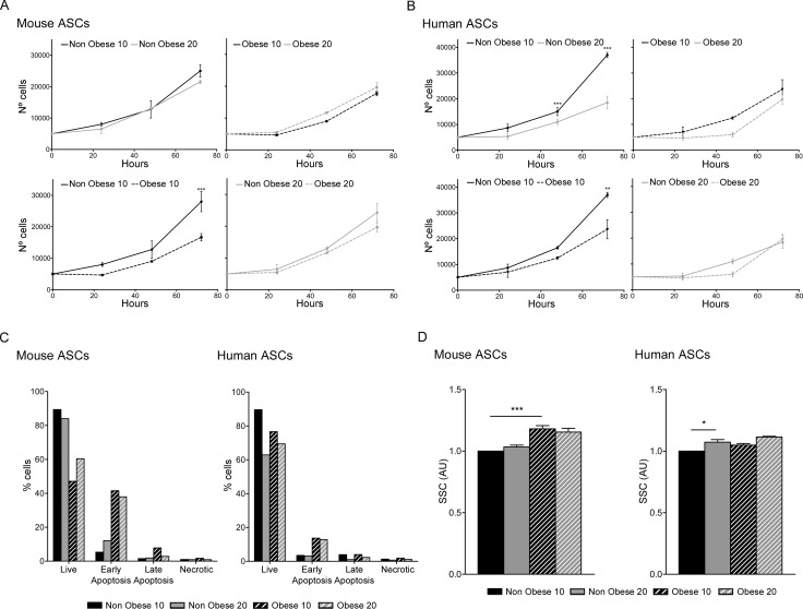
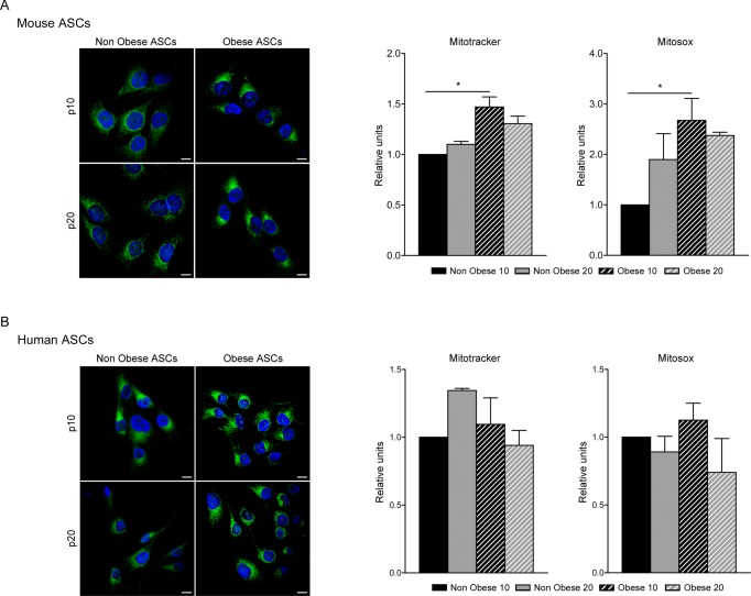
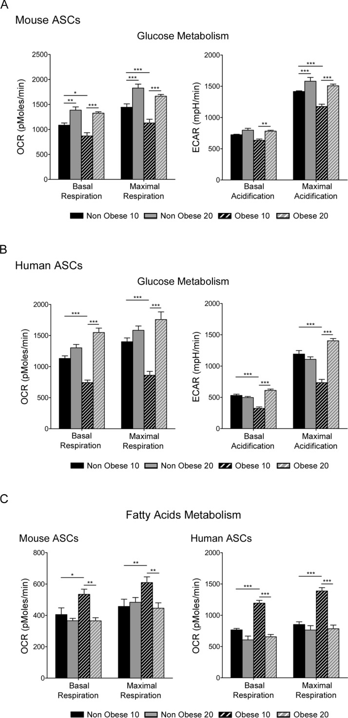
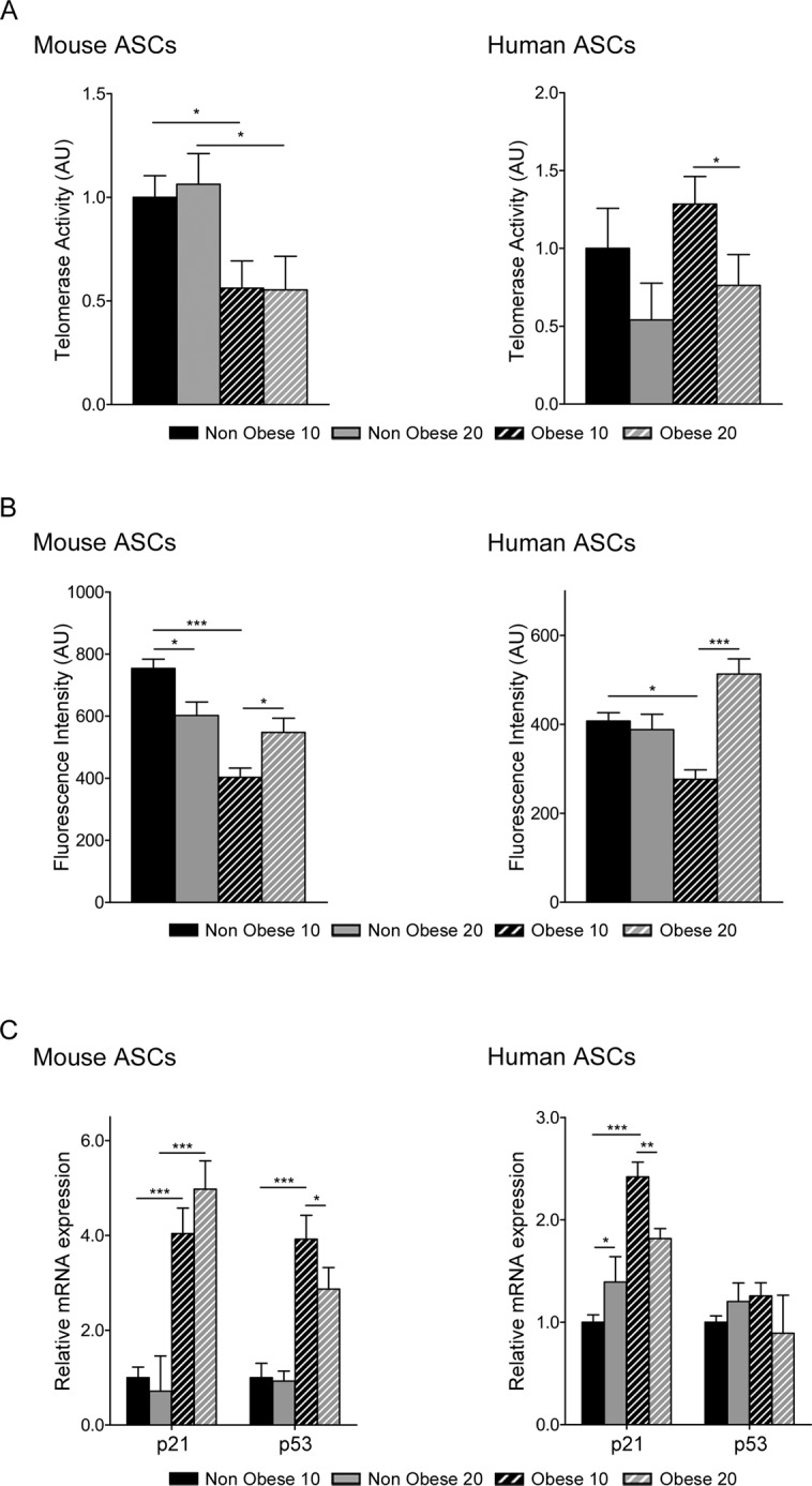
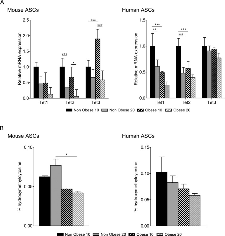
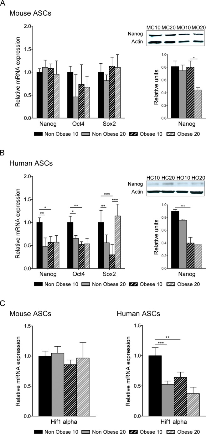
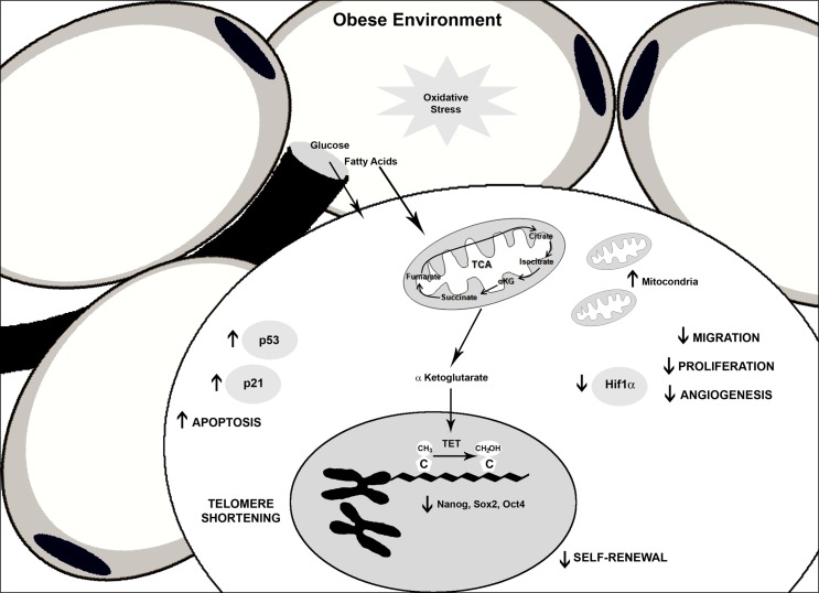
Adipose stem cells (ASCs) are an appealing source of cells for therapeutic intervention; however, the environment from which ASCs are isolated may impact their usefulness. Using a range of functional assays, we have evaluated whether ASCs isolated from an obese environment are comparable to cells from non-obese adipose tissue. Results showed that ASCs isolated from obese tissue have a reduced proliferative ability and a loss of viability together with changes in telomerase activity and DNA telomere length, suggesting a decreased self-renewal capacity. Metabolic analysis demonstrated that mitochondrial content and function was impaired in obese-derived ASCs resulting in changes in favored oxidative substrates. These findings highlight the impact of obesity on adult stem properties. Hence, caution should be exercised when considering the source of ASCs for cellular therapies since their therapeutic potential may be impaired.


Conflict of interest statement

Competing Interests: The authors have declared that no competing interests exist.

Figures

Fig 1
Fig 1. Proliferation and viability of ASCs.
Proliferation was measured in murine (A) and human (B), at passage 10 and 20 nonobese and obese ASCs by cell counting. Data represent mean±SEM from three replicates. Cell death was measured using a radiometric assay kit and quantified by flow cytometry (C). One representative experiment of three independent experiments is shown. Morphological analysis by flow cytometry showing cell size (SSC) (D). Data represent mean±SEM from three independent experiments. * P < 0.05; ** P < 0.01; *** P < 0.001. AU, arbitrary units.
Fig 2
Fig 2. Mitochondrial abundance in ASCs.
Confocal microscopy of Mitotracker Green staining in murine (A) and human (B) ASCs. One representative image out of three independent experiments is shown. Bar, 10 μm. Mitochondria quantification by flow cytometry with Mitotracker Green and ROS production with Mitosox Red in murine (A) and human ASCs (B). Data represent mean±SEM from three independent experiments. * P < 0.05.
Fig 3
Fig 3. Glucose and free fatty acid metabolism in ASCs.
Mitochondrial respiration analysis in murine (A and C) and human (B and C) ASCs. Oxygen consumption rate (OCR) and acidification levels (ECAR) during glucose metabolism (A and B) or fatty acid metabolism (C) were measured. Data represent mean±SEM from one representative experiment with six replicates. * P < 0.05; ** P < 0.01; *** P < 0.001.
Fig 4
Fig 4. Comparison of age related parameters in ASCs from nonobese and obese environments.
Relative telomerase activity in murine and human ASCs measured by qTRAP assay (A). Data represent mean±SEM from three independent experiments. Telomere length was measure by qFISH (B) in murine and human ASCs. Data represent mean fluorescence intensity ± SEM from 20 representative images. Gene expression profile of cell cycle proteins, p53 and p21 (C). Data represent mean±SEM from three independent experiments. * P < 0.05; ** P < 0.01; *** P < 0.001. AU, arbitrary units.
Fig 5
Fig 5. Hydroxymethylation in ASCs.
Quantitative Real Time PCR was performed to evaluate TET family gene expression in murine and human ASCs (A). Data represent mean±SEM from three different experiments. Levels of 5-hydroxymethylcytosine were measured by EpiSeeker hydroxymethylated DNA Quantification Kit in murine and human ASCs (B). * P < 0.05; ** P < 0.01; *** P < 0.001.
Fig 6
Fig 6. Expression analysis of related genes.
Quantitative Real Time PCR was performed on mRNA extracted from nonobese and obese ASCs at passage 10 and 20, to evaluate expression of stem cell markers, Nanog, Oct4 and Sox2 (A, B) and Hif1α (C). Western blotting was performed to measure the relative protein expression of Nanog (A, B). Image representative and graph represent mean±SEM from four different experiments. * P < 0.05; ** P < 0.01; *** P < 0.001. MC10, mouse nonobese ASCs at passage 10; MC20, mouse nonobese ASCs at passage 20; MO10, mouse obese ASCs at passage 10; MO20, mouse obese ASCs at passage 20; HC10, human nonobese ASCs at passage 10; HC20, human nonobese ASCs at passage 20; HO10, human obese ASCs at passage 10; HO20, human obese ASCs at passage 20.
Fig 7
Fig 7. Altered general functions in obese ASCs.
Marked dysfunction in stem cells from obese environments.

References

    1. San Martin N, Galvez BG (2011) A new paradigm for the understanding of obesity: the role of stem cells. Arch Physiol Biochem 117: 188–194. 10.3109/13813455.2011.571700 - DOI - PubMed
    1. Cinti S (2012) The adipose organ at a glance. Dis Model Mech 5: 588–594. 10.1242/dmm.009662 - DOI - PMC - PubMed
    1. Rodeheffer MS, Birsoy K, Friedman JM (2008) Identification of white adipocyte progenitor cells in vivo. Cell 135: 240–249. 10.1016/j.cell.2008.09.036 - DOI - PubMed
    1. Galvez BG, San Martin N, Rodriguez C (2009) TNF-alpha is required for the attraction of mesenchymal precursors to white adipose tissue in Ob/ob mice. PLoS One 4: e4444 10.1371/journal.pone.0004444 - DOI - PMC - PubMed
    1. Panfilov IA, de Jong R, Takashima S, Duckers HJ (2013) Clinical study using adipose-derived mesenchymal-like stem cells in acute myocardial infarction and heart failure. Methods Mol Biol 1036: 207–212. 10.1007/978-1-62703-511-8_16 - DOI - PubMed

Publication types

MeSH terms