. 2015 Apr 13;10(4):e0122348.
doi: 10.1371/journal.pone.0122348. eCollection 2015.

Cajal body proteins differentially affect the processing of box C/D scaRNPs

Affiliations

Cajal body proteins differentially affect the processing of box C/D scaRNPs

Isioma I Enwerem et al. PLoS One. .

Abstract

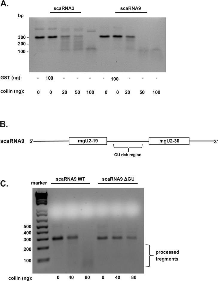
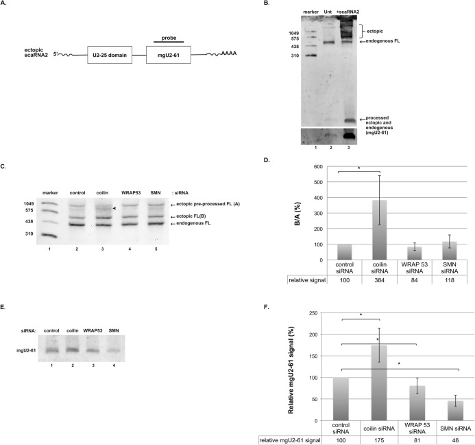
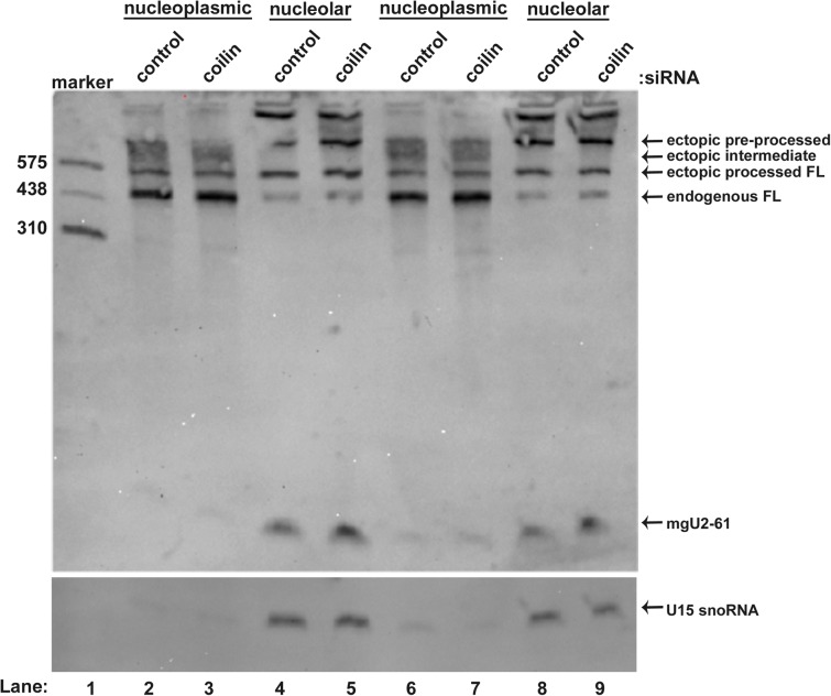
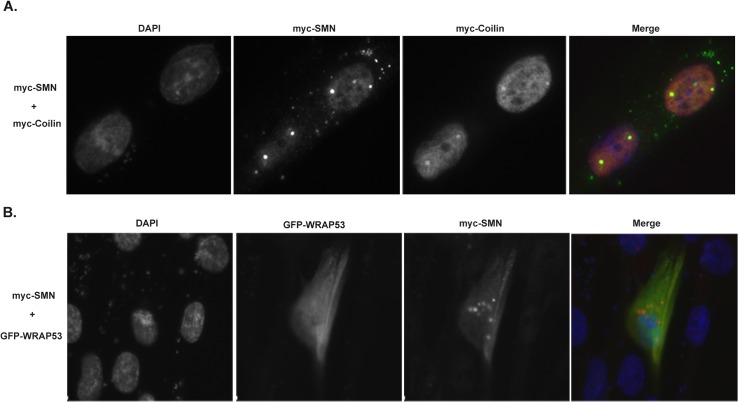
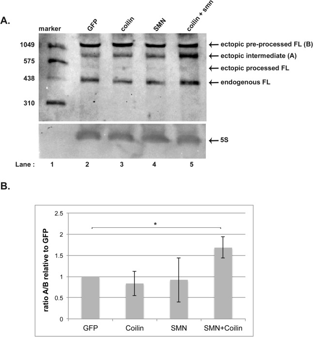
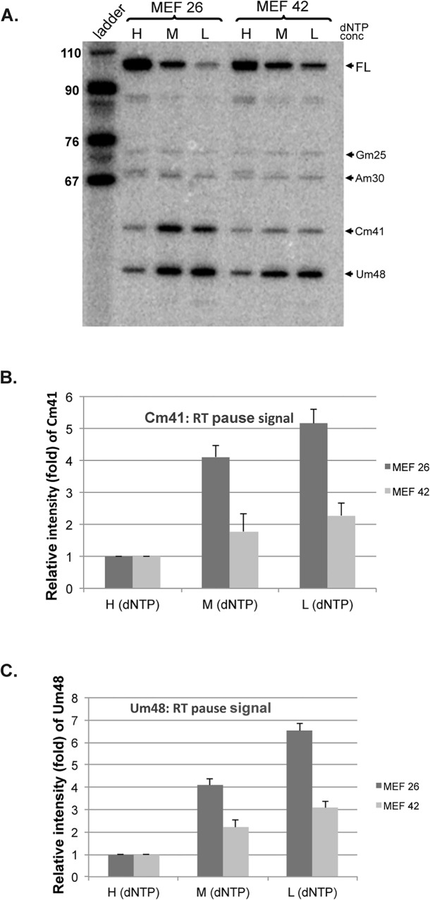
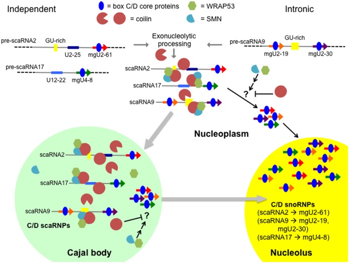
Small nuclear ribonucleoproteins (snRNPs), which are required for pre-mRNA splicing, contain extensively modified snRNA. Small Cajal body-specific ribonucleoproteins (scaRNPs) mediate these modifications. It is unknown how the box C/D class of scaRNPs localizes to Cajal Bodies (CBs). The processing of box C/D scaRNA is also unclear. Here, we explore the processing of box C/D scaRNA 2 and 9 by coilin. We also broaden our investigation to include WRAP53 and SMN, which accumulate in CBs, play a role in RNP biogenesis and associate with coilin. These studies demonstrate that the processing of an ectopically expressed scaRNA2 is altered upon the reduction of coilin, WRAP53 or SMN, but the extent and direction of this change varies depending on the protein reduced. We also show that box C/D scaRNP activity is reduced in a cell line derived from coilin knockout mice. Collectively, the findings presented here further implicate coilin as being a direct participant in the formation of box C/D scaRNPs, and demonstrate that WRAP53 and SMN may also play a role, but the activity of these proteins is divergent to coilin.


Conflict of interest statement

Competing Interests: The authors have declared that no competing interests exist.

Figures

Fig 1
Fig 1. Coilin processing activity shows preference towards scaRNA9 compared to scaRNA2, and the GU region of scaRNA9 impacts coilin processing.
(A) RNA processing assay comparing the processing of in vitro transcribed scaRNA2 and scaRNA9 by bacterially purified coilin. Equivalent amounts of RNA were incubated with no protein, GST (100 ng) or increasing amounts of pure, nucleic acid free coilin (20 ng, 50 ng and 100 ng). Products were resolved on a 2% agarose gel and detected by ethidium bromide staining. (B) Schematic showing full-length scaRNA9 (353 nucleotides in length), the mgU2-19 and mgU2-30 RNAs that are derived from the full-length RNA, and the GU-rich region that lies in between these smaller guide RNAs. (C) RNA processing assay comparing the processing of in vitro transcribed scaRNA9 and scaRNA9 with a deleted GU-rich region (scaRNA9 ΔGU) by purified coilin. Approximately equal amounts of RNA were incubated with no protein, or two amounts of pure, nucleic acid free coilin (40 ng and 80 ng). Products were resolved on a 2% agarose gel and detected by ethidium bromide staining.
Fig 2
Fig 2. Coilin processing of scaRNA2 and scaRNA9 generates smaller, relatively stable, fragments.
(A) Schematic showing full-length scaRNA2 and the guide RNA (mgU2-61) derived from this full-length RNA. Also shown are scaRNA9 and the two guide RNAs (mgU2-19 and mgU2-30) derived from this full-length RNA. The location of the probes used to detect scaRNA2 and scaRNA9, and their processed fragments, are shown. (B and C) Northern blot analysis of RNA processing experiments using in vitro transcribed scaRNA2 or scaRNA9 and increasing amounts of bacterially expressed purified coilin. After incubation, the reactions were subjected to acrylamide electrophoresis, Northern blotting and detection with the indicated probes. Full-length scaRNA 2 and 9 are indicated (FL) and processed fragments for both scaRNA2 and scaRNA9 are bracketed. The mobility of a DIG-labeled RNA marker (in nucleotides) is shown. (D) Quantification of the processed fragments from the scaRNA2 (B) and scaRNA9 (C) experiments.
Fig 3
Fig 3. Reduction of coilin, WRAP53 or SMN dysregulates processing of ectopically expressed scaRNA2.
(A) Schematic showing ectopic pre-processed full-length scaRNA2 (expressed from the pcDNA3.1+ vector) and the locations of the U2-25 domain and the processed guide RNA (mgU2-61). A poly A tail, which does not exist for endogenous scaRNA2, is indicated. The probe used for Northern blots is also shown. (B) Northern blot of HeLa RNA (20 μg/lane) from either non-transfected cells (lane 2) or cells transfected (24 hr) with pcDNA 3.1+ scaRNA2 (lane 3). The probe used is shown in (A) The lower panel shows a longer exposure of the region of the gel with mgU2-61 signal in order to better visualize the endogenous signal. (C) Northern blot of Hela RNA (10 μg/lane) from cells transfected with control (lane 2), coilin (lane 3), WRAP53 (lane 4) or SMN (lane 5) siRNA for 24 hrs then transfected with pcDNA 3.1+ scaRNA2 for 24 hrs. The probe used is shown in (A). Ectopic pre-processed full-length (denoted as A), ectopic processed full-length (denoted as B) and endogenous full-length scaRNA2 is shown. An arrowhead marks an intermediate species often seen in RNA isolated from coilin knockdown cells (lane 3). (D) Histogram quantifying the ratio between ectopic processed full-length and ectopic pre-processed full-length (B/A) from 8 different experimental sets. Standard deviation was used to include error bars. Students t test was used to determine statistical significance, indicated by “*” and corresponding to a p value less than 0.05. (E) Northern blot of Hela RNA (20 μg/lane) from cells transfected with control (lane 1), coilin (lane 2), WRAP53 (lane 3) or SMN (lane 4) siRNA for 24 hrs then transfected with pcDNA 3.1+ scaRNA2 for 24 hrs. The Northern blotting transfer conditions were optimized to retain the smaller, mgU2-61 guide RNA. The probe used is shown in (A). The majority of the signal is coming from ectopically expressed mgU2-61 guide RNA. (F) Histogram quantifying the mgU2-61 guide RNA from 8 different experimental sets. The mobility of a DIG-labeled RNA marker (in nucleotides) is shown. Standard deviation was used to include error bars. Students t test was used to determine statistical significance, indicated by “*” and corresponding to a p value less than 0.05.
Fig 4
Fig 4. The scaRNA2-derived mgU2-61 guide RNA is enriched in the nucleolar fraction upon coilin reduction.
HeLa cells transfected with control or coilin siRNA were fractionated to produce nucleoplasmic and nucleolar fractions. 5 μg/lane of RNA from each fraction was run on a 6% polyacrylamide gel and Northern blotted, followed by detection with a scaRNA2 probe (shown in Fig 3). Ectopic pre-processed, intermediate-processed and processed full-length species are indicated, as is the smaller guide RNA, mgU2-61. The blot was reprobed with an U15 snoRNA probe to verify the fidelity of the nucleolar fractionation and demonstrate that an approximately equal amount of RNA was loaded (lower panel). The mobility of a DIG-labeled RNA marker (in nucleotides) is shown. Duplicate samples are shown in lanes 6–9.
Fig 5
Fig 5. Co-expression of SMN and coilin in the WI-38 primary cell line promotes foci formation.
(A) WI-38 cells were co-transfected with myc-tagged SMN and coilin, followed by fixation and detection of the expressed proteins using anti-SMN or anti-coilin antibodies and appropriate secondary antibodies. DAPI was used to stain the nucleus. In the merged image, the nucleus is blue, SMN is green and coilin signal is red. Foci with co-localized SMN and coilin are yellow. (B) WI-38 cells were co-transfected with myc-SMN and GFP-WRAP53, followed by fixation and detection of the expressed proteins. Anti-SMN antibody was used to detect SMN and the nucleus was stained with DAPI. In the merged image, SMN is red, GFP-WRAP53 is green and the nucleus is blue.
Fig 6
Fig 6. Co-expression of SMN and coilin increases processing of ectopically expressed scaRNA2 in WI-38 cells.
(A) WI-38 cells were co-transfected with pCDNA3.1+scaRNA2 and empty GFP vector (lane 2), myc-coilin (lane 3), myc-SMN (lane 4) or myc-coilin + myc-SMN (lane 5) for 24 hrs followed by RNA isolation. RNA (10 μg/lane) was run on a 6% polyacrylamide gel, Northern blotted and detected using a DIG labeled oligo probe that detects scaRNA2 (shown in Fig 3). The positions of ectopic pre-processed full-length scaRNA2 (indicated as B), ectopic intermediate-processed scaRNA2 (denoted as A), ectopic processed full-length scaRNA2 and endogenous full-length scaRNA2 are shown. The blot was then probed for 5S ribosomal RNA (5S) to verify equivalent loading of RNA (lower panel). (B) Histogram showing quantification of the ectopic intermediate (denoted as A) to ectopic pre-processed FL (denoted as B) ratio for 3 experimental sets represented in panel A. The mobility of a DIG-labeled RNA marker (in nucleotides) is shown.Standard deviation was used to include error bars. Students t test was used to determine statistical significance, indicated by “*” and corresponding to a p value less than or equal to 0.05.
Fig 7
Fig 7. 2′-O-methylation of U2 snRNA is reduced in coilin knockout MEF cells.
(A) Primer extension experiments were done to map the 2’-O-methylation of individually modified nucleotides in U2 snRNA using equal amounts of RNA isolated from MEF 26 (coilin wild-type) and MEF 42 (coilin knock-out) cell lines. High, medium and low (H,M and L) deoxynucleoside triphosphate concentrations were used. The resulting pause signals corresponding to 2′-O-methylated residues in mouse U2 snRNA are indicated by each arrow. (B) Relative levels/intensities of Cm41 and Um48 in MEF26 and MEF 42 cell lines were calculated. For each lane, the percentage of the Cm41 and Um48 signals was calculated. The percentage values of Cm41 and Um48 of the medium and low dNTP lanes were then normalized to that of the high dNTP lanes. The ladder is shown in nucleotides.
Fig 8
Fig 8. Model of box C/D scaRNP 2, 9 and 17 biogenesis.
Box C/D core proteins (blue) are thought to bind the scaRNAs after transcription from independent genes (scaRNA2 and scaRNA17) or a host gene (for scaRNA9). For scaRNA9, the intron lariat is debranched for subsequent exonucleolytic processing to generate the mature scaRNA9. Exonucleolytic processing is also required to produce full-length scaRNA2 and 17 from longer precursor forms (dashed lines). Yellow boxes denote the GU dinucleotide repeats present in scaRNA2 and 9. Also shown are the smaller, nucleolus-enriched guide RNAs derived from scaRNA2 (mgU2-61, red), scaRNA9 (mgU2-19, orange and mgU2-30, purple) and scaRNA17 (mgU4-8, green). In cells without CBs (right pathway), these longer scaRNAs are processed in the nucleoplasm to generate the smaller guide RNAs that accumulate in the nucleolus as snoRNPs. In cells with CBs (left pathway, gray arrow to CB), full-length scaRNP 2, 9 and 17 accumulate in CBs, while the smaller guide RNA derived from these longer forms accumulate in the nucleolus. The mechanism(s) by which the cell governs the ratio of full-length scaRNA to smaller guide RNA is unknown (denoted as “?” in figure). Based on the data presented here, we believe that coilin, WRAP53 and SMN all participate at regulating the flux of scaRNA 2, 9 and 17 distribution and processing. Since coilin can both bind and process scaRNAs, it is possible that this protein contributes to the initial processing events that generate mature full-length scaRNA from the precursor forms containing 5’ and 3’ extensions. Coilin may also take part in the processing steps giving rise to the nucleolus enriched guide RNAs, although our coilin knockdown data (Fig 3) does not support this idea. Alternatively, coilin interaction with WRAP53 and SMN may decrease its processing activities, thereby allowing for the accumulation of full-length scaRNA 2, 9 and 17 in CBs. It is likely that another unknown factor (denoted as ‘?”) also contributes to the generation of the small guide RNAs derived from scaRNA 2, 9 and 17. Based upon the work presented here, we expect that coilin will negatively regulate the activity of this factor but SMN and WRAP53 will promote its processing activity (inhibition and activation arrow in nucleoplasm and CB).

Similar articles

Cited by

References

    1. Yu YT, Shu MD, Steitz JA. Modifications of U2 snRNA are required for snRNP assembly and pre-mRNA splicing. EMBO J. 1998;17: 5783–5795. - PMC - PubMed
    1. Liang XH, Liu Q, Fournier MJ. rRNA modifications in an intersubunit bridge of the ribosome strongly affect both ribosome biogenesis and activity. Mol Cell. 2007;28: 965–977. - PubMed
    1. Kiss T. Biogenesis of small nuclear RNPs. J Cell Sci. 2004;117: 5949–5951. - PubMed
    1. Fischer U, Liu Q, Dreyfuss G. The SMN-SIP1 complex has an essential role in spliceosomal snRNP biogenesis. Cell. 1997;90: 1023–1029. - PubMed
    1. Pellizzoni L, Charroux B, Dreyfuss G. SMN mutants of spinal muscular atrophy patients are defective in binding to snRNP proteins. Proc Natl Acad Sci U S A. 1999;96: 11167–11172. - PMC - PubMed

Publication types

LinkOut - more resources