. 2015;7(3):584-604.
doi: 10.1080/19420862.2015.1029216.

A tetravalent bispecific TandAb (CD19/CD3), AFM11, efficiently recruits T cells for the potent lysis of CD19(+) tumor cells

Affiliations

A tetravalent bispecific TandAb (CD19/CD3), AFM11, efficiently recruits T cells for the potent lysis of CD19(+) tumor cells

Uwe Reusch et al. MAbs. 2015.

Abstract

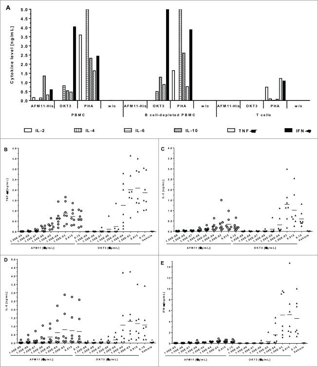
To harness the potent tumor-killing capacity of T cells for the treatment of CD19(+) malignancies, we constructed AFM11, a humanized tetravalent bispecific CD19/CD3 tandem diabody (TandAb) consisting solely of Fv domains. The molecule exhibits good manufacturability and stability properties. AFM11 has 2 binding sites for CD3 and 2 for CD19, an antigen that is expressed from early B cell development through differentiation into plasma cells, and is an attractive alternative to CD20 as a target for the development of therapeutic antibodies to treat B cell malignancies. Comparison of the binding and cytotoxicity of AFM11 with those of a tandem scFv bispecific T cell engager (BiTE) molecule targeting the same antigens revealed that AFM11 elicited more potent in vitro B cell lysis. Though possessing high affinity to CD3, the TandAb mediates serial-killing of CD19(+) cells with little dependence of potency or efficacy upon effector:target ratio, unlike the BiTE. The advantage of the TandAb over the BiTE was most pronounced at lower effector:target ratios. AFM11 mediated strictly target-dependent T cell activation evidenced by CD25 and CD69 induction, proliferation, and cytokine release, notwithstanding bivalent CD3 engagement. In a NOD/scid xenograft model, AFM11 induced dose-dependent growth inhibition of Raji tumors in vivo, and radiolabeled TandAb exhibited excellent localization to tumor but not to normal tissue. After intravenous administration in mice, half-life ranged from 18.4 to 22.9 h. In a human ex vivo B-cell chronic lymphocytic leukemia study, AFM11 exhibited substantial cytotoxic activity in an autologous setting. Thus, AFM11 may represent a promising therapeutic for treatment of CD19(+) malignancies with an advantageous safety risk profile and anticipated dosing regimen.

Keywords: ALL; AUCtot, total area under the curve; B-ALL, B-precursor acute lymphoblastic leukemia; BBB, blood-brain barrier; BiTE, bispecific T cell engager; CAR, chimeric antigen receptor; CCS, cell culture supernatant; CD, cluster of differentiation; CD3; CDR, complementarity determining region; CHO, Chinese hamster ovary; CL, clearance; CLL, chronic lymphocytic leukemia; CNS, central nervous system; Cmax, maximal concentration; DMSO, dimethyl sulfoxide; E:T, effector:target; EC50, half maximal effective concentration; ECL, electrochemiluminescence; F, fluorescence; FACS, fluorescence-activated cell sorting; FCS, fetal calf serum; FR, framework region; Fab, fragment antigen-binding; Fc, fragment crystallizable; FcRn, neonatal Fc receptor; FcgR, Fc gamma receptor; Fv, variable fragment; HMF, high molecular weight forms; HSA, human serum albumin; His, histidine; IFN, interferon; IL, interleukin; IgG, immunoglobulin G; KD, dissociation constant; LMF, low molecular weight forms; MSD, MesoScale Discovery; MWCO, molecular weight cut-off; NHL, non-Hodgkin lymphoma; NK, natural killer; NOD/scid, nonobese diabetic/severe combined immunodeficiency; Non-Hodgkin lymphoma; ORR, overall response rate; PBMC, peripheral blood mononuclear cell; PBS, phosphate buffered saline; PES, polyethersulfone; PHA, phytohemagglutinin; PI, propidium iodide; SABC, standardized antibody binding capacity; SD, standard deviation; SDS-PAGE, sodium dodecyl sulfate polyacrylamide gel electrophoresis; SE-HPLC, size exclusion high-pressure liquid chromatography; SEC, size exclusion chromatography; SPR, surface plasmon resonance; T cells; TNF, tumor necrosis factor; TandAb, tandem diabody; VH, variable heavy; VL, variable light; Vss, volume of distribution at steady state; WBA, whole body autoradiography; bispecific antibodies; ctrl., control; i.v., intravenous; ka, association rate constant; kd, dissociation rate constant; s.c., subcutaneous; scFv, single-chain variable fragment; t1/2, terminal elimination half-life; w/o, without.


Figures

Figure 1.
Figure 1.
Design, production, and purification of recombinant AFM11-His. (A) Scheme of AFM11-His gene design. The gene construct is shown from 5′ to 3′-end encoding the monomeric TandAb subunit with N-terminal signal sequence, VH and VL domains separated by identical (Gly2Ser)2 linkers (L1, L2, L3), and the C-terminal hexa-histidine-tag (His6-tag). Upon expression, 2 polypeptide gene products dimerize in a head-to-tail fashion. (B) AFM11-His was expressed in 10-day fed-batch cultures of stably-transfected CHO cells which grow to high cell densities, maintain high viability, and secrete the AFM11-His product into the cell culture supernatant (CSS). (C) AFM11-His was purified by Ni-NTA Superflow chromatography followed by preparative size exclusion chromatography (SEC) on Superdex 200. High purity of the AFM11-His TandAb homodimer was demonstrated by analytical SEC. Purified AFM11-His runs in SEC as a single peak with a retention time that is consistent with its expected molecular weight of 106 kDa. (D) Integrity of the purified AFM11-His protein was analyzed by SDS-PAGE. Under denaturing, reducing and non-reducing conditions, AFM11-His monomers run at the expected size of ∼50 kDa. (E) Molecular forms of AFM11-His were analyzed by analytical SEC after incubation at 4°C, 21°C, or 37°C for the indicated numbers of days. High Molecular weight Forms (HMF) appear as a mixture of aggregates and species with an apparent molecular weight of ∼200 kDa, consistent with tetrameric forms; AFM11-His homodimer represents the main chromatographic peak under all conditions assayed; Low Molecular weight Forms (LMF) represent a minor fraction under all conditions and are consistent with the monomeric form of the AFM11 with molecular weights <60 kDa.
Figure 2.
Figure 2.
Binding of AFM11 and CD19/CD3 BiTEs to recombinant antigens and CD3+ cells. (A) Recombinant CD19-Fc was directly immobilized on the Sensor Chip (CM5). AFM11-His was injected (100 nM), and during the dissociation phase the second antigen (CD3) was injected. (B) Jurkat cells or (C) Raji cells were stained with the indicated concentrations of AFM11-His, CD19M13-sm/CD3LCHC21-ktay BiTE, and CD19HD37/CD3TR66 BiTE on ice. Cell-surface bound antibodies were detected by anti-His mAb followed by FITC-conjugated goat anti-mouse IgG. Mean fluorescence intensities determined by flow cytometry were modeled as a sigmoidal dose-response by non-linear regression to calculate KD.
Figure 3
Figure 3
(See previous page). Comparison of cytotoxic properties of AFM11-His and the CD19HD37/CD3TR66 BiTE on cell lines. (A) EC50 and (B) specific lysis were determined in cytotoxicity assays with calcein-labeled CD19+ MEC-1 target cells and primary human T cells as effector cells at an E:T ratio of 25:1 with incubation times ranging between 30 min and 300 min. At each time-point the EC50 (calculated by non-linear regression of the data modeled as a sigmoidal curve) and specific lysis for AFM11-His and CD19HD37/CD3TR66 BiTE were plotted as representative experiments. (C) AFM11-His-mediated target cell lysis by CD4+ and CD8+ T cell subsets. 4 and 23 h FACS-based cytotoxicity assays for AFM11-His and CD19HD37/CD3TR66 BiTE were performed with either unfractionated human T cells or enriched human CD4+ or CD8+ T cells as effector cells, and CD19+ NALM-6 cells as target cells at an E:T ratio of 5:1. The plotted values represent the mean and the SD of duplicate measurements. (D, E) Serial killing mediated by AFM11 and CD19HD37/CD3TR66 BiTE was demonstrated by FACS-based cytotoxicity assays. NALM-6 target cells and enriched human T cells were used at the indicated E:T ratios in the presence of increasing concentrations of AFM11-His and CD19HD37/CD3TR66 BiTE. After 23 h incubation, EC50 (D) and percentage of target cell lysis at 10 pM (E) were calculated for each E:T ratio and plotted as representative experiments.
Figure 4.
Figure 4.
Cytotoxic activity of AFM11-His in ex vivo autologous B-CLL cultures. AFM11-induced apoptosis in B-CLL autologous cultures of patient PBMC. PBMC from 4 patients with B-CLL were cultured either in the presence of 100 ng/mL AFM11-His or HSA/CD3 control TandAb. After 24 and 72 h, apoptosis in CD5+/CD23+ CLL blasts was determined by Annexin V staining and flow cytometric analysis. The specific apoptosis in AFM11-His-treated samples was calculated and plotted for all 4 patients (coded with 2 letters and 2 digits). The total fraction of T cells in these samples ranged between 2.0% and 11.1%, the fraction of CD5+CD23+ B-CLL cells ranged between 87.9% and 92.7%, and the E:T ratio ranged between 8 and 46.
Figure 5.
Figure 5.
AFM11 does not facilitate activation of human T cells in the absence of CD19+ target cells. Dose-responsive induction of CD25 by AFM11-His (A) and CD69 (B) expression on human T cells was assayed in cultures of human PBMC, B cell-depleted PBMC, and enriched T cells after 48 h incubation. Unfractionated PBMC contained 3.8% CD19+ cells. B cell-depleted cultures possessed 0.6% CD19+ cells, and the enriched T cell cultures contained 0.1% CD19+ cells. The kinetics of CD25 (C) and CD69 (D) expression induced by AFM11-His (10 ng/mL) were determined in human PBMC cultures containing 3.5% CD19+ cells and in B cell-depleted PBMC cultures containing < 0.1% CD19+ cells. A through D present representative experiments. (E) No induction of T cell proliferation in the absence of target cells. Unfractionated PBMC, B cell-depleted PBMC, and enriched unstimulated human T cells were cultured in the presence of increasing concentrations of AFM11-His or OKT3 for 4 days, and then pulsed overnight with BrdU. Incorporation of BrdU was assayed by a BrdU ELISA. Control (ctrl.) cells were cultured in the absence of antibodies. Mean absorbance, and SD of triplicates, measured at 450 nm are plotted. The relative fractions of CD19+ cells were 8% in unfractionated human PBMC, 0.8% in B cell-depleted PBMC, and < 0.1% in enriched T cells.
Figure 6.
Figure 6.
Cytokine release by AFM11-His is strictly dependent on the presence of CD19+ target cells. (A) Cytokine release in cultures of PBMC, B cell-depleted PBMC, and enriched human T cells. Unfractionated human PBMC, B cell-depleted PBMC, and enriched T cells were cultured in the presence of: i) 10 µg/mL AFM11-His or OKT3, ii) 100 µg/mL PHA, or iii) without antibodies (w/o). After 48 h incubation IL-2, IL-6, IL-10, TNF, and IFN-γ in the cell-free culture supernatant were quantified by multiplexing and the results of representative experiments were plotted. (B) TNF, (C) IL-2, (D) IL-6, and (E) IFN-γ levels in culture supernatants of PBMC from 7 individual donors. Cytokine levels were determined by Luminex upon stimulation with antibodies in solution. PBMC were either stimulated with AFM11 (open circles) or OKT3 (closed triangles) at the indicated concentrations. IL-2 and TNF levels were measured after 24 h, IL-6 and IFN-γ levels were measured after 48 h. Background (asterisks) was measured in cultures supplemented with vehicle (formulation buffer) corresponding to the volume of highest AFM11 concentration used. Horizontal bars indicate mean values across the 7 donors.
Figure 7.
Figure 7.
Xenograft models and tissue-distribution of AFM11 in tumor-bearing mice. (A) Effect of AFM11 on the growth of Raji Burkitt lymphoma in NOD/scid mice. Six experimental groups of immunodeficient NOD/scid mice were xenotransplanted by subcutaneous injection with a suspension of 2.5 × 10⁶ of Raji tumor cells on day 0. Tumor cells were mixed with 1 × 10⁷ human PBMC from healthy donors. To account for potential donor variability, each of the experimental groups was subdivided into 3 cohorts each receiving PBMCs from a single donor. Two groups without PBMC effector cells (tumor cells only, not shown) served as negative controls. One of the control groups was treated only with vehicle; the other received only AFM11. All animals of the experimental groups transplanted with tumor cells and PBMC received an intravenous bolus on days 0, 1, 2, 3 and 4 of either vehicle (control) or AFM11 at 4 different dose levels as indicated (0.005, 0.05, 0.5, and 5 mg/kg). All treatment groups receiving AFM11 exhibited statistically significant tumor growth inhibition, relative to the control group, from day 13 until day 34, excepting the low dose group (0.005 mg/kg) in which statistical significance was restricted to days 17, 20 and 22. (B) No effect of AFM11 dosing regimen on the growth of Raji Burkitt lymphoma in NOD/scid mice. NOD/scid mice were xenotransplanted by subcutaneous injection with a suspension of 2.5 × 10⁶ of tumor cells on day 0. Tumor cells were mixed with 1 × 10⁷ human PBMC from healthy donors. To account for potential donor variability, each of the experimental groups was subdivided into 3 cohorts each receiving PBMCs from a single donor. Animals received an intravenous bolus of AFM11 on days 0, 1, 2, 3, and 4, or a single injection on day 0, at a dose level of 0.5 mg/kg. The control group received 5 injections of vehicle. Both A and B present mean tumor volume and SD. (C) AFM11 tissue distribution in Raji xenograft-bearing NOD/scid mice. Whole body autoradiogram and the corresponding scanned section of a Raji xenograft-bearing NOD/scid mouse sacrificed at 24 h post injection of125I-labeled AFM11.

References

    1. Grillo-López AJ, White CA, Dallaire BK, Varns CL, Shen CD, Wei A, Leonard JE, McClure A, Weaver R, Cairelli S, et al.. Rituximab: the first monoclonal antibody approved for the treatment of lymphoma. Curr Pharm Biotechnol 2000; 1:1-9; PMID:11467356; http://dx.doi.org/2120938010.2174/1389201003379059 - DOI - PubMed
    1. Alduaij W, Illidge TM. The future of anti-CD20 monoclonal antibodies: are we making progress?. Blood 2011; 117:2993-3001; PMID:21209380; http://dx.doi.org/10.1182/blood-2010-07-298356 - DOI - PubMed
    1. Illidge T, Cheadle EJ, Donaghy C, Honeychurch J. Update on obinutuzumab in the treatment of B-cell malignancies. Expert Opin Biol Ther 2014; 14:1507-17; PMID:25190612; http://dx.doi.org/10.1517/14712598.2014.948414 - DOI - PubMed
    1. Remer M, Al-Shamkhani A, Glennie M, Johnson P. Mogamulizumab and the treatment of ccr4-positive t-cell lymphomas. Immunotherapy 2014; 6:1187-206; PMID:25496334; http://dx.doi.org/10.2217/imt.14.94 - DOI - PubMed
    1. Horton HM, Bernett MJ, Pong E, Peipp M, Karki S, Chu SY, Richards JO, Vostiar I, Joyce PF, Repp R, et al.. Potent in vitro and in vivo activity of an Fc-engineered anti-CD19 monoclonal antibody against lymphoma and leukemia. Cancer Res 2008; 68:8049-57; PMID:18829563; http://dx.doi.org/10.1158/0008-5472.CAN-08-2268 - DOI - PubMed

MeSH terms