

Specific uptake and genotoxicity induced by polystyrene nanobeads with distinct surface chemistry on human lung epithelial cells and macrophages
- PMID: 25875304
- PMCID: PMC4398494
- DOI: 10.1371/journal.pone.0123297
Specific uptake and genotoxicity induced by polystyrene nanobeads with distinct surface chemistry on human lung epithelial cells and macrophages
Abstract
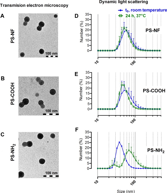
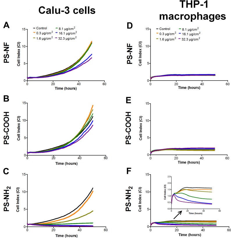
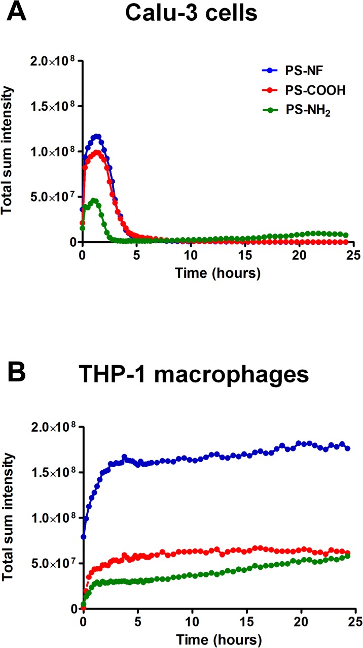
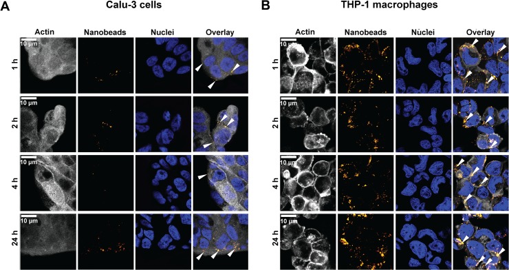
Nanoparticle surface chemistry is known to play a crucial role in interactions with cells and their related cytotoxic effects. As inhalation is a major route of exposure to nanoparticles, we studied specific uptake and damages of well-characterized fluorescent 50 nm polystyrene (PS) nanobeads harboring different functionalized surfaces (non-functionalized, carboxylated and aminated) on pulmonary epithelial cells and macrophages (Calu-3 and THP-1 cell lines respectively). Cytotoxicity of in mass dye-labeled functionalized PS nanobeads was assessed by xCELLigence system and alamarBlue viability assay. Nanobeads-cells interactions were studied by video-microscopy, flow cytometry and also confocal microscopy. Finally ROS generation was assessed by glutathione depletion dosages and genotoxicity was assessed by γ-H2Ax foci detection, which is considered as the most sensitive technique for studying DNA double strand breaks. The uptake kinetic was different for each cell line. All nanobeads were partly adsorbed and internalized, then released by Calu-3 cells, while THP-1 macrophages quickly incorporated all nanobeads which were located in the cytoplasm rather than in the nuclei. In parallel, the genotoxicity study reported that only aminated nanobeads significantly increased DNA damages in association with a strong depletion of reduced glutathione in both cell lines. We showed that for similar nanoparticle concentrations and sizes, aminated polystyrene nanobeads were more cytotoxic and genotoxic than unmodified and carboxylated ones on both cell lines. Interestingly, aminated polystyrene nanobeads induced similar cytotoxic and genotoxic effects on Calu-3 epithelial cells and THP-1 macrophages, for all levels of intracellular nanoparticles tested. Our results strongly support the primordial role of nanoparticles surface chemistry on cellular uptake and related biological effects. Moreover our data clearly show that nanoparticle internalization and observed adverse effects are not necessarily associated.
Conflict of interest statement
Figures
Similar articles
-
Aminated polystyrene and DNA strand breaks in A549, Caco-2, THP-1 and U937 human cell lines.Mutat Res Genet Toxicol Environ Mutagen. 2025 Apr;903:503865. doi: 10.1016/j.mrgentox.2025.503865. Epub 2025 Mar 5. Mutat Res Genet Toxicol Environ Mutagen. 2025. PMID: 40185540
-
Human cell line-dependent WC-Co nanoparticle cytotoxicity and genotoxicity: a key role of ROS production.Toxicol Sci. 2015 Feb;143(2):385-97. doi: 10.1093/toxsci/kfu238. Epub 2014 Nov 14. Toxicol Sci. 2015. PMID: 25398624
-
Differential bioreactivity of neutral, cationic and anionic polystyrene nanoparticles with cells from the human alveolar compartment: robust response of alveolar type 1 epithelial cells.Part Fibre Toxicol. 2015 Jul 2;12:19. doi: 10.1186/s12989-015-0091-7. Part Fibre Toxicol. 2015. PMID: 26133975 Free PMC article.
-
Silver Nanoparticle-Mediated Cellular Responses in Various Cell Lines: An in Vitro Model.Int J Mol Sci. 2016 Sep 22;17(10):1603. doi: 10.3390/ijms17101603. Int J Mol Sci. 2016. PMID: 27669221 Free PMC article. Review.
-
Functionalized polystyrene nanoparticles as a platform for studying bio-nano interactions.Beilstein J Nanotechnol. 2014 Dec 15;5:2403-12. doi: 10.3762/bjnano.5.250. eCollection 2014. Beilstein J Nanotechnol. 2014. PMID: 25671136 Free PMC article. Review.
Cited by
-
Role of MicroRNAs in regulation of DNA damage in monocytes exposed to polystyrene and TiO2 nanoparticles.Toxicol Rep. 2020 Jun 3;7:743-751. doi: 10.1016/j.toxrep.2020.05.007. eCollection 2020. Toxicol Rep. 2020. PMID: 32579136 Free PMC article.
-
Antioxidant Defense in Primary Murine Lung Cells following Short- and Long-Term Exposure to Plastic Particles.Antioxidants (Basel). 2023 Jan 18;12(2):227. doi: 10.3390/antiox12020227. Antioxidants (Basel). 2023. PMID: 36829786 Free PMC article.
-
A review on enhanced microplastics derived from biomedical waste during the COVID-19 pandemic with its toxicity, health risks, and biomarkers.Environ Res. 2023 Jan 1;216(Pt 1):114434. doi: 10.1016/j.envres.2022.114434. Epub 2022 Oct 7. Environ Res. 2023. PMID: 36209789 Free PMC article. Review.
-
Microplastics and Oxidative Stress-Current Problems and Prospects.Antioxidants (Basel). 2024 May 8;13(5):579. doi: 10.3390/antiox13050579. Antioxidants (Basel). 2024. PMID: 38790684 Free PMC article. Review.
-
Derivatives of Plastics as Potential Carcinogenic Factors: The Current State of Knowledge.Cancers (Basel). 2022 Sep 24;14(19):4637. doi: 10.3390/cancers14194637. Cancers (Basel). 2022. PMID: 36230560 Free PMC article. Review.
References
-
- ISO (2008) Nanotechnologies—Terminology and definitions for nano-objects—Nanoparticle, nanofibre and nanoplate. ISO/TS 27687:2008.
-
- Tetley TD. Health effects of nanomaterials. Biochem Soc Trans 2007; 35: 527–531. - PubMed
Publication types
MeSH terms
Substances
LinkOut - more resources
Full Text Sources
Other Literature Sources
Research Materials
