. 2015 Aug;236(4):433-44.
doi: 10.1002/path.4548. Epub 2015 May 12.

Macrophage PPARγ and impaired wound healing in type 2 diabetes

Affiliations

Macrophage PPARγ and impaired wound healing in type 2 diabetes

Rita E Mirza et al. J Pathol. 2015 Aug.

Abstract

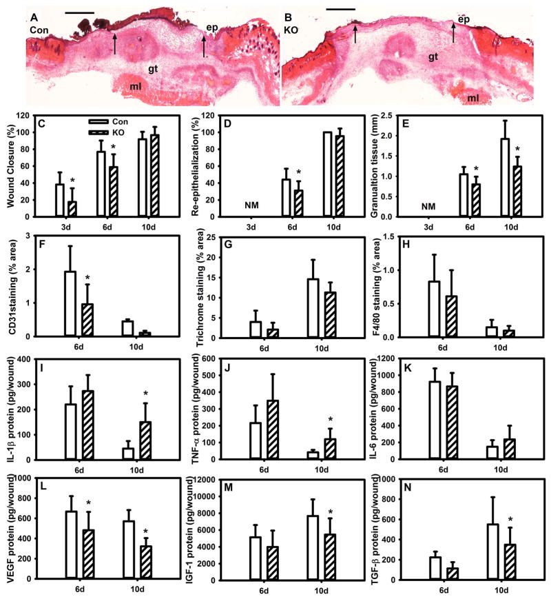
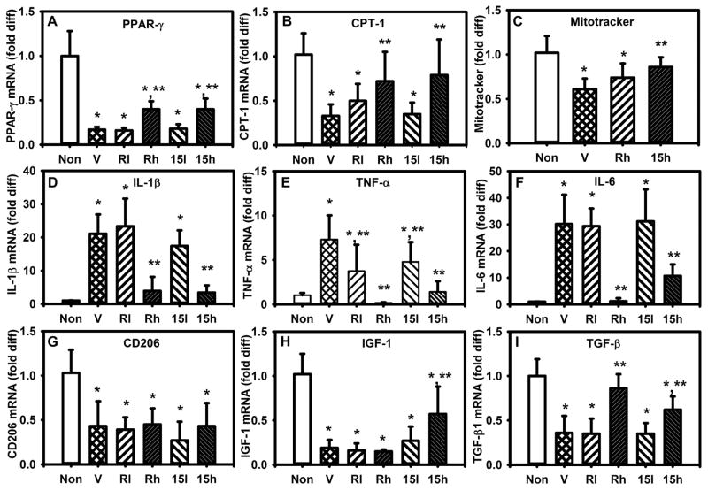
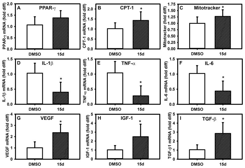
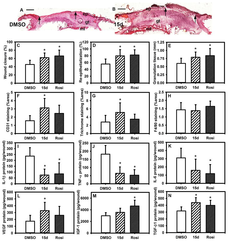
Macrophages undergo a transition from pro-inflammatory to healing-associated phenotypes that is critical for efficient wound healing. However, the regulation of this transition during normal and impaired healing remains to be elucidated. In our studies, the switch in macrophage phenotypes during skin wound healing was associated with up-regulation of the peroxisome proliferator-activated receptor (PPAR)γ and its downstream targets, along with increased mitochondrial content. In the setting of diabetes, up-regulation of PPARγ activity was impaired by sustained expression of IL-1β in both mouse and human wounds. In addition, experiments with myeloid-specific PPARγ knockout mice indicated that loss of PPARγ in macrophages is sufficient to prolong wound inflammation and delay healing. Furthermore, PPARγ agonists promoted a healing-associated macrophage phenotype both in vitro and in vivo, even in the diabetic wound environment. Importantly, topical administration of PPARγ agonists improved healing in diabetic mice, suggesting an appealing strategy for down-regulating inflammation and improving the healing of chronic wounds.

Keywords: diabetes; inflammation; macrophage; resolution of inflammation; wound healing.


Conflict of interest statement

Conflict of Interest Statement: The authors have no conflicting financial interests.

Figures

Figure 1
Figure 1. Impaired PPAR-γ activity in diabetic wound macrophages
(A–D) Macrophages (Mp) were isolated from chronic wound biopsies and expression of PPAR-γ, PGC-1β and downstream targets CD36 and CPT-1 assessed by real time PCR. For comparison, blood-derived Mp from healthy volunteers were either left non-stimulated (Non) or stimulated with IL-1β. (EH) Expression of PPAR-γ, PGC-1β and downstream targets in Mp isolated from wounds of non-diabetic (ND) and diabetic (DB) mice on days 5, 10 and 20 post-injury. (I–L) Expression of PPAR-γ, PGC-1β and downstream targets in bone marrow-derived Mp from wild-type mice stimulated with day 5 or 10 wound conditioned medium (CM) from ND or DB mice. (M) Representative flow cytogram of MitoTracker green labeling in Mp (F4/80+ cells) isolated from day 10 ND and DB wounds. (N) Summary data for median fluorescence intensity (MFI) for MitoTracker green in Mp isolated from day 5 and 10 wounds of ND and DB mice. (O) Representative flow cytogram of MitoTracker green labeling in cultured Mp treated with CM from day 10 wounds of ND or DB mice. (P) Summary data for MFI for MitoTracker green in cultured Mp treated with recombinant IL-1β or with CM from day 10 wounds of ND or DB mice. For all graphs, bars = mean + SD, n = 6 for human data and n = 6–8 for mouse data. *mean value significantly different from that for Non controls or 5d values, **mean value for DB significantly different from that for ND at same time point, p < 0.05.
Figure 2
Figure 2. IL-1β in the diabetic wound environment inhibits Mp PPAR-γ activity
(A–D) Correlation between expression of IL-1β and expression of PPAR-γ, PGC-1β or downstream targets in macrophages (Mp) isolated from chronic wound biopsies; expression of each gene assessed by real time PCR. (E–H) Expression of PPAR-γ, PGC-1β and downstream targets in Mp isolated on day 10 after injury from wounds in diabetic (DB) mice treated topically with control IgG (Ig) or IL-1β blocking antibody (ab) compared with non-diabetic (ND) mice. (I–L) Expression of PPAR-γ, PGC-1β and downstream targets in cultured bone marrow-derived Mp from wild-type (WT) mice either left non-stimulated (Non) or stimulated with day 10 DB wound-conditioned medium (CM) along with Ig or ab. (M–P) Expression of PPAR-γ, PGC-1β and downstream targets in bone marrow-derived Mp from WT and IL-1R1 KO mice stimulated with CM from wounds of day 10 wounds of DB mice. For all graphs, bars = mean ± SD, n = 6. *mean value significantly different from that for ND mice for in vivo experiments and for non-stimulated controls for in vitro experiments, **mean value for ab-treated condition significantly different from that for Ig-treated condition. #mean value significantly different from that for Non controls of same strain, ##mean value significantly different from that for CM-treated WT macrophages, p < 0.05.
Figure 3
Figure 3. Myeloid cell-specific knockout of PPAR-γ impairs resolution of inflammation and wound healing in non-diabetic mice
(A,B) Excisional wounds in control (Con) PPAR-γflox mice and Lyz2Cre/PPAR-γflox knockout (KO) mice were harvested on day 6 after injury, sectioned and stained with H&E. Note the reduced re-epithelialization and granulation tissue formation in the KO mice. ep: epithelium, gt: granulation tissue, ml: muscle layer; arrows indicate ends of epithelial tongues, scale bar = 0.5 mm. (C) Wound closure assessed in digital images of wound surface on days 3, 6 and 10 after injury; (D,E) re-epithelialization and granulation tissue thickness measured in H&E stained cryosections; (F–H) CD31 staining, Trichrome staining and F4/80 staining measured in cryosections as % area stained. (I–N) Levels of cytokines in wound homogenates measured using ELISA, including pro-inflammatory cytokines IL-1β, TNF-α, IL-6 and healing-associated cytokines VEGF, IGF-1, TGF-β. For all graphs, bars = mean ± SD, n = 8. *mean value significantly different from that for Con at same time point, p < 0.05.
Figure 4
Figure 4. PPAR-γ agonists reverse effects of a simulated diabetic wound environment
(A,B) Expression of PPAR-γ and downstream target CPT-1 in cultured bone marrow-derived Mp from non-diabetic (ND) mice either left non-stimulated (Non) or stimulated with day 10 DB wound conditioned medium (CM) plus vehicle (V), CM plus low or high doses of rosiglitazone (Rl, Rh; 10, 50 μM) or CM plus low or high doses of 15d-PGJ2 (15l, 15h; 1, 10 μM). (C) Median fluorescence (MFI) of MitoTracker staining, (D–F) expression of pro-inflammatory cytokines, (G–I) expression of healing-associated factors, with stimulation as indicated. For all graphs, bars = mean ± SD, n = 6. *mean value significantly different from that for Non controls, **mean value significantly different from that for CV + V treated samples, p < 0.05.
Figure 5
Figure 5. PPAR-γ agonists induce switch to healing-associated wound Mp phenotype in diabetic mice
Excisional wounds in diabetic (DB) mice were treated topically with 15d-PGJ2 (10 μM) or DMSO vehicle and Mp isolated from day 10 wounds. (A,B) Wound Mp expression of PPAR-γ and downstream target CPT-1, (C) median fluorescence (MFI) of MitoTracker staining, (D–F) expression of pro-inflammatory cytokines, (G–I) expression of healing-associated factors, with stimulation as indicated. For all graphs, bars = mean ± SD, n = 8. *mean value significantly different from that for DMSO controls, p < 0.05.
Figure 6
Figure 6. PPAR-γ agonists improve wound healing in diabetic mice
Excisional wounds in diabetic (DB) mice were treated topically with 15d-PGJ2 (10 μM), rosiglitazone (50 μM) or DMSO vehicle and harvested on day 10 after injury. (A,B) Examples of wounds treated with 15d-PGJ2 and assessed by H&E staining. Note the increased re-epithelialization and granulation tissue formation in the PPAR-γ agonist treated mice. ep: epithelium, gt: granulation tissue, ml: muscle layer; arrows indicate ends of epithelial tongues, scale bar = 0.5 mm. (C) Wound closure assessed in digital images of wound surface, (D,E) re-epithelialization and granulation tissue thickness measured in H&E-stained cryosections (F–H) CD31 staining, Trichrome staining and F4/80 staining measured in cryosections as % area stained. (I–N) Levels of cytokines in wound homogenates measured using ELISA, including pro-inflammatory cytokines IL-1β, TNF-α, IL-6 and healing-associated cytokines VEGF, IGF-1, TGF-β. For all graphs, bars = mean ± SD, n = 8. *mean value significantly different from that for DMSO treatment, p < 0.05.

References

    1. Ramasamy R, Yan SF, Schmidt AM. Receptor for AGE (RAGE): signaling mechanisms in the pathogenesis of diabetes and its complications. Ann N Y Acad Sci. 2011;1243:88–102. doi: 10.1111/j.1749-6632.2011.06320.x. - DOI - PMC - PubMed
    1. Hill MJ, Metcalfe D, McTernan PG. Obesity and diabetes: lipids, ‘nowhere to run to’. Clin Sci (Lond) 2009;116:113–123. doi: 10.1042/CS20080050. CS20080050 [pii] - DOI - PubMed
    1. Jin C, Flavell RA. Innate sensors of pathogen and stress: linking inflammation to obesity. J Allergy Clin Immunol. 2013;132:287–294. doi: 10.1016/j.jaci.2013.06.022. S0091-6749(13)00990-1 [pii] - DOI - PubMed
    1. Maskrey BH, Megson IL, Whitfield PD, et al. Mechanisms of resolution of inflammation: a focus on cardiovascular disease. Arterioscler Thromb Vasc Biol. 2011;31:1001–1006. doi: 10.1161/ATVBAHA.110.213850. 31/5/1001 [pii] - DOI - PubMed
    1. Spite M, Claria J, Serhan CN. Resolvins, specialized proresolving lipid mediators, and their potential roles in metabolic diseases. Cell Metab. 2014;19:21–36. doi: 10.1016/j.cmet.2013.10.006. S1550-4131(13)00418-X [pii] - DOI - PMC - PubMed

Publication types

MeSH terms