. 2015 Apr 13;10(4):e0122799.
doi: 10.1371/journal.pone.0122799. eCollection 2015.

A mathematical model of collective cell migration in a three-dimensional, heterogeneous environment

Affiliations

A mathematical model of collective cell migration in a three-dimensional, heterogeneous environment

David P Stonko et al. PLoS One. .

Abstract

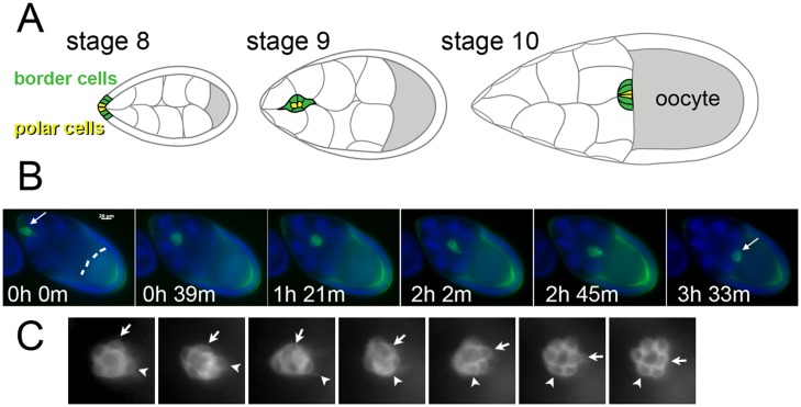
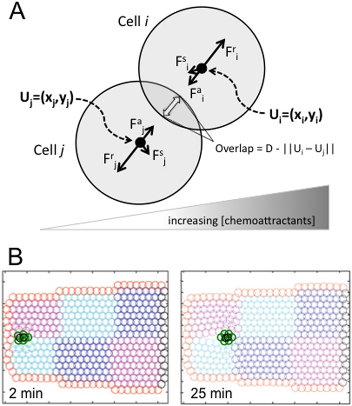
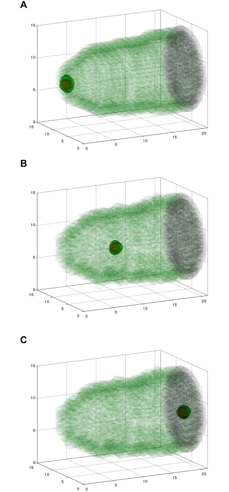
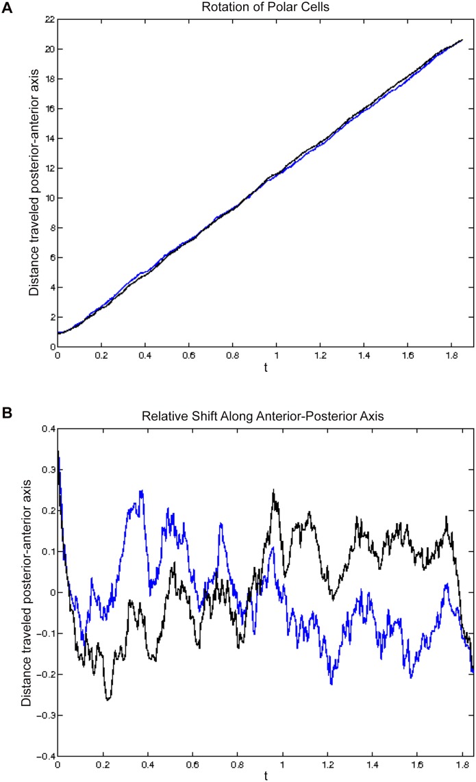
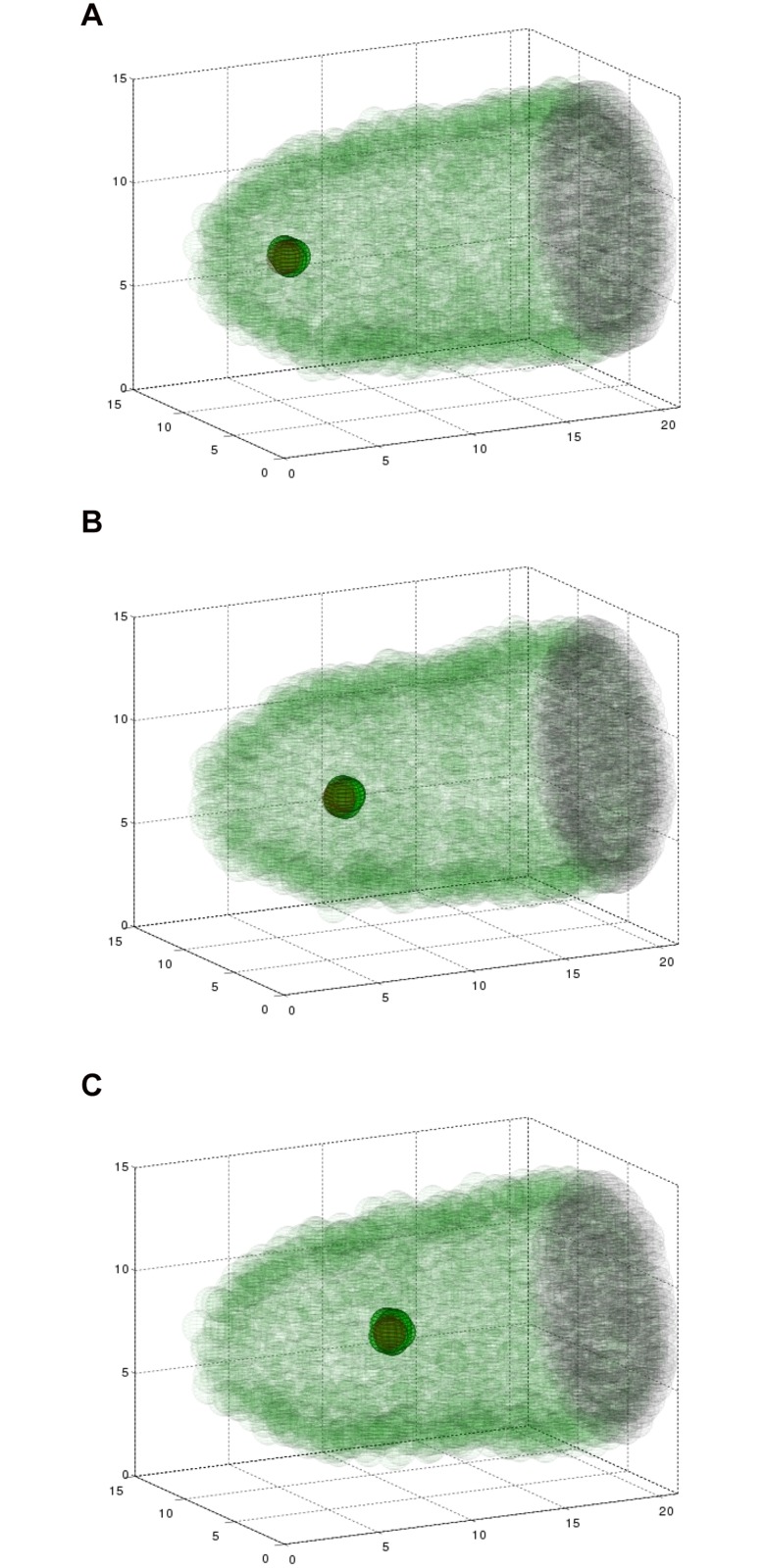
Cell migration is essential in animal development, homeostasis, and disease progression, but many questions remain unanswered about how this process is controlled. While many kinds of individual cell movements have been characterized, less effort has been directed towards understanding how clusters of cells migrate collectively through heterogeneous, cellular environments. To explore this, we have focused on the migration of the border cells during Drosophila egg development. In this case, a cluster of different cell types coalesce and traverse as a group between large cells, called nurse cells, in the center of the egg chamber. We have developed a new model for this collective cell migration based on the forces of adhesion, repulsion, migration and stochastic fluctuation to generate the movement of discrete cells. We implement the model using Identical Math Cells, or IMCs. IMCs can each represent one biological cell of the system, or can be aggregated using increased adhesion forces to model the dynamics of larger biological cells. The domain of interest is filled with IMCs, each assigned specific biophysical properties to mimic a diversity of cell types. Using this system, we have successfully simulated the migration of the border cell cluster through an environment filled with larger cells, which represent nurse cells. Interestingly, our simulations suggest that the forces utilized in this model are sufficient to produce behaviors of the cluster that are observed in vivo, such as rotation. Our framework was developed to capture a heterogeneous cell population, and our implementation strategy allows for diverse, but precise, initial position specification over a three- dimensional domain. Therefore, we believe that this model will be useful for not only examining aspects of Drosophila oogenesis, but also for modeling other two or three-dimensional systems that have multiple cell types and where investigating the forces between cells is of interest.


Conflict of interest statement

Competing Interests: The authors have declared that no competing interests exist.

Figures

Fig 1
Fig 1. Border cell migration in Drosophila melanogaster egg development.
(a) At the beginning of stage eight, the polar cells (yellow) and border cells (green) lie in the follicular epithelium of the developing egg chamber. In stage nine, these cells coalesce to form a cluster that detaches from the epithelium. The cluster then translocates between large nurse cells through the egg chamber to reach the developing oocyte (gray) by stage ten. The border cell cluster migrates about 150μm over approximately 4–6 hours. (b) Still images from a time-lapse movie of wild-type border cell migration. The motile cells are marked in green by expression of Slbo-life-Act-GFP. The oocyte, which autofluoresces, is indicated by the dashed line. The nuclei of all cells, including the large, polyploid nurse cells, are seen in blue. In the image at time 0, border cells have already clustered (arrow) and begun moving towards the oocyte. In this example, the border cells reached the oocyte border by 3.5h (arrow on right-most panel). (c) Still images at a higher magnification from a time-lapse movie of a different egg chamber. Images differ by 30 minute intervals. The border cells are marked by a membrane-tethered GFP, and show wild-type behavior. Cells can be observed to change relative positions with respect to the front of the cluster as they move toward the right (arrow and arrowhead indicate the same cell over time). See also Supplemental S1 and S2 Movies.
Fig 2
Fig 2. A force-based biophysical model comprised of discrete Individual Math Cells.
(a) The forces between two adjacent IMCs, i and j. The repulsive force acts to separate contacting IMCs, while the adhesive force acts to attract IMC i and j when they are within an ε distance from one another. If one of the IMCs is migratory, it produces a migratory force perpendicular to the axis of interaction due to signal from the gradient of chemoattractants. There is stochastic fluctuation in the position of each IMC. These forces balance and produce overlap between i and j of D − ‖U iU j2, where D is the diameter of the IMC. Adhesion force between IMCs creates the integrity of a large single cell or cluster of individual cells with special affinity. The central cluster with a higher adhesion coefficient is closer or more tightly bound to one another than to outside cells, or than outside cells are to one another. (b) The IMC- based domain and simulation in two dimensions. The anterior half of the egg chamber is represented by IMCs with different properties. The epithelial cells are equated to IMCs (red), while the nurse cells are formed from many IMCs (blue, pink, and cyan). The line of black IMCs to the right is the surface of the oocyte at the mid-point of the whole egg chamber. The migratory cluster is formed of tightly bound border cells (green) and polar cells (black). 25 minutes into a 2D simulation, the model shows penetration of the cluster into the egg chamber between malleable nurse cells.
Fig 3
Fig 3. Simulating the three dimensional model results in collective migration.
A simulation showing six border cells (green), two polar cells (red), the epithelium (transparent green), and the surface of the oocyte (black, right) at three time points during the migration. Fifteen nurse cells are situated inside the egg chamber, but are not plotted so as to maintain clarity of this three dimensional structure. Polar cells are surrounded by border cells, making them hard to distinguish. (A) At 2 minutes, cells are beginning to invade between nurse cells. (B) At 2.4 hours, the cluster is about halfway to its destination. (C) At 5.6 hours, the border cell cluster has reached the edge of the oocyte. See also Supplemental S3 Movie.
Fig 4
Fig 4. Polar cell positions along main axis of migration.
(A) The distance of the polar cells from the anterior of the egg chamber versus time. (B) The relative positions of the two polar cells to one another, along the axis that runs from anterior to posterior through the egg chamber. Each line corresponds to one of the polar cells. As the cluster moves forward, we observe that the polar cells are changing position with respect to one another along this axis, including a complete switch at 0.8 hours. This simulation modeled six border cells.
Fig 5
Fig 5. Simulations with four, six, and eight border cells at the same time point (t = 1.8 hours) during migration.
The cluster with four border cells (A) has moved significantly less distance than the cluster with six (B) or eight (C) border cells.

References

    1. Starz-Gaiano M, Melani M, Wang X, Meinhardt H, Montell DJ. Feedback Inhibition of JAK/STAT Signaling by Apontic Is Required to Limit an Invasive Cell Population. Developmental Cell. 2008;14(5):726–738. Available from: http://www.sciencedirect.com/science/article/pii/S1534580708001135 . doi: http://www.sciencedirect.com/science/article/pii/S153458070800113510.1016/j.devcel.2008.03.005. doi: 10.1016/j.devcel.2008.03.005 - DOI - PubMed
    1. Ridley AJ, Schwartz MA, Burridge K, Firtel RA, Ginsberg MH, Borisy G, et al. Cell Migration: Integrating Signals from Front to Back. Science. 2003;302(5651):1704–1709. Available from: http://www.sciencemag.org/content/302/5651/1704.abstract . doi: http://www.sciencemag.org/content/302/5651/1704.abstract10.1126/science.1092053. doi: 10.1126/science.1092053 - DOI - PubMed
    1. Montell DJ, Yoon WH, Starz-Gaiano M. Group choreography: mechanisms orchestrating the collective movement of border cells. Nature Reviews Molecular Cell Biology. 2012;13(10):631–645. 10.1038/nrm3433 - DOI - PMC - PubMed
    1. Wright N, Hoffmann W, Otto W, Rio MC, Thim L. Rolling in the clover: trefoil factor family (TFF)-domain peptides, cell migration and cancer. FEBS Letters. 1997;408(2):121–123. Available from: http://www.sciencedirect.com/science/article/pii/S0014579397004249 . doi: http://www.sciencedirect.com/science/article/pii/S001457939700424910.1016/S0014-5793(97)00424-9. doi: 10.1016/S0014-5793(97)00424-9 - DOI - PubMed
    1. Ridley AJ. Rho GTPases and cell migration. Journal of Cell Science. 2001;114(15):2713–2722. Available from: http://jcs.biologists.org/content/114/15/2713.abstract - PubMed

Publication types

LinkOut - more resources