. 2015 Apr 13;10(4):e0119559.
doi: 10.1371/journal.pone.0119559. eCollection 2015.

Soluble T-cell receptors produced in human cells for targeted delivery

Affiliations

Soluble T-cell receptors produced in human cells for targeted delivery

Even Walseng et al. PLoS One. .

Abstract

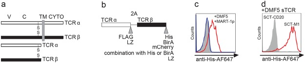
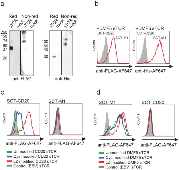
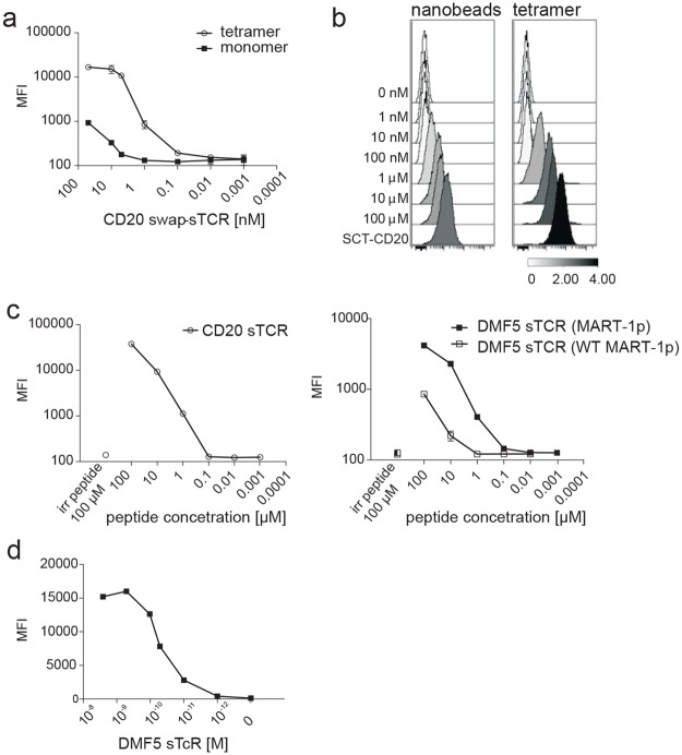
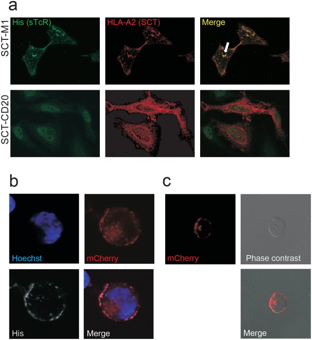
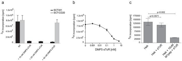
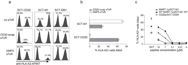
Recently, technology has become available to generate soluble T-cell receptors (sTCRs) that contain the antigen recognition part. In contrast to antibodies, sTCRs recognize intracellular in addition to extracellular epitopes, potentially increasing the number of applications as reagents for target detection and immunotherapy. Moreover, recent data show that they can be used for identification of their natural peptide ligands in disease. Here we describe a new and simplified expression method for sTCRs in human cells and show that these sTCRs can be used for antigen-specific labeling and elimination of human target cells. Four different TCRs were solubilized by expression of constructs encoding the TCR alpha (α) and beta (β) chains lacking the transmembrane and intracellular domains, linked by a ribosomal skipping 2A sequence that facilitates equimolar production of the chains. Cell supernatants containing sTCRs labeled target cells directly in a peptide (p)-human leukocyte antigen (HLA)-specific manner. We demonstrated that a MART-1p/HLA-A*02:01-specific sTCR fused to a fluorescent protein, or multimerized onto magnetic nanoparticles, could be internalized. Moreover, we showed that this sTCR and two sTCRs recognizing CD20p/HLA-A*02:01 could mediate selective elimination of target cells expressing the relevant pHLA complex when tetramerized to streptavidin-conjugated toxin, demonstrating the potential for specific delivery of cargo. This simple and efficient method can be utilized to generate a wide range of minimally modified sTCRs from the naturally occurring TCR repertoire for antigen-specific detection and targeting.


Conflict of interest statement

Competing Interests: The authors have declared that no competing interests exist.

Figures

Fig 1
Fig 1. Design of the sTCR and its use as a labeling reagent.
(a) TCRα/β chains in full-length (upper part) and in soluble form (lower part). V: Variable domain, C: Constant domain, TM: transmembrane, CYTO: cytosolic domain. Interchain cystein bridges are indicated with S. (b) Design of the expression construct in which truncated TCRα and β chains are separated by a ribosome skipping sequence (2A) and tagged on their 3’-end. The tags used in this study are listed at their respective position. To increase interchain stability, a high affinity leucine zipper (LZ) was added to some sTCR constructs. (c) HLA-A2pos T2 cells were loaded O/N with 10 μM MART-1p (red, blue) or 10 μM of an irrelevant peptide (CD20p188–196) (filled grey). Ten μL of supernatant from HEK293 cells producing the DMF5 sTCR (red), or from mock transfected HEK 293 (blue), was added to the T2 cells and incubated for 15 minutes at RT followed by labeling with anti-His-647 antibodies and flow cytometric analysis (d) SupT1 cells expressing SCT-M1 (red) or SCT-CD20 (filled grey) were incubated with the DMF5 sTCR supernatant as described in (c) and stained using anti-His-647. The results in (c) and (d) are representative of at least two experiments and histograms are gated on viable cells, displayed as FSChi, SSChi events.
Fig 2
Fig 2. The sTCR is produced as a heterodimer and recognizes the correct pMHC.
(a) Supernatant from HEK293 cells expressing FLAG- and His-tagged DMF5 sTCR or not (mock) was loaded on SDS-PAGE under reducing (Red) or non-reducing (Non-red) conditions. The membrane was subsequently stained with anti-His or anti-FLAG antibodies. (b) The same sTCR as in (a) was used to stain SupT1 cells expressing SCT-M1 (red) or SCT-CD20 (filled grey) and stained with anti-FLAG-647 (left panel) or anti-His-AF647 (right panel). The results in (a) and (b) are representative of at least 2 experiments. (c) SupT1 cells expressing SCT-CD20 (left panel) or SCT-M1 (right panel, negative control) were incubated with unmodified (green), cys modified (blue) or sTCR LZ modified (red) CD20 swap-sTCR. They were visualized using anti-FLAG-647 antibody. EBV-sTCR (filled grey) served as a negative control. (d) SupT1 cells expressing SCT-M1 (left panel) or SCT-CD20 (right panel, negative control) were incubated with unmodified DMF5 sTCR (green), DMF5 sTCR with a cys modification (blue) or DMF5 sTCR LZ (red), and subsequently visualized using an anti-FLAG-647 antibody. As a negative control, the SCT-M1 cells were also incubated with an irrelevant EBV-sTCR (filled grey). In (b-d), histograms are gated on viable cells, displayed as FSChi, SSChi events.
Fig 3
Fig 3. Multimerization of the sTCR increases sensitivity of antigen detection by flow cytometry.
(a) SupT1 cells expressing SCT-CD20 were incubated with increasing concentrations of CD20 swap-sTCR monomer or tetramer, as indicated, and indirectly labeled with anti-FLAG AF647. Results shown are representative of two experiments, and error bars represent SD of duplicates (b) HLA-A2+ SupT1 cells were loaded with indicated concentrations of peptide and subsequently labeled with saturating amounts of either sTCR tetramer or nanobeads conjugated to sTCR monomers, followed by staining with anti-FLAG-AF647. Results shown are representative of two experiments. SupT1 cells expressing SCT-CD20 were used as a positive control. The scale shows the calculation of Arcsinh ratio of the Median. (c) SupT1 cells were loaded with increasing concentrations of CD20p (left panel), MART-1p or WT MART-1p (right panel), followed by labeling with saturating amounts of indicated sTCR tetramer, and indirectly stained with anti-FLAG AF647. Irrelevant peptide control (irr peptide) was used at 100 μM (disconnected symbols). Results shown are from one experiment representative of 3 (CD20p and MART-1p) or 2 (WT MART-1p) experiments. Error bars indicate SD of duplicates. (d) SupT1 cells expressing SCT-M1 were incubated with increasing concentrations of PE-conjugated sTCR tetramer.
Fig 4
Fig 4. DMF5 sTCR is internalized upon specific ligand binding.
(a) HeLa cells transfected with either SCT-M1-mCherry or SCT-CD20-mCherry (red) were incubated with His-tagged DMF5 sTCR labeled with an anti-His antibody and visualized using anti-mouse AF488 (green). Co-localization of the sTCR and SCT-M1 is shown in yellow (arrow). (b) Sup-T1 cells expressing SCT-M1 were incubated with DMF5 supernatants containing monomeric sTCR mCherry-His at 37°C for 30 minutes. The cells were subsequently put on ice to block endocytosis and stained with anti-His-AF647 (His). The nucleus was visualized by Hoechst stain (blue). (c) Sup-T1 cells expressing SCT-M1 were incubated with biotinylated DMF5 sTCR-mCherry bound to SA-Miltenyi nanobeads at 37°C.
Fig 5
Fig 5. Antigen-specific elimination of target cells by sTCR-Saporin as measured by 3H-thymidine incorporation.
(a) SupT1 cells constitutively expressing SCT-M1 or SCT-CD20 were incubated with or without (NT) 10–40 nM DMF5 sTCR-Sap for 3 days. Cells were then labeled with 3H-thymidine and radioactive incorporation was measured (cpm). (b) SupT1 cells expressing SCT-M1 were incubated with increasing amounts of DMF5 sTCR-Sap for 3 days and 3H-thymidine incorporation was plotted as in (a). (c) HEK293 cells were pulsed with or without 10 μM MART-1p in the presence or absence of 20 nM DMF5 sTCR-Sap for 3 days, and 3H-thymidine incorporation was determined. The experiments shown in (a-c) are each representative of two performed and error bars represent SD from triplicates.
Fig 6
Fig 6. Antigen-specific elimination of target cells by sTCR-Saporin as measured by flow cytometry.
(a) SupT1 cells expressing the indicated SCT were mixed at a 1:1 ratio with HLA-A2neg SupT1 cells and co-cultured for 3 days in the presence of relevant or irrelevant sTCR conjugated to Saporin, as indicated. At end of culture, cells were stained with anti-HLA-A2 AF647 to determine the frequencies of HLA-A2 positive and negative target cells among FSChi,SSChi events using flow cytometry. (b) Assay was performed as described in (a) and the percentage of SupT1 cells expressing indicated SCT that was eliminated by addition of sTCR is shown, calculated as described in materials and methods. Error bars indicate SEM of n = 3. (c) HLA-A2pos SupT1 cells mixed at a ratio of 1:1 with HLA-A2neg SupT1 cells were incubated overnight with indicated concentrations of peptide, washed, and indicated sTCR-Sap was added. SupT1 cells expressing SCT were used as positive controls (disconnected symbols). Culture and analysis to determine the percentage of eliminated HLA-A2pos targets was performed as described in (a,b). Results shown are representative of two experiments performed, and error bars represent SD of duplicates.

References

    1. Davis MM, Bjorkman PJ. T-cell antigen receptor genes and T-cell recognition. Nature. 1988;334(6181):395–402.. - PubMed
    1. Gregoire C, Rebai N, Schweisguth F, Necker A, Mazza G, Auphan N, et al. Engineered secreted T-cell receptor alpha beta heterodimers. Proc Natl Acad Sci U S A. 1991;88(18):8077–81. . - PMC - PubMed
    1. Garcia KC, Degano M, Stanfield RL, Brunmark A, Jackson MR, Peterson PA, et al. An alphabeta T cell receptor structure at 2.5 A and its orientation in the TCR-MHC complex. Science. 1996;274(5285):209–19. . - PubMed
    1. Molloy PE, Sewell AK, Jakobsen BK. Soluble T cell receptors: novel immunotherapies. Curr Opin Pharmacol. 2005;5(4):438–43. . - PubMed
    1. Richman SA, Kranz DM. Display, engineering, and applications of antigen-specific T cell receptors. Biomol Eng. 2007;24(4):361–73. . - PubMed

Publication types

MeSH terms