. 2015 Apr 15;11(4):e1005169.
doi: 10.1371/journal.pgen.1005169. eCollection 2015 Apr.

A missense change in the ATG4D gene links aberrant autophagy to a neurodegenerative vacuolar storage disease

Affiliations

A missense change in the ATG4D gene links aberrant autophagy to a neurodegenerative vacuolar storage disease

Kaisa Kyöstilä et al. PLoS Genet. .

Abstract

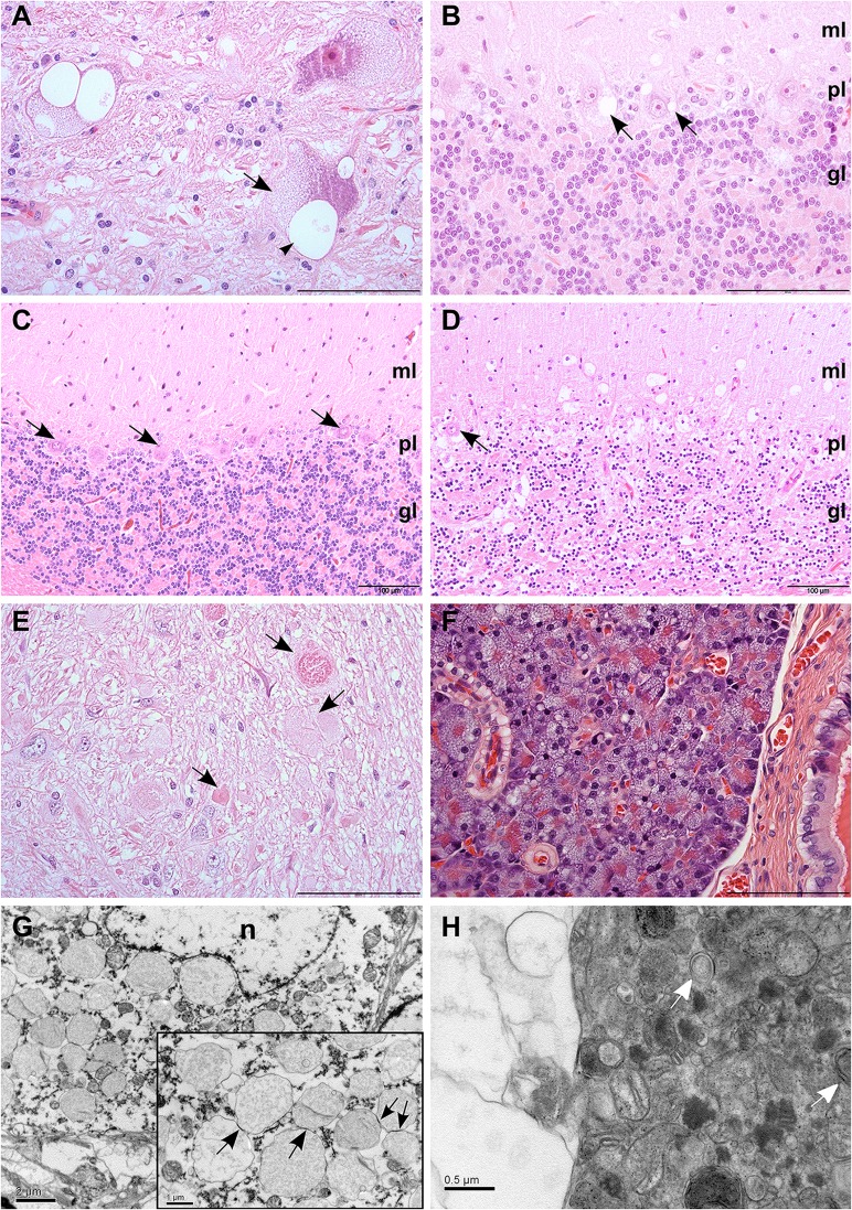
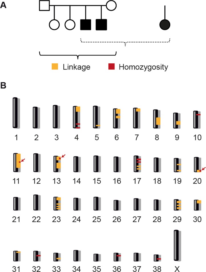
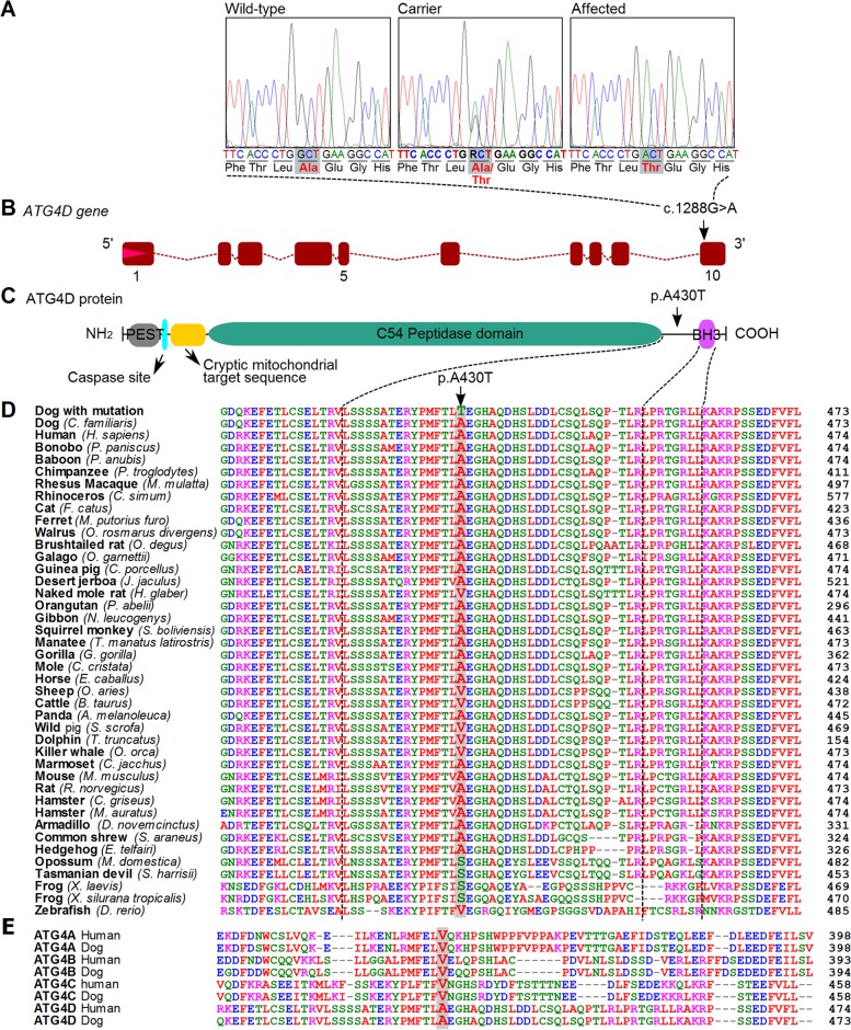
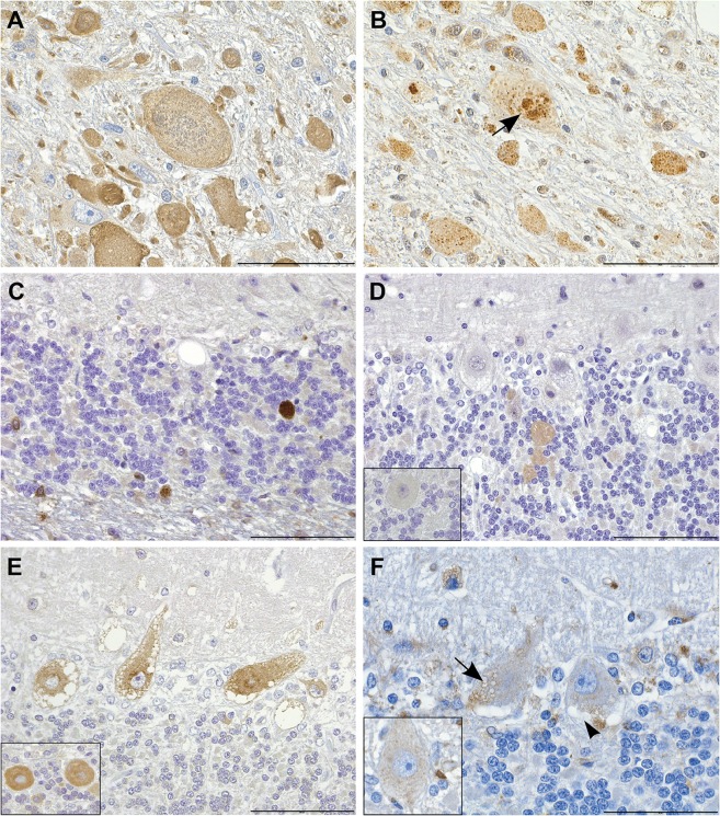
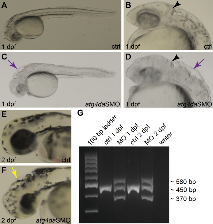
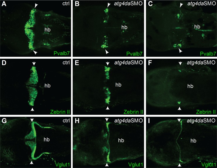
Inherited neurodegenerative disorders are debilitating diseases that occur across different species. We have performed clinical, pathological and genetic studies to characterize a novel canine neurodegenerative disease present in the Lagotto Romagnolo dog breed. Affected dogs suffer from progressive cerebellar ataxia, sometimes accompanied by episodic nystagmus and behavioral changes. Histological examination revealed unique pathological changes, including profound neuronal cytoplasmic vacuolization in the nervous system, as well as spheroid formation and cytoplasmic aggregation of vacuoles in secretory epithelial tissues and mesenchymal cells. Genetic analyses uncovered a missense change, c.1288G>A; p.A430T, in the autophagy-related ATG4D gene on canine chromosome 20 with a highly significant disease association (p = 3.8 x 10-136) in a cohort of more than 2300 Lagotto Romagnolo dogs. ATG4D encodes a poorly characterized cysteine protease belonging to the macroautophagy pathway. Accordingly, our histological analyses indicated altered autophagic flux in affected tissues. The knockdown of the zebrafish homologue atg4da resulted in a widespread developmental disturbance and neurodegeneration in the central nervous system. Our study describes a previously unknown canine neurological disease with particular pathological features and implicates the ATG4D protein as an important autophagy mediator in neuronal homeostasis. The canine phenotype serves as a model to delineate the disease-causing pathological mechanism(s) and ATG4D function, and can also be used to explore treatment options. Furthermore, our results reveal a novel candidate gene for human neurodegeneration and enable the development of a genetic test for veterinary diagnostic and breeding purposes.


Conflict of interest statement

I have read the journal's policy and the authors of this manuscript have the following competing interests: A genetic test will be available later from Genoscoper Ltd, which is partly owned by HL.

Figures

Fig 1
Fig 1. Histological findings in neurons and pancreas.
(A) Swelling of neurons in the vestibular nucleus due to fine vesiculation (arrows) and clear vacuolization (arrowhead) of the cytoplasm. HE, scale bar 100 μm. (B) Clear cytoplasmic vacuolization (arrows) in cerebellar cortical Purkinje cells. HE, scale bar 100 μm. (C) Normal cerebellar cortex of an unaffected dog shows viable Purkinje cells (arrows) and a dense granular cell layer. HE, scale bar 100 μm. (D) Marked neuronal loss is present the cerebellar cortex of an affected dog. The number of neurons in the granular cell layer is reduced and only scattered Purkinje cells remain (arrow). HE, scale bar 100 μm. (E) Axonal spheroids of varying quality were seen in the white matter (arrows) of cerebellum and brainstem. HE, scale bar 100 μm. (F) Diffuse cytoplasmic vacuolization of the exocrine pancreatic acinar cells. HE, scale bar 100 μm. (G) Purkinje cell with numerous single-membrane bound, cytoplasmic vacuoles tethering to each other (arrows, inset). Electronmicrograph, scale bar 2 μm. Inset: scale bar 1 μm. (H) Axonal spheroid containing aggregated degenerated mitochondria, occasional double-membrane-bound autophagosomes (arrows) and free electron dense material, compressed by a peripheral clear vacuolar space. Electronmicrograph, scale bar 0.5 μm. Abbreviations: n, nucleus; ml, molecular layer; pl, Purkinje cell layer; gl, granular cell layer.
Fig 2
Fig 2. Linkage analysis and homozygosity mapping.
(A) A family comprising six animals and one very distantly related case were available for the initial mapping of the disease locus. We performed parametric linkage analysis for a recessive trait in the family and homozygosity analysis across the three cases. (B) The analyses yielded 25 linked genome segments (orange) and 11 homozygous genome segments (red). Only three regions on chromosomes 11, 13, and 20 showed both linkage and homozygosity and were considered the critical intervals (arrows).
Fig 3
Fig 3. A missense variant in the ATG4D gene.
(A) Chromatograms showing the c.1288G>A variant in a wild-type, carrier and an affected dog. (B) A schematic presentation of the canine ATG4D gene. The missense variant is positioned in the last exon of the gene. (C) The domain structure of the ATG4D protein. The p.A430T change is situated between the functional domains near the carboxy-terminus. (D) The canine ATG4D 430 alanine residue shows a moderate degree of conservation across the animal kingdom. (E) Conservation of the 430 alanine in the ATG4 protein family in human and dog.
Fig 4
Fig 4. Immunohistochemistry indicates disturbed autophagic flow in neurons.
(A) Axonal spheroids stain diffusely positive for LC3B. IHC LC3B, scale bar 100 μm. (B,C) The granular cores of the spheroids are positive for (B) ubiquitin (arrow) and (C) p62. IHC ubiquitin and p62, scale bars 20 and 100 μm, respectively. (D) Smooth axonal swellings in the cerebellar cortex contain ATG4D. Inset: control. IHC ATG4D, scale bar 100 μm. (E) Affected neurons show increased perinuclear granular LC3B positivity. Inset: control. IHC LC3B, scale bar 100 μm. (F) Neuronal vacuoles are partially LAMP2 positive (arrow) and partially negative (arrow head). Inset: control. IHC LAMP2, scale bar 20 μm.
Fig 5
Fig 5. Phenotype of atg4da morphant zebrafish embryos.
(A-F) Lateral views of control and morphant zebrafish embryos. (A,B) Control embryos appear normal at 1 dpf. (C,D) Morphant embryos show severe abnormalities in different regions of the brain at 1 dpf. Black arrowheads denote the developing cerebellum. Purple arrows indicate the hindbrain irregularities. (E) Control embryo at 2 dpf. (F) A morphant embryo at 2 dpf displaying hydrocephalus (yellow arrow) and small head and eye. (G) RT-PCR assay showing the efficiency of the atg4daSMO.
Fig 6
Fig 6. Suppression of atg4da in zebrafish leads to loss of cerebellar neurons.
Immunostaining with (A-C) anti-Pvalb7 and (D-F) anti-zebrinII antibody show loss of cerebellar Purkinje cells in atg4da morphants. (A,D) Control embryos at 4.5 dpf. (B,E) Morphants with mild phenotype show partial loss of cerebellar Purkinje cells. (C,F) Morphants with strong phenotype show either total loss of Purkinje cells or presence of few differentiated neurons, which are laterally located in the cerebellum. (G-I) Labeling of cerebellar granule cells with anti-Vglut1 antibody in control and morphant embryos. (H) Mildly affected morphants show reduced expression of Vglut1 in the cerebellum. (I) Vglut1 expression is strongly reduced in embryos showing severe phenotype. White arrowheads indicate the region of cerebellum. Abbreviations: hb, hindbrain.

References

    1. Jellinger KA. Basic mechanisms of neurodegeneration: A critical update. J Cell Mol Med. 2010;14(3): 457–487. 10.1111/j.1582-4934.2010.01010.x - DOI - PMC - PubMed
    1. Murrow L, Debnath J. Autophagy as a stress-response and quality-control mechanism: Implications for cell injury and human disease. Annu Rev Pathol. 2013;8: 105–137. 10.1146/annurev-pathol-020712-163918 - DOI - PMC - PubMed
    1. Amm I, Sommer T, Wolf DH. Protein quality control and elimination of protein waste: The role of the ubiquitin-proteasome system. Biochim Biophys Acta. 2014;1843(1): 182–196. 10.1016/j.bbamcr.2013.06.031 - DOI - PubMed
    1. Korolchuk VI, Menzies FM, Rubinsztein DC. Mechanisms of cross-talk between the ubiquitin-proteasome and autophagy-lysosome systems. FEBS Lett. 2010;584(7): 1393–1398. 10.1016/j.febslet.2009.12.047 - DOI - PubMed
    1. Schreiber A, Peter M. Substrate recognition in selective autophagy and the ubiquitin-proteasome system. Biochim Biophys Acta. 2014;1843(1): 163–181. 10.1016/j.bbamcr.2013.03.019 - DOI - PubMed

Publication types

Substances