. 2015;31(2):221-7.
doi: 10.1080/08927014.2015.1026337.

Interfacial pH during mussel adhesive plaque formation

Affiliations

Interfacial pH during mussel adhesive plaque formation

Nadine R Martinez Rodriguez et al. Biofouling. 2015.

Abstract

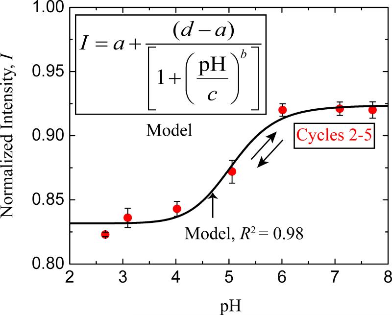
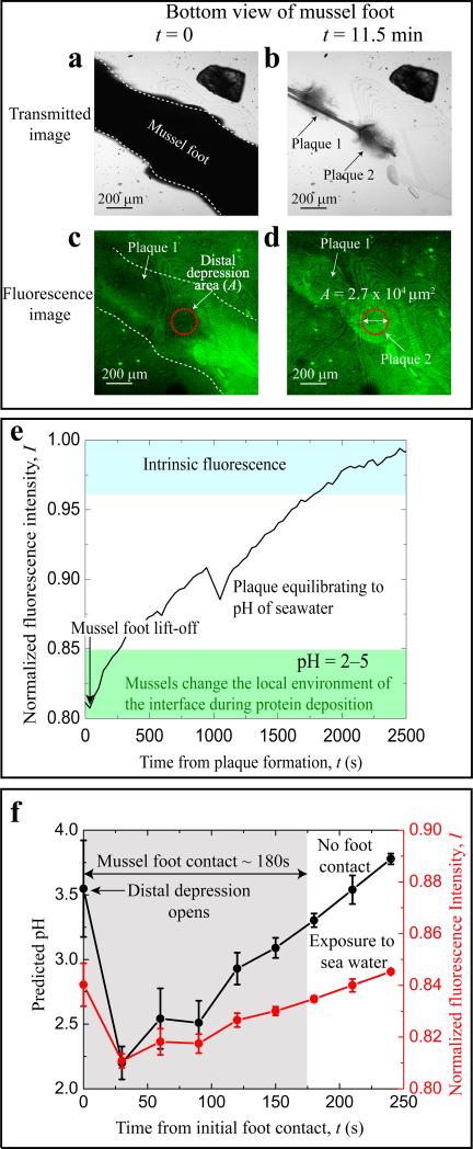
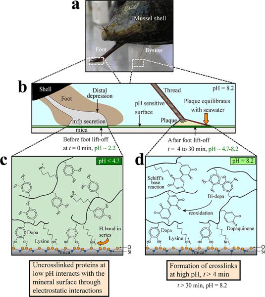
Mussel (Mytilus californianus) adhesion to marine surfaces involves an intricate and adaptive synergy of molecules and spatio-temporal processes. Although the molecules, such as mussel foot proteins (mfps), are well characterized, deposition details remain vague and speculative. Developing methods for the precise surveillance of conditions that apply during mfp deposition would aid both in understanding mussel adhesion and translating this adhesion into useful technologies. To probe the interfacial pH at which mussels buffer the local environment during mfp deposition, a lipid bilayer with tethered pH-sensitive fluorochromes was assembled on mica. The interfacial pH during foot contact with modified mica ranged from 2.2 to 3.3, which is well below the seawater pH of ~ 8. The acidic pH serves multiple functions: it limits mfp-Dopa oxidation, thereby enabling the catecholic functionalities to adsorb to surface oxides by H-bonding and metal ion coordination, and provides a solubility switch for mfps, most of which aggregate at pH ≥ 7-8.

Keywords: Dopa; Oregon Green® 488 DHPE; mussel interfacial pH; pH sensitive surface.


Figures

Figure 1
Figure 1
The fluorochrome, Oregon Green 488, tethered to a bilayer adsorbed to mica shows a reversible response to pH change. The correlation of fluorescence intensity with pH was initiated by decreasing the ambient pH incrementally from pH 7.7 to 2.7 (cycle #1 not shown see Figure S2). The pH was then titrated back and forth between pH 2.7 and pH 7.7 for five cycles (Figure 1). Fluorescent yield underwent significant hysteresis between cycles 1 and 2, but followed a similar sigmoidal trajectory for cycles 2 to 5 (red solid circles). The error bars on the red solid circles indicate the SD in the intensities of the fluorescent dye (n=4). The black solid line in the plot of Normalized Intensity vs pH represents a ‘4 parameter logistic nonlinear regression’ model fit to the experimental data points (red solid circles). The equation used for modeling is denoted in the graph inset. <>
Figure 2
Figure 2
Fluorescent images and intensities of the plaque substratum interface during plaque formation by juvenile mussels (length < 10mm). Transmitted light images taken at t = 0 (30 s after initial contact) and t = 11.5 min, respectively, of an Oregon Green DHPE/DMPC-labeled mica surface during foot contact (a) and following foot disengagement (b) from the new plaques. Corresponding fluorescence images are in (c) and (d). Distal depression of the foot is highlighted by a red circle (A = 2.7 × 104μm2, diameter ~209 μm). (e) Normalized fluorescence intensity (I) after disengagement of foot from plaque and direct equilibration with seawater. (f) Normalized fluorescent intensity (right axis) and pH (left axis) during actual mussel foot-surface contacts (shaded gray area) which typically lasted ≤180s in juvenile mussels. Equation 1 was used to convert the fluorescent intensity to pH. <>
Figure 3
Figure 3
pH and mussel adhesive plaque formation on a pH-sensitive mica surface depicting chemistry under reducing (acidic pH) and oxidizing (neutral to slightly alkaline pH) conditions. (a) M. californianus with extended foot and a single completed plaque and thread. (b) Foot contact with a mica surface evicts seawater from the distal depression and lowers the pH to ~2.2 (c) The foot disengages from the surface and a plaque is deposited. The uncross-linked proteins at low pH interact with the mineral surface through bidentate catechol-mediated interactions. (d) The foot has disengaged from the plaque allowing its equilibration with the ambient seawater. The pH increase to pH 8 is linked directly and indirectly (via catecholoxidase) to formation of cross-links within the plaque. <>

References

    1. Ahn BK, Lee DW, Israelachvili JN, Waite JH. Surface-initiated self-healing of polymers in aqueous media. Nature materials. 2014;13:867–872. - PubMed
    1. Carrington E, Waite JH, Sarà G, Sebens KP. Mussels as a model system for integrative ecomechanics. Marine Science. 2015;7 - PubMed
    1. Danner EW, Kan Y, Hammer MU, Israelachvili JN, Waite JH. Adhesion of mussel foot protein Mefp-5 to mica: an underwater superglue. Biochemistry. 2012;51:6511–6518. - PMC - PubMed
    1. Dorsett LC, Hawkins CJ, Grice JA, Lavin MF, Merefield PM, Parry DL, Ross IL. Ferreascidin: a highly aromatic protein containing 3, 4-dihydroxyphenylalanine from the blood cells of a stolidobranch ascidian. Biochemistry. 1987;26:8078–8082.
    1. Draper NR, Smith H, Pownell E. Applied regression analysis. Wiley; New York: 1966.

Publication types