. 2015 Apr 15;10(4):e0123607.
doi: 10.1371/journal.pone.0123607. eCollection 2015.

Use of a conformational switching aptamer for rapid and specific ex vivo identification of central nervous system lymphoma in a xenograft model

Affiliations

Use of a conformational switching aptamer for rapid and specific ex vivo identification of central nervous system lymphoma in a xenograft model

Joseph F Georges et al. PLoS One. .

Abstract

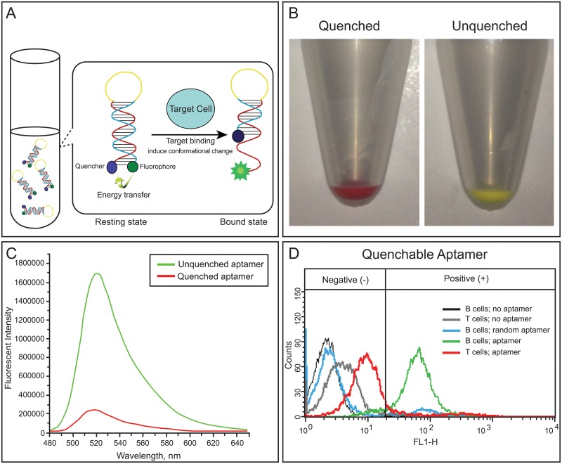
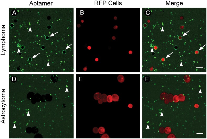
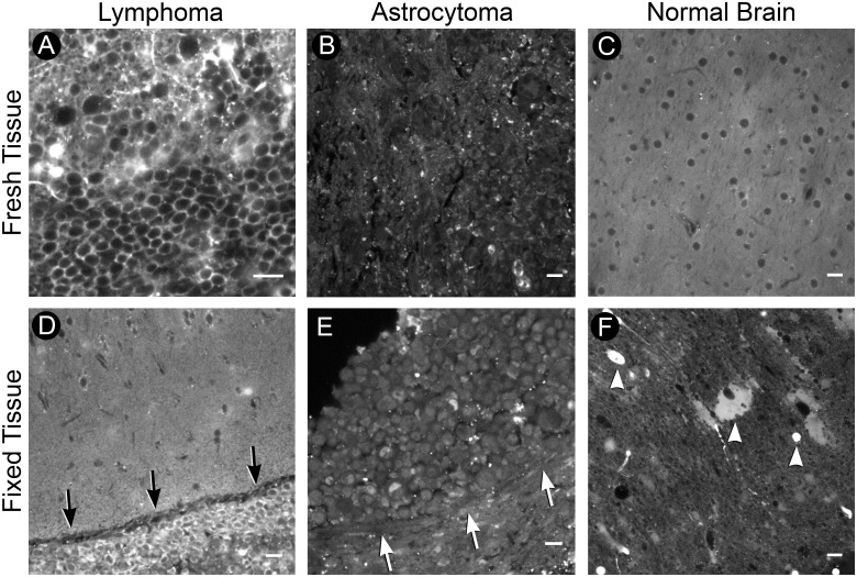
Improved tools for providing specific intraoperative diagnoses could improve patient care. In neurosurgery, intraoperatively differentiating non-operative lesions such as CNS B-cell lymphoma from operative lesions can be challenging, often necessitating immunohistochemical (IHC) procedures which require up to 24-48 hours. Here, we evaluate the feasibility of generating rapid ex vivo specific labeling using a novel lymphoma-specific fluorescent switchable aptamer. Our B-cell lymphoma-specific switchable aptamer produced only low-level fluorescence in its unbound conformation and generated an 8-fold increase in fluorescence once bound to its target on CD20-positive lymphoma cells. The aptamer demonstrated strong binding to B-cell lymphoma cells within 15 minutes of incubation as observed by flow cytometry. We applied the switchable aptamer to ex vivo xenograft tissue harboring B-cell lymphoma and astrocytoma, and within one hour specific visual identification of lymphoma was routinely possible. In this proof-of-concept study in human cell culture and orthotopic xenografts, we conclude that a fluorescent switchable aptamer can provide rapid and specific labeling of B-cell lymphoma, and that developing aptamer-based labeling approaches could simplify tissue staining and drastically reduce time to histopathological diagnoses compared with IHC-based methods. We propose that switchable aptamers could enhance expeditious, accurate intraoperative decision-making.


Conflict of interest statement

Competing Interests: The authors have declared that no competing interests exist.

Figures

Fig 1
Fig 1. Aptamer fluorescence unquenching.
(A, B). A) Illustration of aptamer fluorescence unquenching with binding to molecular target. B) Macrofluorescence change in fluorescence emission between quenched and unquenched aptamers. Target-binding induced change in fluorescence (C, D). C) Fluorescence emission intensity of the unquenched probe versus the quenched. Note a near 8-fold change in fluorescence intensity. D) Fluorescence intensity of the quenchable aptamer tested on negative control T cells and positive control B cells. Note increased fluorescence and number of labeled B cells versus T cells.
Fig 2
Fig 2. Quenchable aptamer staining of cultured human fluorescent lymphoma and astrocytoma cells.
(A-C) Lymphoma. A) B cell lymphoma cells incubated with the quenchable aptamer; note ring-like staining pattern (arrows) and fluorescent artifacts (arrowheads). B) Lymphoma cells expressing RFP. C) Merged image of RFP-lymphoma cells and aptamer staining. (D-F) Astrocytoma. D) Astrocytoma cells incubated with the quenchable aptamer; note fluorescent artifacts (arrowheads) and lack of ring-like staining. E) Astrocytoma cells expressing RFP. F) Merged image of RFP-astrocytoma cells and aptamer staining. Scale bars equal 20um. © 2015, Barrow Neurological Institute, provided under CC BY 4.0.
Fig 3
Fig 3. Xenograft acute slices.
(A-C) Fresh tissue. A) Tumor region of B cell lymphoma acute slice incubated with the quenchable aptamer. Note the ring-like staining pattern. B) Tumor region of astrocytoma acute slice; note lack of ring-like staining pattern. C) Contralateral normal brain from lymphoma acute slice. Note hypo-fluorescent regions indicating location of cell-bodies. (D-F) Fixed tissue. D) Fixed acute slice containing B cell lymphoma; note positive cells in hypercellular tumor region (arrows). E) Fixed astrocytoma tissue lacking positive staining; note absence of positive cells within hypercellular tumor (arrows). F) Contralateral normal brain from lymphoma fixed slice; note additional fluorescent artifacts (arrowheads). Scale bars equal 20um. © 2015, Barrow Neurological Institute, provided under CC BY 4.0.
Fig 4
Fig 4. Fluorescent xenograft acute slices.
(A-C) Lymphoma. A) Aptamer staining of lymphoma acute slice with positive staining regions (arrowheads). B) RFP-expressing lymphoma cells within tissue slice. C) Merge (D-F) Astrocytoma. D) Aptamer staining of astrocytoma acute slice; note lack of positively stained cells. E) RFP-expressing astrocytoma cells within tissue slice. F) Merge. (G-I) Normal brain. G) Aptamer staining of normal brain lacking aptamer-positive cells. H) Absence of ring-like staining and RFP-expressing cells in normal brain. I) Merge. J) Q-TD05 labels 80.75 ± 2.52% of RFP-expressing lymphoma cells and 8.25 ± 1.51% of non-RFP cells, p<0.001. K) Coefficient of correlation between RFP-lymphoma cells and aptamer staining (R2) = 0.92 with 95% confidence intervals (dotted lines) of 0.87–0.99, p<0.001. Scale bars equal 20um. © 2015, Barrow Neurological Institute, provided under CC BY 4.0.

References

    1. Haybaeck J, von Campe G, Hainfellner JA. [Rapid frozen sections in neuropathology]. Pathologe. 2012;33(5): 379–88. Der Schnellschnitt in der Neuropathologie. 10.1007/s00292-012-1604-x - DOI - PubMed
    1. Schiffer D, Giordana MT, Mauro A, Migheli A. Immunohistochemistry in neuro-oncology. Basic Appl Histochem. 1986;30(2): 253–65. - PubMed
    1. Plesec TP, Prayson RA. Frozen section discrepancy in the evaluation of central nervous system tumors. Arch Pathol Lab Med. 2007;131(10): 1532–40. - PubMed
    1. Hokfelt T. Neurobiology thanks to microbiology: the legacy of Albert H. Coons (1912–1978). Brain Res Bull. 1999;50(5–6): 371–2. - PubMed
    1. Safi MA. An overview of various labeled assays used in medical laboratory diagnosis. Immune and non-immune assays. Saudi Med J. 2010;31(4): 359–68. - PubMed

MeSH terms