. 2015 Feb 7:12:25.
doi: 10.1186/s12974-015-0246-3.

MiR-27a ameliorates inflammatory damage to the blood-spinal cord barrier after spinal cord ischemia: reperfusion injury in rats by downregulating TICAM-2 of the TLR4 signaling pathway

MiR-27a ameliorates inflammatory damage to the blood-spinal cord barrier after spinal cord ischemia: reperfusion injury in rats by downregulating TICAM-2 of the TLR4 signaling pathway

Xiao-Qian Li et al. J Neuroinflammation. .

Abstract

Background: Spinal cord ischemia reperfusion (IR) injury causes inflammation and subsequently increases blood-spinal cord barrier leakage and Toll-like receptor 4 (TLR4) pathway activation. MicroRNAs (miRs) effectively regulate numerous target mRNAs during ischemia. However, their roles during IR injury are poorly understood. We investigated miRs involvement, particularly miR-27a, in TLR4 pathway-mediated inflammatory responses after IR.

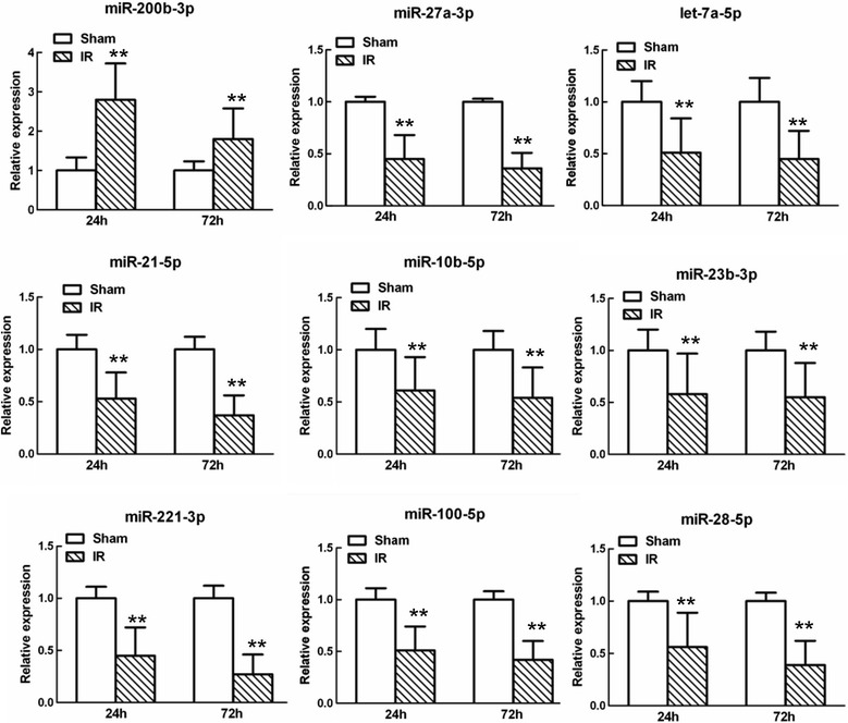
Method: We used a genomics approach to examine changed miRs of rats that had undergone 14 minutes of ischemia, followed by 24 or 72 hours of reperfusion. Quantitative RT-PCR was used to identify and confirm the miRs involved in regulating TLR4 pathway activation. We scanned miR databases for potential miR targets and confirmed these targets by quantitative RT-PCR. The miR mimic and anti-miR oligonucleotides (AMOs) were intrathecally injected at 12-hour intervals beginning three days before the ischemia. The effects of miRs on the TLR4 pathway and downstream cytokines were analyzed by PCR, western blotting, and ELISA. Double immunofluorescence staining was perfumed to determine the relationship between the targets and TLR4. Blood-spinal cord barrier (BSCB) permeability was examined using Evans blue (EB) dye.

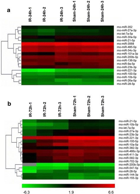
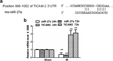
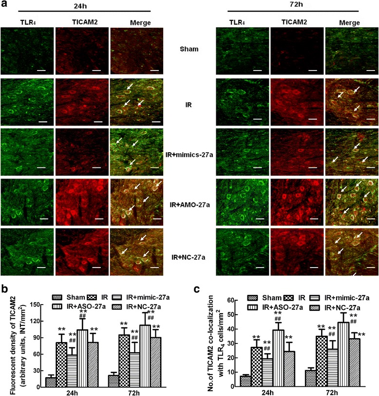
Results: A microarray analysis revealed that at 24 hours post-injury, three miRs were upregulated (>2.0 fold) and 15 miRs were downregulated (<0.5 fold), and at 72 hours, four miRs were upregulated and 14 were downregulated compared to their levels in sham-operated controls. We focused on miR-27a, which is predicted to contain sequences complementary to the 3'-untranslated region (UTR) of Toll-like receptor adaptor molecule 2 (TICAM-2). Double immunostaining indicated that TLR4 activation correlated with changes in TICAM-2 expression. Compared to the rats in the IR and negative control groups, intrathecal infusion of the miR-27a mimic attenuated IR-induced TLR4 activation and inflammatory damage to the BSCB, which was shown as decreased EB extravasation and lower levels of nuclear factor kappa-B (NF-κB) and lnterleukin (IL)-1β at 24 and 72 hours after reperfusion, whereas pretreatment with miR-27a AMO aggravated these injuries.

Conclusions: We present the first evidence that miRs play an important role in spinal cord IR injury. We identified TICAM-2 as a novel target of miR-27a. miR-27a upregulation attenuates IR-induced inflammatory damage to the BSCB by negatively regulating TICAM-2 of the TLR4 signaling pathway and inhibiting the NF-κB/IL-1β pathway. These results provide new therapeutic targets for IR injury treatment.


Figures

Figure 1
Figure 1
Hierarchical cluster analyses of altered microRNAs (miRs) after spinal cord ischemia reperfusion (IR) injury. (a) Differentially expressed miRs in the sham-operated and IR groups at 24 hours post-injury (n = three per group). (b) Differentially expressed miRs in the sham-operated and IR groups at 72 hours post-injury (n = three per group).Each row represents an miR and each column represents a sample. The color code shown at the bottom of the heat maps is linear, with green as the lowest and red as the highest. The miRs that were upregulated are shown in green to red, whereas the miRs that were downregulated are shown from red to green. Among all the significantly changed miRs, miR-27a was the most significantly downregulated in injured spinal cords at the abovementioned time points.
Figure 2
Figure 2
Quantitative real-time polymerase chain reaction (qRT-PCR) analysis confirming the microarray analyses of the nine miRs that were abnormally expressed at both 24 and 72 hours after spinal cord ischemia reperfusion (IR). Relative expression is the change in expression compared to the sham-operated group at either 24 or 72 hours after reperfusion. Data are expressed as mean ± SEM. **P <0.05 versus the sham group.
Figure 3
Figure 3
A putative target site of miR-27a located in the 3′-UTR of TICAM-2 mRNA was predicted by bioinformatics analysis. (a) The predicted miR-27a binding site in the 3′-untranslated region (UTR) of TICAM-2 in rats. (b) PCR analysis confirming TICAM-2 mRNA was abnormally expressed at both 24 and 72 hours after spinal cord ischemia reperfusion (IR). The profiles of TICAM-2 were expressed in an opposite manner as those of miR-27a. Relative expression is the change in expression compared to the sham-operated group at either 24 or 72 hours after reperfusion. Data are expressed as mean ± SEM. **P <0.05 versus the sham group. Results shown are representative data from three separate experiments.
Figure 4
Figure 4
Effects of intrathecal injection of mimic-27a and AMO-27a on the expression of TICAM-2 and the TLR 4 /NF- κB/IL-1β pathway in rats after spinal cord ischemia reperfusion (IR). (a) Representative immunoblots were probed with antibodies against TICAM-2, TLR4, and nuclear NF-κB p65, and an antibody against GAPDH served as a loading control. (b–g)Quantification of the densities of the TICAM-2 (b), TLR 4 (d), and NF-κB p65 (f) bands in nuclear extracts under different conditions. Protein expression is presented in relative units. Real-time PCR analyses of TICAM-2 (c), TLR 4 (e), and NF-κB p65 (g) were performed in duplicate and normalized to GAPDH mRNA levels. (h) Quantification of IL-1β production in the spinal cord at 24 and 72 hours after IR injury, as assessed by ELISA. IR caused significant increases in TICAM-2 expression, and intrathecal injection of mimic-27a and AMO-27a altered TICAM-2 expression after IR. In accordance with the decreased TICAM-2 expression, increased miR-27a levels induced by intrathecal injection of mimic-27a also prevented the increases in TLR4 and nuclear NF-κB p65 expression, as well as the increased IL-1β levels, whereas suppression of miR-27a by treatment with AMO-27a reversed the above changes, indicating that the TLR4/NF-κB/IL-1β pathway is involved in the miR-27a-mediated regulation of inflammation in the spinal cord through TICAM-2 targeting. All data are presented as mean ± SEM. **P <0.05 versus the sham group; ## P <0.05 versus the IR group.
Figure 5
Figure 5
Effects of intrathecal injection of mimic-27a and AMO-27a on the expression and colocalization of TLR 4 and TICAM-2 in vivo after spinal cord ischemia reperfusion (IR). (a) Representative micrographs showing the colocalization of TLR4 (green) and TICAM-2 (Red) at 24 and 72 hours after IR injury. Arrows show their co-localization. Scale bars = 100 μm. (b) Quantification of TICAM-2 immunoreactivity is presented as the average fluorescence intensity (FI) of three independent experiments. (c) Histogram for the quantification of co-localized cells (cells with yellow signals). Double fluorescence immunohistochemistry showing that membrane-bound TLR4 has an expression profile that is similar to that of TICAM-2 in the cytoplasm of neurons and glial cells from injured regions of the spinal cord at 24 and 72 hours after surgery. Increasing the levels of miR-27a by intrathecal pretreatment with mimic-27a significantly decreased TICAM-2 immunoreactivity after IR and the number of TICAM-2-TLR4 positive cells, whereas these effects were reversed by intrathecal injection of AMO-27a. All data are presented as mean ± SEM. **P <0.01 versus the sham group; ## P <0.05 versus the IR group.
Figure 6
Figure 6
Effects of intrathecal injection of mimic-27a and AMO-27a on blood-spinal cord barrier (BSCB) integrity after ischemia reperfusion (IR). (a) Effects of intrathecal injection of mimic-27a and AMO-27a on BSCB permeability measured by Evans blue (EB) extravasation. Almost no red fluoresce was seen in the spinal cord of the sham group at 24 and 72 hours after IR. Compared with the IR group, EB extravasation (red) was significantly lower in rats who received an intrathecal injection of mimic-27a. Conversely, at 24 hours after injury, much more red fluorescence, especially in the gray matter, was seen in rats who received an intrathecal injection of AMO-27a, which was even stronger at 72 hours post-injury. (b) The EB content of the spinal cord (μg/g). (c) Quantification of EB fluorescence density (INT/mm2) . Original magnification, 100×; Scale bar = 50 μm. **P <0.05 versus the sham group; ## P <0.05 versus the IR group.

References

    1. Lim KH, Connolly M, Rose D, Siegman F, Jacobowitz I, Acinapura A, et al. Prevention of reperfusion injury of the ischemic spinal cord: use of recombinant superoxide dismutase. Ann Thorac Surg. 1986;42(3):282–6. doi: 10.1016/S0003-4975(10)62735-X. - DOI - PubMed
    1. Fan L, Wang K, Shi Z, Die J, Wang C, Dang X. Tetramethylpyrazine protects spinal cord and reduces inflammation in a rat model of spinal cord ischemia-reperfusion injury. J Vasc Surg. 2011;54(1):192–200. doi: 10.1016/j.jvs.2010.12.030. - DOI - PubMed
    1. Zhu P, Li JX, Fujino M, Zhuang J, Li XK. Development and treatments of inflammatory cells and cytokines in spinal cord ischemia-reperfusion injury. Mediators Inflamm. 2013;2013:701970. - PMC - PubMed
    1. Ni B, Cao Z, Liu Y. Glycyrrhizin protects spinal cord and reduces inflammation in spinal cord ischemia-reperfusion injury. Int J Neurosci. 2013;123(11):745–51. doi: 10.3109/00207454.2013.796551. - DOI - PubMed
    1. Bell MT, Puskas F, Agoston VA, Cleveland JC, Jr, Freeman KA, Gamboni F, et al. Toll-like receptor 4-dependent microglial activation mediates spinal cord ischemia-reperfusion injury. Circulation. 2013;128(11 Suppl 1):S152–6. doi: 10.1161/CIRCULATIONAHA.112.000024. - DOI - PubMed

Publication types

MeSH terms