. 2014:79:29-39.
doi: 10.1101/sqb.2014.79.024794. Epub 2015 Apr 15.

The Brainstem Oscillator for Whisking and the Case for Breathing as the Master Clock for Orofacial Motor Actions

Affiliations

The Brainstem Oscillator for Whisking and the Case for Breathing as the Master Clock for Orofacial Motor Actions

David Kleinfeld et al. Cold Spring Harb Symp Quant Biol. 2014.

Abstract

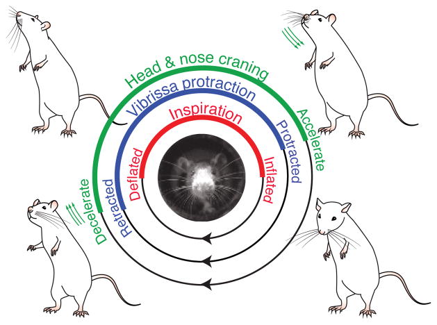
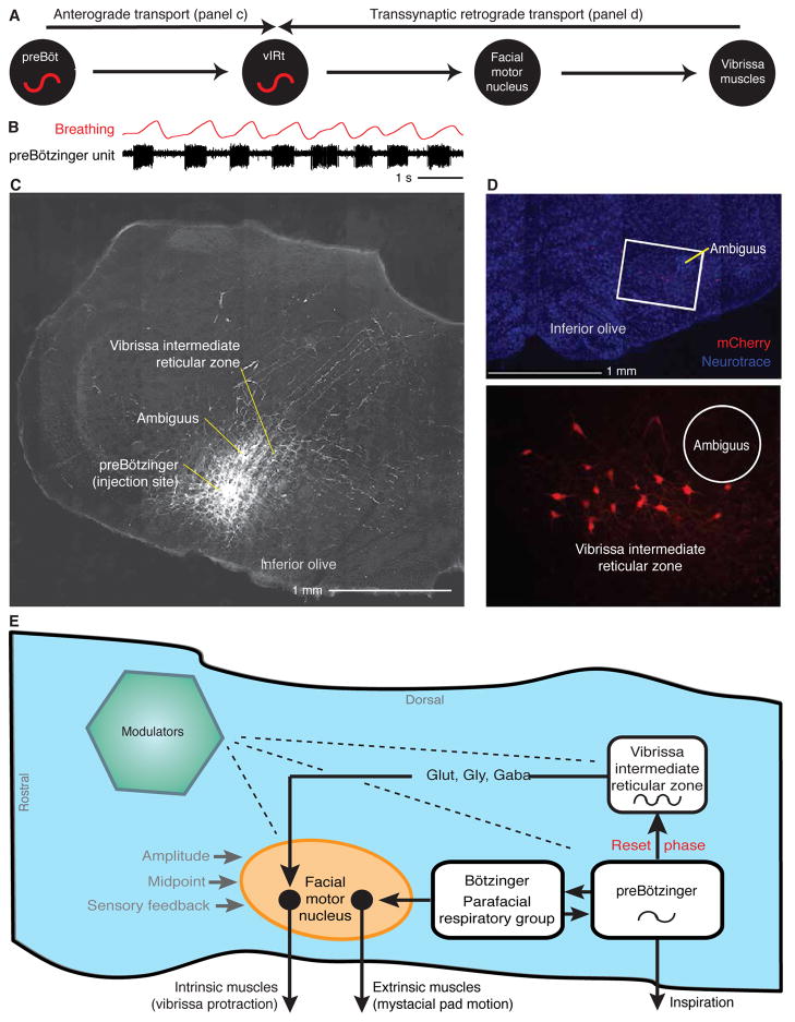
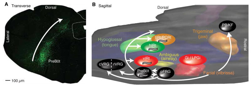
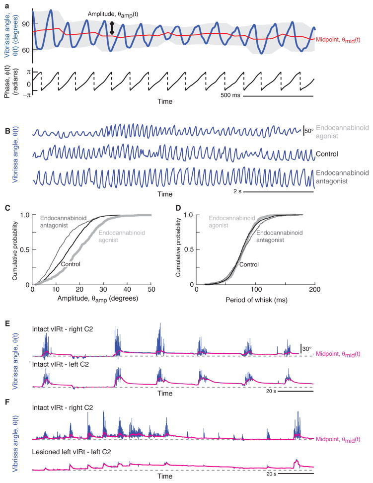
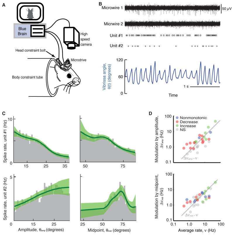
Whisking and sniffing are predominant aspects of exploratory behavior in rodents. We review evidence that these motor rhythms are coordinated by the respiratory patterning circuitry in the ventral medulla. A recently described region in the intermediate reticular zone of the medulla functions as an autonomous whisking oscillator, whose neuronal output is reset upon each breath by input from the pre-Bötzinger complex. Based on similarities between this neuronal circuit architecture and that of other orofacial behaviors, we propose that the pre-Bötzinger complex, which projects broadly to premotor regions throughout the intermediate reticular zone of the medulla, functions as a master clock to coordinate multiple orofacial actions involved in exploratory and ingestive behaviors. We then extend the analysis of whisking to the relatively slow control of the midpoint of the whisk. We conjecture, in a manner consistent with breathing as the "master clock" for all orofacial behaviors, that slow control optimizes the position of sensors whereas the breathing rhythm provides a means to perceptually bind the inputs from different orofacial modalities.


Figures

Figure 1
Figure 1. A cycle of exploratory behavior locked to sniffing
Three motor actions, whisking, head bobbing, and nose twitching, are linked to a fourth, sniffing, during orofacial exploration. Abstracted from the claims of Welker (1964).
Figure 2
Figure 2. The nature of the motor plant for whisking
(A) A tangential slice containing a whole set of intrinsic muscles that form slings about the follicles that house the vibrissae. Most of the muscle slings are only partially represented in this plane. (B) Drawing of intrinsic musculature and follicular anatomy. The intrinsic muscles join adjacent follicles of a single row. Each muscle attaches medially and laterally to the superior part of the caudal follicle while forming a sling around the lower third of the rostral follicle. The skin on the outside and the plate on the inside provide a passive restoring force. Superficial extrinsic muscles run just below the skin. Asterisks indicate the approximate locations of the exposed tips of EMG microwires (Hill et al. 2008). Adapted from Dorfl (1982). (C) Drawing of extrinsic musculature. Four extrinsic muscles invade the mystacial pad while maintaining external attachment points. The protractor m. nasalis attaches rostral to the pad at the nasal septum and runs under the skin as it extends caudally. The retractor m. nasolabialis attaches dorsal-caudal to the pad and runs superficially below the skin. The retractor m. maxillolabialis attaches ventral-caudal to the pad and fuses with the fibers of m. nasolabialis as they invade the pad. m. transversus nasi lies transverse to the snout and runs superficially through the pad. The recording sites for each muscle are indicated by asterisks. Adapted from Dorfl (1982). (D) Muscle activity from head-restrained rats. Electromyogram signals are abbreviated as INT (intrinsic muscles), NA (m. nasalis), NL (m. nasolabialis). The signals are high-pass filtered, full-wave rectified, and low-pass filtered. Adapted from Hill et al. (2008). (E) Schematic of a biomechanical model that represents one row of vibrissae in the maxillary compartment of the rat mystacial pad in resting position. Intrinsic whisker protractors (IP) represent intrinsic muscles. The protractor (CP) of the mystacial pad represents the Pars media inferior of the m. nasalis extrinsic muscle, which attached to the skin. The retractor (CR) of the mystacial pad represents the m. maxillolabialis and m. nasolabialis extrinsic muscles, which also attach to the skin. The whisker retractor (PR) represents the Pares maxillares superficialis and profunda of the m. nasalis extrinsic muscle, which attach to the plate. Green dots in the skin and blue dots in the plate rows represent attachment sites of the extrinsic muscles. Large black dots represent the centers of mass of the vibrissae. Empty circles represent springs coupled with dampers that symbolize the elasticity of the tissue. Anchors represent non-elastic sites in the mystacial pad. Adapted from Haidarliu et al. (2010), after Simony et al. (2010) and Hill et al. (2008). (F) Vibrissa and mystacial pad motion from head-restrained rats recorded concurrently with the EMG data of panel D. The black curve was calculated from a reduced model of the motor plant using the data of panel d as the sole input. Adapted from Hill et al. (2008).
Figure 3
Figure 3. Simultaneous measurements of vibrissa angular position (blue) and breathing (red) in head-fixed rats
All panels adapted from Moore and Deschênes et al. (2013). (A) Measurement showing epochs of breathing (red) without whisking (blue) and synchronous sniffing and whisking in a head-fixed rat. Insert is a schematic for simultaneous measurements of vibrissa angular position through videography and breathing through the change in temperature of a thermocouple connected to a tube inserted into the nasal passage. (B) Measurement showing an epoch of whisking without breathing, where inspiration was halted by introduction of ammonia into the air mixture. (C) Measurement showing breathing with intervening whisks between inspirations, along with concurrent recordings of multiunit activity (black) in the preBötzinger complex. (D) Measurement showing breathing with intervening whisks between inspirations, along with concurrent recordings of multiunit activity (black) in the vibrissa zone of the intermediate reticular formation. (E) The recording sites for all data imposed on a three dimensional reconstruction of the medulla. Whisking units (blue) are located in the IRt dorsomedial to inspiratory units (red) in the preBötzinger complex.
Figure 4
Figure 4. Necessity and sufficiency of the vIRt as the rhythm generator for whisking
All panels adapted from Moore and Deschênes et al. (2013). (A, B) Lesion of the vIRt impairs ipsilateral whisking. Panel A shows an example of whisking bout following an electrolytic lesion. In panel B, all lesion sites were mapped onto a three dimensional reconstruction of the medulla and selected anatomical substructures. The lesion centroids are denoted with symbols, with circles for electrolytic lesions, triangles for lesion via transport of Sindbis virus, and squares for chemical lesion by ibotenic acid. (C, D) Injection of the glutamate channel agonist kainic acid activates the vIRt, which drives facial motoneurons and induces whisking. Panel C shows vibrissa motion (blue), breathing (red), and extrinsic (green) EMG. Panel D is a polar plot of the coherence between spiking activity and vibrissa motion at the peak frequency of whisking. Open circles represent multiunit activity and closed circles represent single units. The green bar represents the coherence of the EMG for the intrinsic muscle (panel b) with vibrissa motion. (Inserts) Spiking activity of neuronal units in the vIRt (black) in relation to vibrissa motion (blue).
Figure 5
Figure 5. Anatomy and circuit for the resetting of whisking by breathing
(A) Proposed connectivity from the preBötzinger complex, through the vIRt, and onward to the follicle muscles. Resolution of this connection pattern makes use of anterograde labeling to define the path from the preBötzinger complex to the vIRt and retrograde transsynaptic labeling to define the pathway from the vIRt to the mystacial pad. (B) Recording of a single inspiratory unit in the preBötzinger complex, together with breathing. Adapted from Moore and Deschênes et al. (2013). (C) Tracing via injection of the anterograde tracer biotinylated dextran amine through the same pipette used to record (panel a) leads to labeling of axons and terminals in the vIRt from cells in the preBötzinger complex. Adapted from Moore and Deschênes et al. (2013). (D) Retrograde labeling of neurons in the vIRt by modified Rabies virus, ΔG-RV, that was pressure injected into the facial musculature of juvenile transgenic mice engineered to express the glycoprotein under the premotor for choline acetyltransferase. Monosynaptically-connected premotor neurons in the vIRt are labeled by ΔG-RV (red) and counterstained with Neurotrace fluorescent Nissl stain (Life Technologies) (blue). Ambiguus refers to nucleus ambiguus. Adapted from Takatoh et al. (2013). (E) Model of the circuitry that generates whisking that is coordinated with breathing. Dashed lines indicate diffuse synaptic input from modulatory brain nuclei. GLUT, glutamate; GLY, glycine; GABA, γ-aminobutyric acid. Adapted from Moore and Deschênes et al. (2013).
Figure 6
Figure 6. The preBötzinger complex forms a “backplane” of collateral projections across all of the intermediate reticular zone
(A) Injection of adeno-associated virus 2, which labels neurons by using the somatostatin promoter to drive the expression of enhanced green fluorescent protein (EGFP) (Tan et al. 2008), forms a band of EGFP-expressing axons along the intermediate reticular zone and revealed labeled axons in parahypoglossal nucleus/nucleus of the solitary tract regions (top) and ventral respiratory group caudal to preBötzinger complex (bottom). No EGFP-expressing cell bodies were observed. Adapted from Tan et al. (2010). (B) Three-dimensional reconstruction of the pons and medulla, showing the pools of cranial motoneurons that control the jaws (orange), face (red), airway (yellow), and tongue (green), as well as the approximate locations of known pre-motor nuclei to each of the motoneuron pools. Premotor nuclei are color coded according to the primary motor nucleus that they innervate. Breathing-related regions are shown in black, and the projections of the preBötzinger complex are shown as white arrows. Putative rhythmic oscillators for whisking, licking, and chewing are denoted “~”. Abbreviations: cVRG, caudal ventral respiratory group; Gi, gigantocellular reticular formation; hIRt, hypoglossal intermediate reticular formation; LPGi, lateral paragigantocellular reticular formation; PCRt, parvocellular reticular formation; pFRG, parafacial respiratory group; PreBöC, preBötzinger complex; rVRG, rostral ventral respiratory group; tIRt, trigeminal intermediate reticular formation; vIRt, vibrissa intermediate reticular formation. Adapted from Moore et al. (2014b).
Figure 7
Figure 7. Evidence for the separation of slow processes, i.e., changes in amplitude and midpoint of whisking, from the fast rhythmic oscillation
(A) Decomposition of whisking into rapidly and slowly varying parameters via the Hilbert transform. Top panel shows vibrissa position along with its decomposition into the amplitude, θamp, and midpoint, θmid, of the envelope (gray band). The lower trace shows the phase, ϕ, along with broken vertical lines indicate wrapping of phase from π to –π. Adapted from Hill et al. (2011). (B–D) Effect of a cannabinoid receptor type 1 agonist and an antagonist on whisking amplitude but not frequency. Panel B shows typical traces of vibrissa angle executed by an animal four hours after administration of either Δ9-THC, an agonist (dark gray), vehicle (black), or SR141716A, an antagonist (light gray). Vertical calibration bar corresponds to 50°. Panel C shows the cumulative probability distribution function of protraction amplitudes across all animals; note the significant effect of agonists and antagonist. Panel D shows the cumulative probability distribution function of the period (inverse of frequency) of whisking across all animals; note the lack of a significant effect of agonists and antagonist. Panels adapted from Pietr et al. (2010). (E, F) Effect of lesioning the vIRt in one hemisphere (Fig. 4a,b) quenches rhythmic whisking yet does not interfere with changes in midpoint. In panel E, the midpoint of the motion (red) is seen to track between the two sides of the face. In panel F, lesion of the vIRt of the left side only prevents whisking on that side. Yet the midpoint of the vibrissae of the right and left side of the face continue to track each other. New data obtained with the methods in Moore and Deschênes et al. (2013).
Figure 8
Figure 8. The vibrissa area of motor cortex codes the slowly varying midpoint and amplitude of whisking
All panels adapted from Hill et al. (2011). (A) The scheme used to measure the spiking activity of units in agranular motor cortex as body constrained animals rhythmically whisk in air. Vibrissa position is determined from videography. Single units, indicative of spiking by an individual neuron, are extracted from extracellular micro-wire recordings. (B) Example records. (C) Firing rate profiles for two example units as a function of amplitude and midpoint of vibrissa motion. The two rows are profiles of units that show different relative modulation. Each plot is calculated by dividing the distribution of the respective signal at spike time by the distribution of that signal over the entire behavioral session. Green lines are fits from a smoothing algorithm along with the 95 % confidence band. (D) Composite data across units illustrates that, on average, the rate is unaffected by whisking, consistent with the presence of units that both increase (green) and decrease (red) their rate with increasing angle.

Similar articles

Cited by

References

    1. Berg RW, Kleinfeld D. Rhythmic whisking by rat: Retraction as well as protraction of the vibrissae is under active muscular control. Journal of Neurophysiology. 2003;89:104–117. - PubMed
    1. Bieger D, Hopkins DA. Viscerotopic representation of the upper alimentary tract in the medulla oblongata in the rat: The nucleus ambiguus. Journal of Comparative Neurology. 1987;262:546–562. - PubMed
    1. Bouvier J, Thoby-Brisson M, NR, Dubreuil V, Ericson J, Champagnat J, Pierani A, AC, Fortin G. Hindbrain interneurons and axon guidance signaling critical for breathing. Nature Neuroscience. 2010;13:1066–1074. - PubMed
    1. Brecht M, Krauss A, Muhammad S, Sinai-Esfahani L, Bellanca S, Margrie TW. Organization of rat vibrissa motor cortex and adjacent areas according to cytoarchitectonics, microstimulation, and intracellular stimulation of identified cells. Jounal of Comparative Neurology. 2004;479:360–373. - PubMed
    1. Carvell GE, Simons DJ. Biometric analyses of vibrissal tactile discrimination in the rat. Journal of Neuroscience. 1990;10:2638–2648. - PMC - PubMed

Publication types

LinkOut - more resources