

Tubular cross talk in acute kidney injury: a story of sense and sensibility
- PMID: 25877507
- PMCID: PMC4469890
- DOI: 10.1152/ajprenal.00030.2015
Tubular cross talk in acute kidney injury: a story of sense and sensibility
Abstract
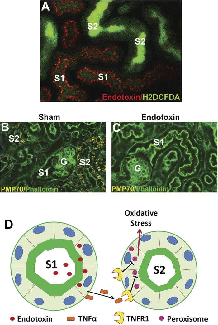
The mammalian kidney is an organ composed of numerous functional units or nephrons. Beyond the filtering glomerulus of each nephron, various tubular segments with distinct populations of epithelial cells sequentially span the kidney from cortex to medulla. The highly organized folding of the tubules results in a spatial distribution that allows intimate contact between various tubular subsegments. This unique arrangement can promote a newly recognized type of horizontal epithelial-to-epithelial cross talk. In this review, we discuss the importance of this tubular cross talk in shaping the response of the kidney to acute injury in a sense and sensibility model. We propose that injury-resistant tubules such as S1 proximal segments and thick ascending limbs (TAL) can act as "sensors" and thus modulate the responsiveness or "sensibility" of the S2-S3 proximal segments to injury. We also discuss new findings that highlight the importance of tubular cross talk in regulating homeostasis and inflammation not only in the kidney, but also systemically.
Keywords: Tamm-Horsfall protein; acute kidney injury; sepsis; tubular cross talk; uromodulin.
Figures


Similar articles
-
Tamm-Horsfall protein translocates to the basolateral domain of thick ascending limbs, interstitium, and circulation during recovery from acute kidney injury.Am J Physiol Renal Physiol. 2013 Apr 15;304(8):F1066-75. doi: 10.1152/ajprenal.00543.2012. Epub 2013 Feb 6. Am J Physiol Renal Physiol. 2013. PMID: 23389456 Free PMC article.
-
Tamm-Horsfall protein-deficient thick ascending limbs promote injury to neighboring S3 segments in an MIP-2-dependent mechanism.Am J Physiol Renal Physiol. 2011 Apr;300(4):F999-1007. doi: 10.1152/ajprenal.00621.2010. Epub 2011 Jan 12. Am J Physiol Renal Physiol. 2011. PMID: 21228114 Free PMC article.
-
Tamm-Horsfall protein protects the kidney from ischemic injury by decreasing inflammation and altering TLR4 expression.Am J Physiol Renal Physiol. 2008 Aug;295(2):F534-44. doi: 10.1152/ajprenal.00083.2008. Epub 2008 May 21. Am J Physiol Renal Physiol. 2008. PMID: 18495803 Free PMC article.
-
The role of metabolic reprogramming in tubular epithelial cells during the progression of acute kidney injury.Cell Mol Life Sci. 2021 Aug;78(15):5731-5741. doi: 10.1007/s00018-021-03892-w. Epub 2021 Jun 29. Cell Mol Life Sci. 2021. PMID: 34185125 Free PMC article. Review.
-
Evolving Concepts in Uromodulin Biology, Physiology, and Its Role in Disease: a Tale of Two Forms.Hypertension. 2022 Nov;79(11):2409-2418. doi: 10.1161/HYPERTENSIONAHA.122.18567. Epub 2022 Aug 12. Hypertension. 2022. PMID: 35959659 Free PMC article. Review.
Cited by
-
Crosstalk between proximal tubular epithelial cells and other interstitial cells in tubulointerstitial fibrosis after renal injury.Front Endocrinol (Lausanne). 2024 Jan 8;14:1256375. doi: 10.3389/fendo.2023.1256375. eCollection 2023. Front Endocrinol (Lausanne). 2024. PMID: 38260142 Free PMC article. Review.
-
The proximal tubule is the primary target of injury and progression of kidney disease: role of the glomerulotubular junction.Am J Physiol Renal Physiol. 2016 Jul 1;311(1):F145-61. doi: 10.1152/ajprenal.00164.2016. Epub 2016 May 18. Am J Physiol Renal Physiol. 2016. PMID: 27194714 Free PMC article. Review.
-
The Presence of Testis Determines Aristolochic Acid-Induced Nephrotoxicity in Mice.Toxins (Basel). 2023 Feb 1;15(2):118. doi: 10.3390/toxins15020118. Toxins (Basel). 2023. PMID: 36828432 Free PMC article.
-
Tissue Cytometry With Machine Learning in Kidney: From Small Specimens to Big Data.Front Physiol. 2022 Mar 4;13:832457. doi: 10.3389/fphys.2022.832457. eCollection 2022. Front Physiol. 2022. PMID: 35309077 Free PMC article. Review.
-
Vascular endothelial cadherin shedding is more severe in sepsis patients with severe acute kidney injury.Crit Care. 2019 Jan 18;23(1):18. doi: 10.1186/s13054-019-2315-y. Crit Care. 2019. PMID: 30658667 Free PMC article.
References
-
- Aggarwal S, Ghilardi N, Xie MH, de Sauvage FJ, Gurney AL. Interleukin-23 promotes a distinct CD4 T cell activation state characterized by the production of interleukin-17. J Biol Chem 278: 1910–1914, 2003. - PubMed
-
- Aregger F, Pilop C, Uehlinger DE, Brunisholz R, Carrel TP, Frey FJ, Frey BM. Urinary proteomics before and after extracorporeal circulation in patients with and without acute kidney injury. J Thorac Cardiovasc Surg 139: 692–700, 2010. - PubMed
-
- Bachmann S, Koeppen-Hagemann I, Kriz W. Ultrastructural localization of Tamm-Horsfall glycoprotein (THP) in rat kidney as revealed by protein A-gold immunocytochemistry. Histochemistry 83: 531–538, 1985. - PubMed
-
- Brenner BM, Rector FC. Brenner & Rector's The Kidney. Philadelphia, PA: Saunders Elsevier, 2008, p. 2.
Publication types
MeSH terms
Substances
Grants and funding
LinkOut - more resources
Full Text Sources
Other Literature Sources
Miscellaneous