Review
. 2015 Sep;1353(1):72-88.
doi: 10.1111/nyas.12758. Epub 2015 Apr 15.

MicroRNAs in diabetic nephropathy: functions, biomarkers, and therapeutic targets

Affiliations
Review

MicroRNAs in diabetic nephropathy: functions, biomarkers, and therapeutic targets

Mitsuo Kato et al. Ann N Y Acad Sci. 2015 Sep.

Abstract

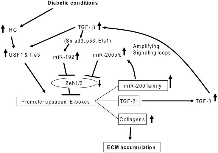
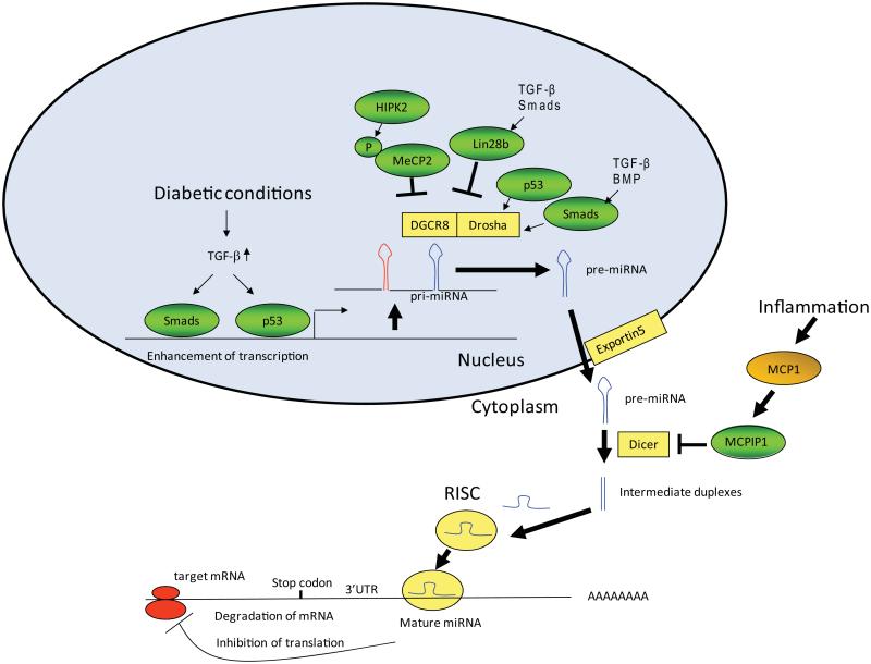
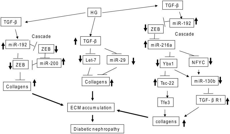
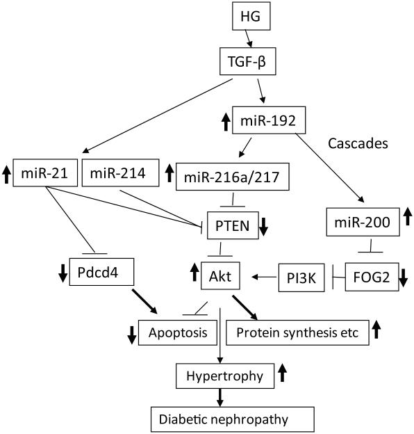
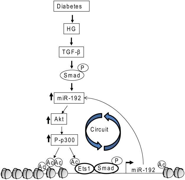
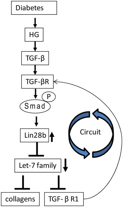
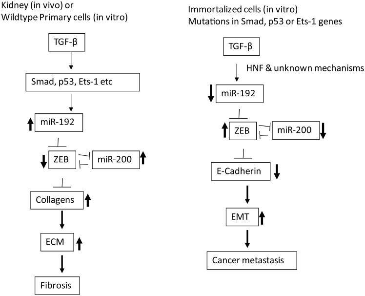
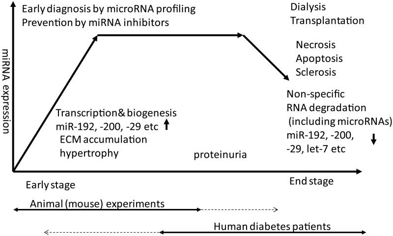
MicroRNAs (miRNAs) are short noncoding RNAs that regulate gene expression by posttranscriptional and epigenetic mechanisms and thereby affect many cellular processes and disease states. Over 2,000 human mature miRNAs have been identified, and at least 60% of all human protein-coding genes are known to be regulated by miRNAs. MicroRNA biogenesis involves classical transcription regulation and processing by key ribonucleases, as well as other protein factors and epigenetic mechanisms. Diabetic nephropathy (DN), a severe microvascular complication frequently associated with diabetes mellitus, is a major cause of renal failure. Although several mechanisms of regulation of key renal genes implicated in DN pathogenesis have been identified, a greater understanding is needed to develop better treatment modalities. Recent studies show that miRNAs induced in renal cells in vivo and in vitro under diabetic conditions can promote the accumulation of extracellular matrix proteins related to fibrosis and glomerular dysfunction. In this review, we highlight the significance of the expression of miRNAs in various stages of DN and emerging approaches to exploit them as biomarkers for early detection or novel therapeutic targets to prevent progression of DN.

Keywords: biomarkers; diabetic nephropathy; microRNAs; noncoding RNA; signal transduction; therapeutics; transforming growth factor β1.


Figures

Figure 1
Figure 1
TGF-β1 induced by high-glucose conditions via upstream transcription factors (USFs) creates signaling circuits mediated by miRNAs. Diabetic conditions increase the expression of TGF-β1 initially through USFs that bind to E-boxes in the TGF-β1 promoter. Upregulated TGF-β1 induces miR192 (inhibitor of E-box repressors Zeb1/2) via mechanisms involving the actions of Smad3, Ets-1, and p53. MiR-200b/c are also upregulated due to the downregulation of Zeb1/2 by miR-192. MiR-200b/c targeting Zeb1/2 can lead to a further decrease of Zeb1/2 and thus further augments the expression of miR-200b/c, TGF-β1, Col1a2, and Col4a1 through a loss of repression, along with a gain of activation (via Tfe3 and/or USF1) at their E-boxes. These signaling loops accelerate TGF-β1 signaling and downstream ECM gene expression involved in the progression of chronic kidney diseases such as DN. Modified from Kato et al.
Figure 2
Figure 2
Biogenesis and actions of microRNAs. MicroRNAs (miRNAs) are initially transcribed in the nucleus as pri-miRNAs, which are processed into pre-miRNAs by the Drosha enzyme. Pre-miRNAs are further cleaved by Dicer to result in double-stranded miRNA duplexes. Drosha processing can be regulated by p53 or Smads (activated by TGF-β or BMPs). This process can also be inhibited via interactions of phosphorylated MeCP2 (by HIPK2) with DGCR8, a cofactor of Drosha. In addition, Dicer processing can be inhibited by MCPIP1, which is induced by MCP-1 (related to inflammation). Processing of let-7 family members is inhibited by Lin28b, which can be induced by TGF-β/Smad signaling. The miRNA duplexes are then unwound by the action of Dicer, and the mature miRNA guide strand is loaded into the RISC complex. Please refer to the main text for details. RISC, RNA-induced silencing complex; UTR, untranslated region. Modified from Kato et al.
Figure 3
Figure 3
Extracellular matrix (ECM) protein accumulation by microRNA cascades in the early stages of DN. High-glucose (HG) conditions induce TGF-β1, which causes DN through increased fibrosis and ECM protein accumulation. Signaling cascades are depicted to delineate key intermediate black boxes in TGF-β1–induced signaling in DN. TGF-β1 increases ECM genes, such as collagens, through miR-192 and miR-200 and downregulation of their targets, E-box repressors (ZEB1/2). One cascade shown runs from miR-192 to miR-200 family members. In another, HG and TGF-β1 also induce collagens by inhibiting the expression of miR-29 family and let-7 family members that target several collagens. In a third cascade, miR-192 regulation of collagens is via the intermediate actions of miR-216a and miR-130b and their targets, including Ybx1, NFYC, and TGF-β1R1. These miRNA cascades can amplify the initial signal transduction events to accelerate chronic kidney diseases such as DN.
Figure 4
Figure 4
MicroRNA cascades related to hypertrophy and apoptosis in the early stages of DN. High-glucose (HG) conditions induce TGF-β1, which causes DN through increased hypertrophy via activation of Akt kinase. TGF-β1 activates Akt kinase through the miR-216a/217/200 family, which target and downregulate PTEN or FOG2 and thereby promoter hypertrophy. HG and TGF-β1 also activate Akt by increasing miR-21 and miR-214 that target PTEN. Two cascades from miR-192 are depicted: to the miR-216a/217 cluster and to the miR-200 family. Increased protein synthesis and inhibition of apoptosis through Akt activation can lead to hypertrophy. Increased miR-21 can also affect apoptosis directly through its apoptosis-related targets, such as Pdcd4. These miRNA cascades can amplify the signaling events initiated by hyperglycemia and TGF-β1 and thereby promote chronic kidney diseases such as DN.
Figure 5
Figure 5
Epigenetic regulation of miRNA expression (with miR-192 as an example) and circuitry mediated by Akt/p300 signaling. Autoactivation of the miR-192 promoter by acetylation of chromatin histones and transcription factors (such as Ets-1) through p300. Activation of Akt is mediated by key miRNAs downstream of miR-192 that target and downregulate PTEN or FOG2 (Fig. 4). Continuous and uncontrolled activation of these signaling pathways in this circuitry may induce chronic kidney diseases such as DN.
Figure 6
Figure 6
Regulation of pro-fibrotic genes by diabetic conditions through Lin28-mediated inhibition of biogenesis of the let-7 family. TGF-β1 induced by diabetic conditions upregulates Lin28b through activation of Smad 2/3. Lin28b downregulates let-7 family miRNAs by inhibiting the processing of let-7. Decreased levels of let-7 members result in the upregulation of collagens (let-7 targets), leading to glomerular ECM protein accumulation and progression of DN, illustrating another circuit for signal perpetuation.
Figure 7
Figure 7
Differential effects of miRNAs in kidney fibrosis and epithelial to mesenchymal transition (EMT) in cancer metastasis. In the early stage of DN, TGF-β1 induced by diabetic conditions upregulates miR-192 in mesangial and other renal cells through the activation of Smad 2/3, p53, or via Ets-1-mediated mechanisms. MiR-192 induces collagens by inhibiting E-box repressors (Zeb1/2) and also via increases in miR-200 family members. The miR-200 family also enhances collagen expression by targeting Zeb1/2 to amplify the signaling. On the other hand, in epithelial cancer cell lines or immortalized epithelial cell lines that have mutation in genes such as p53, Smads or Ets-1, TGF-β1 decreases miR-192. This also leads to decreases miR-200 family and E-Cadherin genes through E-box repressors (Zeb1/2) to induce EMT. Renal cell–specific transcription factors (such as HNF) can also be critical for cell-specific regulation of miRNAs in response to TGF-β1.
Figure 8
Figure 8
DN stage–specific regulation of miRNAs: humans (~ late stage) versus animal models (~ early stage). Usually, renal biopsies are obtained from diabetic patients at the middle or late stage of DN because they present with micro- or macro-albuminuria, glomerular filtration rate (GFR) decline, or other clear symptoms of renal dysfunction. When compared, most RNAs (including coding and noncoding) are likely to depict lower expression in the end stage of DN relative to the intermediate stage. It might be interpreted that downregulation of RNA is associated with renal failure; however, decreases in RNA expression could be due to poor quality of RNAs in end-stage biopsies. In experimental models of DN, animals can be systematically monitored from the healthy to more advanced stages of renal disease, although most mouse models used to study miRNAs do not depict features of human DN. Therefore, some miRNAs found to be upregulated in the early stage of DN in animal experiments may appear to be downregulated in samples from humans who are in much later stages of DN, due to poor sample quality or non-comparable stages. Thus, miRNAs that are induced in early DN in animal models can be potentially assessed as therapeutic targets in humans because their inhibition might slow down progression to late- or end-stage disease even before clear symptoms are evident.

References

    1. Fineberg D, Jandeleit-Dahm KA, Cooper ME. Diabetic nephropathy: diagnosis and treatment. Nat Rev Endocrinol. 2013;9:713–723. - PubMed
    1. Jones CA, et al. Epidemic of end-stage renal disease in people with diabetes in the United States population: do we know the cause? Kidney Int. 2005;67:1684–1691. - PubMed
    1. Sarnak MJ, et al. Kidney disease as a risk factor for development of cardiovascular disease: a statement from the American Heart Association Councils on Kidney in Cardiovascular Disease, High Blood Pressure Research, Clinical Cardiology, and Epidemiology and Prevention. Circulation. 2003;108:2154–2169. - PubMed
    1. de Boer IH, et al. Intensive diabetes therapy and glomerular filtration rate in type 1 diabetes. N Engl J Med. 2011;365:2366–2376. - PMC - PubMed
    1. Nathan DM, et al. Diabetes control and complications trial/epidemiology of diabetes interventions and complications study at 30 years: advances and contributions. Diabetes. 2013;62:3976–3986. - PMC - PubMed

Publication types