. 2015 Apr 15;7(283):283ra52.
doi: 10.1126/scitranslmed.aaa4306.

STING agonist formulated cancer vaccines can cure established tumors resistant to PD-1 blockade

Affiliations

STING agonist formulated cancer vaccines can cure established tumors resistant to PD-1 blockade

Juan Fu et al. Sci Transl Med. .

Abstract

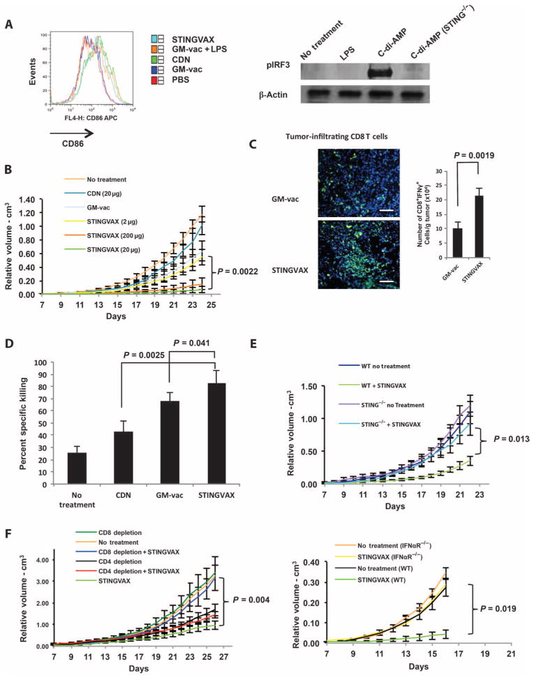
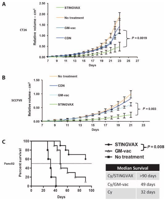
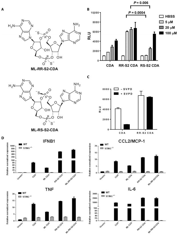
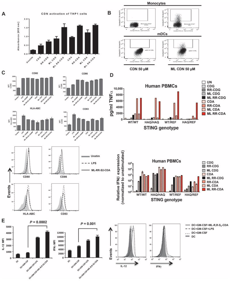
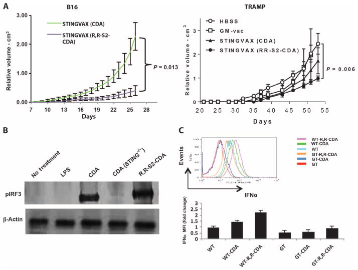
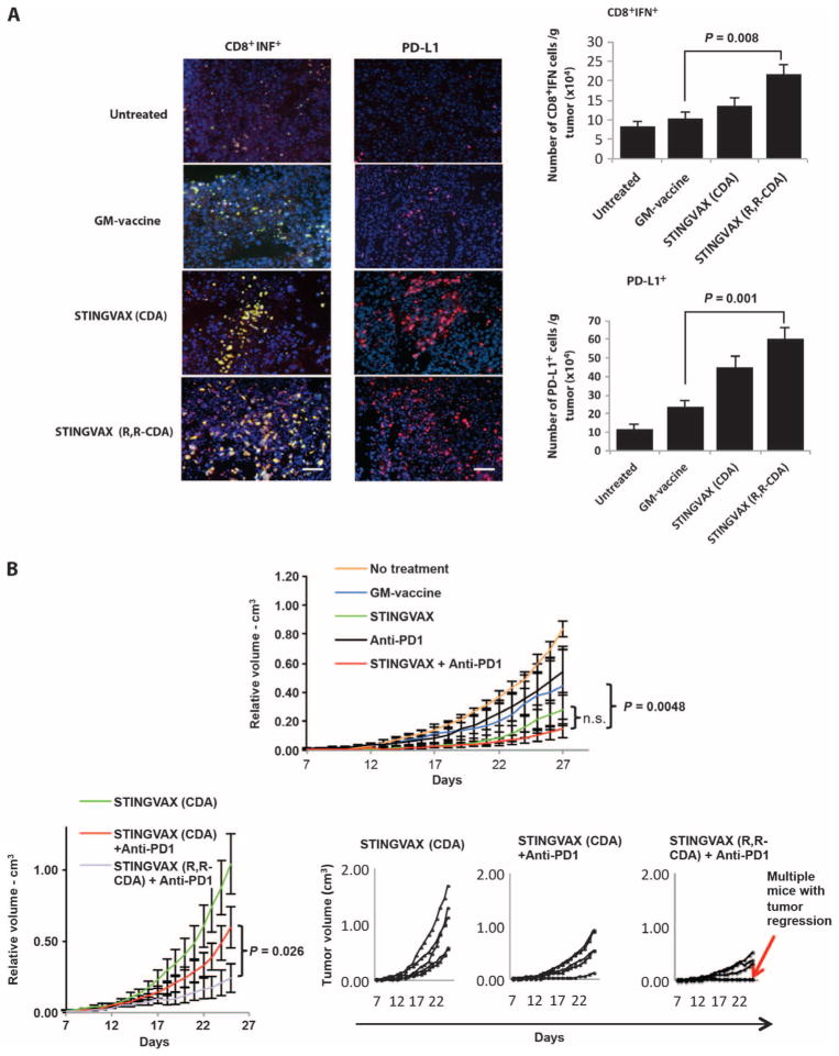
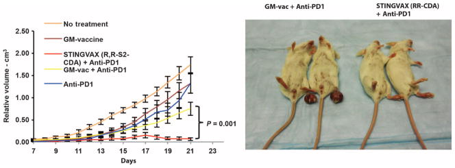
Stimulator of interferon genes (STING) is a cytosolic receptor that senses both exogenous and endogenous cytosolic cyclic dinucleotides (CDNs), activating TBK1/IRF3 (interferon regulatory factor 3), NF-κB (nuclear factor κB), and STAT6 (signal transducer and activator of transcription 6) signaling pathways to induce robust type I interferon and proinflammatory cytokine responses. CDN ligands were formulated with granulocyte-macrophage colony-stimulating factor (GM-CSF)-producing cellular cancer vaccines--termed STINGVAX--that demonstrated potent in vivo antitumor efficacy in multiple therapeutic models of established cancer. We found that rationally designed synthetic CDN derivative molecules, including one with an Rp,Rp dithio diastereomer and noncanonical c[A(2',5')pA(3',5')p] phosphate bridge structure, enhanced antitumor efficacy of STINGVAX in multiple aggressive therapeutic models of established cancer in mice. Antitumor activity was STING-dependent and correlated with increased activation of dendritic cells and tumor antigen-specific CD8(+) T cells. Tumors from STINGVAX-treated mice demonstrated marked PD-L1 (programmed death ligand 1) up-regulation, which was associated with tumor-infiltrating CD8(+)IFNγ(+) T cells. When combined with PD-1 (programmed death 1) blockade, STINGVAX induced regression of palpable, poorly immunogenic tumors that did not respond to PD-1 blockade alone.


Conflict of interest statement

Competing Interests: D.M.P. is an uncompensated consultant for Amplimmune, Aduro Biotech, and ImmuneXcite. D.A.P. has a consulting relationship with and a financial interest in Aduro Biotech, and Y.K., D.A.P., and the company stand to benefit from the commercialization of the results of this research.

Figures

Fig. 1
Fig. 1. STINGVAX is a potent cancer vaccine
(A) STINGVAX can activate DLN DC via IRF3 phosphorylation. CDNs, STINGVAX, GM-vaccine (GM-vac), LPS with GM-vaccine, or phosphate-buffered saline (PBS) control was inoculated into naïve C57BL/6 mice, and DLNs from the site of injection were harvested after 3 days. DCs gated with CD11c and MHCII were examined for CD86 expression [% mean fluorescence intensity (MFI)]. Bone marrow–derived DCs were treated with C-di-AMP [CDNs (20 μg/ml) and LPS (10 μg/ml) for 24 hours], and the Western blot was probed with antibodies to pIRF3 and β-actin. STING−/− labeled samples were from goldenticket mice that have a functionally defective STING gene. (B) STINGVAX treatment of established B16 melanoma demonstrated a significant antitumor effect in vivo, and this effect was dose-dependent. Mice bearing palpable B16 melanoma were treated with a single injection of STINGVAX [using 2 to 200 μg of CDNs (CDA) per vaccine], with STINGVAX showing a better anti-tumor effect than GM-vaccine or CDNs at doses greater than 20 μg. (C) STINGVAX increased tumor-infiltrating CD8+ T lymphocytes. B16 tumors treated with either GM-vac or STINGVAX were stained with αCD8-FITC (fluorescein isothiocyanate) and counterstained with DAPI (4′,6-diamidino-2-phenylindole). Scale bar, 100 μm. CD8+IFNγ+ TILs from these treated mice were harvested and quantitated relative to tumor mass. Data are means ±SEM from five tumor samples. (D) STINGVAX increased the number of tumor-specific CTL in treated mice. In vivo CTL assay was performed on day 20. Splenocytes were isolated from each of the treated groups, and p15E peptide–specific in vivo T cell activity (y axis labeled as “% specific killing”) was measured. Data are means ±SEM, n = 3. (E) The in vivo efficacy of STINGVAX was STING-dependent. Palpable B16 tumors were treated in wild-type (WT) and goldenticket (STING−/−) mice. The animals were treated with STINGVAX (20 μg of CDA), and the tumors were compared as in (B). (F) STINGVAX (CDA) was used to treat tumor-bearing mice subjected to CD4 (GK1.5) or CD8 (c2.43) depletion (left panel). STINGVAX (CDA) antitumor efficacy was abrogated in IFNαR mice (right panel). For (B), (E), and (F), each group had 10 mice, and data represent means ± SEM. Each graph is representative of five experiments.
Fig. 2
Fig. 2. STINGVAX induced antitumor responses in all the tumor models tested
(A) Balb/c mice bearing palpable CT26 colon carcinoma were treated with STINGVAX (CDA), GM-vac, CDNs (CDA), and PBS controls. STINGVAX significantly decreased CT26 tumor growth compared to GM-vaccine or CDNs (CDA) alone in vivo. (B) C3H/HeOUJ mice bearing palpable SCCFVII tongue carcinomas were treated with STINGVAX, GM-vac (from SCCFVII–GM-vac), CDNs, and PBS controls. (C) STINGVAX was used in a liver metastasis model of Panc02 pancreatic carcinoma inoculated into a hemispleen. Cyclophosphamide (Cy) was injected to reduce regulatory T cells for all the groups. Survival was monitored for 90 days. STINGVAX-treated mice demonstrated significantly improved overall survival compared to control groups (log-rank test). (A) to (C) were replicated five times with 10 mice per group. Data are means ± SEM for (A) and (B).
Fig. 3
Fig. 3. RR-S2 CDNs have increased activity compared to canonical CDNs
(A) Structure of RR-S2 and RS-S2 CDA diastereomers showing alternative positions of sulfur (S) atom substitutions for nonbridging oxygens. (B) Induction of IFNβ in mouse DC2.4 cells. IFNβ was measured using L929-ISRE IFN reporter cells. Data are means ± SD of five samples. HBSS, Hanks’ balanced salt solution. (C) The indicated CDN (100 μM) was incubated overnight with snake venom phosphodiesterase (SVPD; 0.1 μg/μl). After boiling and centrifugation, the capacity for the samples to induce IFNβ in DC2.4 cells was tested as in (B). (D) CDNs mixed with Effectene reagent (Qiagen) were added to BMMs isolated from WT or goldenticket (STING−/−) mice at 5 μM. After 6 hours of incubation, induced downstream expression of IRF3-dependent (IFNB1), NF-κB–dependent (TNF, IL-6), and STAT6-dependent (CCL2/MCP-1) proinflammatory cytokines was assessed by qRT-PCR, and relative expression was determined by comparison with unstimulated BMMs and Gapdh and Yhwaz reference genes.
Fig. 4
Fig. 4. ML–RR-S2–CDA potently activates human APCs
(A) Human THP1-Blue cells with IRF3 reporter gene were stimulated with 50 μM of CDNs, and the concentration of secreted embryonic alkaline phosphatase (SEAP) reporter was measured by spectrophotometry. Histogram represents mean ± SD of triplicate samples. (B) PBMCs from a WT STING human donor were stimulated with 50 μM of the indicated CDNs. After 24 hours, intracellular IFNα on gated CD14+ monocytes or on CD11c+HLA-DR+ DCs (mDCs) was measured by fluorescence-activated cell sorting (FACS). (C) Cultured human DCs were stimulated with 50 μM synthetic CDNs or LPS (1 μg/ml) as a positive control. After 48 hours, MHCI (HLA-ABC), CD80, CD83, and CD86 were measured on the gated CD11c+ DCs. Bar graphs indicate the average MFI (top panel); representative histograms show CD80, CD86, CD83, and MHCI expression in human DCs. Filled histograms correspond to unstimulated cells, the dotted line represents LPS stimulation, and the solid line represents stimulation with ML–RR-S2–CDA (bottom). (D) Synthetic noncanonical CDNs can stimulate all the hSTING alleles with increased potency. Human PBMCs from four donors, each with a different STING genotype (STINGWT, STINGHAQ, STINGWT/REF, or STINGHAQ/REF), were stimulated with 10 μM of the indicated CDNs. After 6 hours of incubation, the supernatants were harvested for analysis of TNFα protein (top), and the cells were harvested for analysis of IFNβ induction by qRT-PCR (bottom). (E) CDN-stimulated DCs can activate TH1 response in human T cells. Human DCs (CD11c+ cells) were probed for IL-12 expression after treatment with GM-CSF, LPS, or ML–RR-S2–CDA. Human DCs treated with GM-CSF, LPS, or ML–RR-S2–CDA were used in MLRs to stimulate human T cells (gated for CD8+ cells). Intracellular IFNγ was measured in CD8+-gated cells. Left panels display mean MFI (IL-12, left; IFNγ, right), and right panels show representative histograms from gated flow analysis. Data are means ± SD of five samples.
Fig. 5
Fig. 5. Synthetic RR-S2 CDA increased STINGVAX’s potency
(A) STINGVAX was formulated with either canonical CDA or RR-S2 CDA, with equimolar CDN amounts per mouse (20 μg per vaccine), and used to treat palpable B16 and TRAMP tumors. To improve the sensitivity of the in vivo treatment assay between the different STINGVAX formulations, we increased the initial tumor burden for the B16 tumor to 105 cells per inoculation for these experiments. Each group had 10 mice, and data represent means ± SEM. Each graph is representative of three to five experiments. (B) WT and goldenticket (STING−/−) bone marrow–derived murine DCs were incubated with CDA, and the cell lysates were probed with pIRF3 antibody. (C) DCs harvested from lymph nodes of WT and goldenticket (GT) mice were incubated with CDNs overnight, and their IFNα levels were quantitated by flow cytometry. Shown are the data for CD11c+B220+-gated DCs and the MFI change relative to untreated WT DC controls (WT). Bottom panel illustrates the IFNα MFI change relative to WT untreated DC (WT), and the top panel is a representative histogram. Three replicates were performed, and data represent means ± SEM.
Fig. 6
Fig. 6. STINGVAX can up-regulate PD-L1
(A) STINGVAX treatment induces up-regulation of PD-L1 on tumors, correlating with increased infiltration of IFNγ+CD8+ T cells. Scale bars, 100 μm. Harvested tumor tissues from treated mice were probed with CD8, IFNγ, and PD-L1 conjugates, followed by DAPI counter-stain. Colocalized CD8 and IFNγ costaining positive cells are shown in yellow; CD8, green (FITC); IFNγ, red [phycoerythrin (PE)]. PD-L1–PE conjugate (red) was used for the right panel. DAPI (blue) was used as a counterstain. The absolute number of CD8+IFNγ+ and PD-L1+ cells per mass of tumor tissue is shown in the right panels. Data are means ±SEM from five samples. (B) Combination of STINGVAX (with CDA) and PD-1–blocking antibody slightly improved the antitumor response of established B16 tumors compared to the STINGVAX, but did not reach statistical significance (top panel). However, the combination of STINGVAX (RR-S2 CDA) + anti–PD-1 significantly improved the antitumor response when compared against the combination of STINGVAX (CDA) + anti–PD-1. The smaller panels on the bottom right are volume plots of individual mice (overlapping curves represent multiple mice with regression). Each group had 10 mice, and data represent means ± SEM. The graph is representative of five experiments. n.s., not significant.
Fig. 7
Fig. 7. STINGVAX with PD-1 blockade can induce tumor regression
Combination of STINGVAX (RR-S2 CDA) and PD-1–blocking antibody can cure all of the mice with established CT26 tumors. The right panel depicts the mice and tumors at day 20. Each group had 10 mice, and data represent means ± SEM. The graph is representative of five experiments.

References

    1. Morgan RA, Dudley ME, Wunderlich JR, Hughes MS, Yang JC, Sherry RM, Royal RE, Topalian SL, Kammula US, Restifo NP, Zheng Z, Nahvi A, de Vries CR, Rogers-Freezer LJ, Mavroukakis SA, Rosenberg SA. Cancer regression in patients after transfer of genetically engineered lymphocytes. Science. 2006;314:126–129. - PMC - PubMed
    1. Scholler J, Brady TL, Binder-Scholl G, Hwang WT, Plesa G, Hege KM, Vogel AN, Kalos M, Riley JL, Deeks SG, Mitsuyasu RT, Bernstein WB, Aronson NE, Levine BL, Bushman FD, June CH. Decade-long safety and function of retroviral-modified chimeric antigen receptor T cells. Sci Transl Med. 2012;4:132ra53. - PMC - PubMed
    1. Steinman RM. Decisions about dendritic cells: Past, present, and future. Annu Rev Immunol. 2012;30:1–22. - PubMed
    1. Mellman I. Dendritic cells: Master regulators of the immune response. Cancer Immunol Res. 2013;1:145–149. - PubMed
    1. Dubensky TW, Jr, Reed SG. Adjuvants for cancer vaccines. Semin Immunol. 2010;22:155–161. - PubMed

Publication types

MeSH terms