Observational Study
. 2015 Apr 15;7(283):283ra54.
doi: 10.1126/scitranslmed.aaa1408.

Clonal status of actionable driver events and the timing of mutational processes in cancer evolution

Affiliations
Observational Study

Clonal status of actionable driver events and the timing of mutational processes in cancer evolution

Nicholas McGranahan et al. Sci Transl Med. .

Abstract

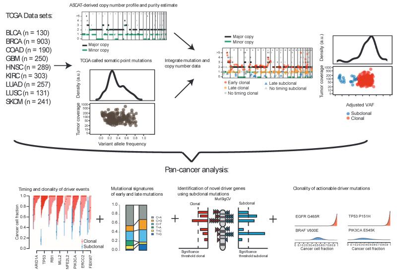
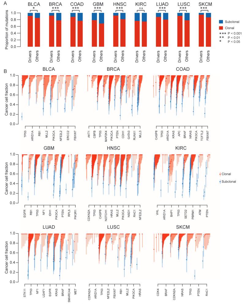
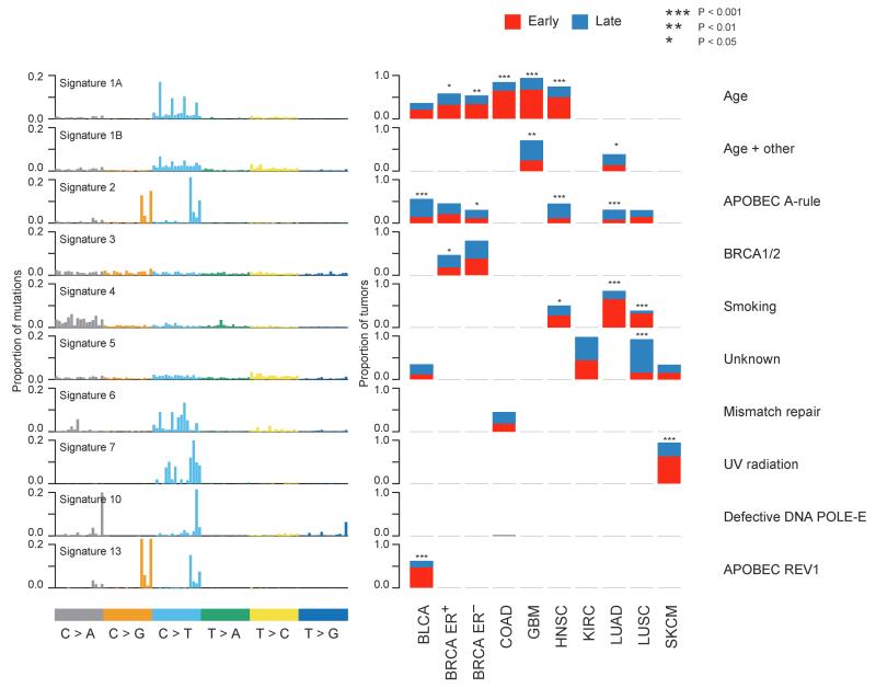
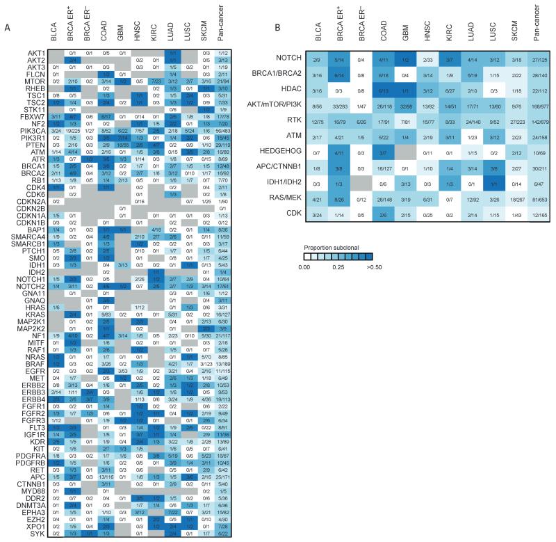
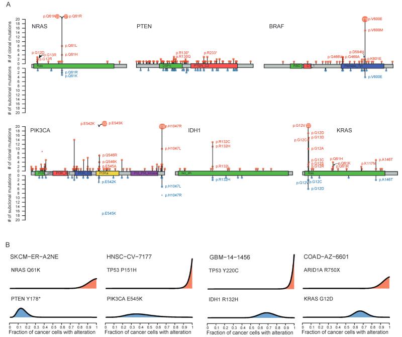
Deciphering whether actionable driver mutations are found in all or a subset of tumor cells will likely be required to improve drug development and precision medicine strategies. We analyzed nine cancer types to determine the subclonal frequencies of driver events, to time mutational processes during cancer evolution, and to identify drivers of subclonal expansions. Although mutations in known driver genes typically occurred early in cancer evolution, we also identified later subclonal "actionable" mutations, including BRAF (V600E), IDH1 (R132H), PIK3CA (E545K), EGFR (L858R), and KRAS (G12D), which may compromise the efficacy of targeted therapy approaches. More than 20% of IDH1 mutations in glioblastomas, and 15% of mutations in genes in the PI3K (phosphatidylinositol 3-kinase)-AKT-mTOR (mammalian target of rapamycin) signaling axis across all tumor types were subclonal. Mutations in the RAS-MEK (mitogen-activated protein kinase kinase) signaling axis were less likely to be subclonal than mutations in genes associated with PI3K-AKT-mTOR signaling. Analysis of late mutations revealed a link between APOBEC-mediated mutagenesis and the acquisition of subclonal driver mutations and uncovered putative cancer genes involved in subclonal expansions, including CTNNA2 and ATXN1. Our results provide a pan-cancer census of driver events within the context of intratumor heterogeneity and reveal patterns of tumor evolution across cancers. The frequent presence of subclonal driver mutations suggests the need to stratify targeted therapy response according to the proportion of tumor cells in which the driver is identified.


Figures

Fig. 1
Fig. 1
Temporal dissection of mutations and mutational processes in TCGA samples. Integration of copy number, purity estimates, and VAF of each somatic mutation permits calculation of the cancer cell fraction, describing the fraction of cancer cells with an alteration. The mutation copy number can also be estimated, allowing further timing of mutations in the case of a genome-doubling or amplification event. These data can reveal the clonality of driver events and shifts in mutational spectra and mutational signatures over time, as well as permitting identification of driver genes using subclonal mutations. a.u., arbitrary unit.
Fig. 2
Fig. 2
Clonal and subclonal mutations in nine cancer types. (A) The proportion of aggregated driver mutations versus other mutations that are clonal/subclonal is indicated for each cancer type. Red represents clonal mutations, and blue represents subclonal mutations. Notably, there is a higher proportion of clonal driver mutations compared to other clonal mutations. Significance from Fisher’s exact test is indicated. Exact P values are as follows: BLCA, P= 0.0292; BRCA, P= 8.19× 10⁻²¹; COAD, P= 1.45× 10⁻⁹; GBM, P= 0.000791; HNSC, P= 2.58× 10⁻⁸; KIRC, P= 0.619; LUAD, P= 0.00311; LUSC, P= 1.89× 10⁻⁵; SKCM, P= 2.71 × 10⁻⁵. (B) The cancer cell fraction of mutations in driver genes within each cancer type is depicted. Each symbol represents a somatic mutation in an individual tumor. On the basis of the probability distributions of the cancer cell fraction, mutations were determined to be either clonal (red circles, upper bound of confidence interval ≥1) or subclonal (blue circles, upper band of confidence interval <1). Error bars represent the 95% confidence interval.
Fig. 3
Fig. 3
Temporal dissection of mutational signatures. For each mutational signature identified in at least one cancer type, the proportion of patients with either a higher fraction of early (red) or late (blue) mutations corresponding to a signature is indicated. A sample is classified as harboring a mutational signature if more than 100 mutations or more than 25%of mutations in that sample correspond to the signature. The factors responsible for the mutational processes are indicated in the right panel. The names of the signatures correspond to those used by Alexandrov et al. (4).
Fig. 4
Fig. 4
Clonal heterogeneity of mutations in genes linked to therapies. (A) Heatmap showing the proportion of nonsilent mutations that are subclonal for each potentially actionable gene across nine cancer types. For each mutation, the number of subclonal mutations identified is indicated for each cancer type and the combined pan-cancer data set. Gray indicates the absence of a mutation. (B) The clonality of actionable pathways is depicted for each cancer type and the combined pan-cancer data set. Pathways are ordered according to subclonality in the pan-cancer data set, with pathways that have a higher proportion of subclonal mutations at the top of the heatmap. Genes related to CDKs (cyclin-dependent kinases) have very few subclonal mutations. RTK, receptor tyrosine kinases. For details of all the genes within each pathway, see table S4.
Fig. 5
Fig. 5
Clonal and subclonal actionable mutations in known cancer genes. (A) The distribution of nonsilent clonal and subclonal mutations in known cancer genes across the entire pan-cancer cohort. Red lollipops indicate clonal mutations, whereas blue lollipops represent subclonal mutations. Square lollipops indicate loss-of-function mutations (such as stop codon or frameshift). Hotspot sites harbor both clonal and subclonal mutations. For hotspot sites that harbor more than 20 mutations, the number of mutations is indicated inside the lollipop. (B) In many cases, mutations in known cancer genes occur in tumors that also harbor clonal driver mutations. Probability distributions over the cancer cell fraction for individual mutations are shown for specific tumors.

Comment in

Similar articles

Cited by

References

    1. Lawrence MS, Stojanov P, Mermel CH, Robinson JT, Garraway LA, Golub TR, Meyerson M, Gabriel SB, Lander ES, Getz G. Discovery and saturation analysis of cancer genes across 21 tumour types. Nature. 2014;505:495–501. - PMC - PubMed
    1. Kandoth C, McLellan MD, Vandin F, Ye K, Niu B, Lu C, Xie M, Zhang Q, McMichael JF, Wyczalkowski MA, Leiserson MD, Miller CA, Welch JS, Walter MJ, Wendl MC, Ley TJ, Wilson RK, Raphael BJ, Ding L. Mutational landscape and significance across 12 major cancer types. Nature. 2013;502:333–339. - PMC - PubMed
    1. Tamborero D, Gonzalez-Perez A, Perez-Llamas C, Deu-Pons J, Kandoth C, Reimand J, Lawrence MS, Getz G, Bader GD, Ding L, Lopez-Bigas N. Comprehensive identification of mutational cancer driver genes across 12 tumor types. Sci. Rep. 2013;3:2650. - PMC - PubMed
    1. Alexandrov LB, Nik-Zainal S, Wedge DC, Aparicio SA, Behjati S, Biankin AV, Bignell GR, Bolli N, Borg A, Borresen-Dale AL, Boyault S, Burkhardt B, Butler AP, Caldas C, Davies HR, Desmedt C, Eils R, Eyfjord JE, Foekens JA, Greaves M, Hosoda F, Hutter B, Ilicic T, Imbeaud S, Imielinski M, Jager N, Jones DT, Jones D, Knappskog S, Kool M, Lakhani SR, Lopez-Otin C, Martin S, Munshi NC, Nakamura H, Northcott PA, Pajic M, Papaemmanuil E, Paradiso A, Pearson JV, Puente XS, Raine K, Ramakrishna M, Richardson AL, Richter J, Rosenstiel P, Schlesner M, Schumacher TN, Span PN, Teague JW, Totoki Y, Tutt AN, Valdes-Mas R, VanBuuren MM, van ’t Veer L, Vincent-Salomon A, Waddell N, Yates LR, Zucman-Rossi J, Futreal PA, McDermott U, Lichter P, Meyerson M, Grimmond SM, Siebert R, Campo E, Shibata T, Pfister SM, Campbell PJ, Stratton MR. Signatures of mutational processes in human cancer. Nature. 2013;500:415–421. - PMC - PubMed
    1. Helleday T, Eshtad S, Nik-Zainal S. Mechanisms underlying mutational signatures in human cancers. Nat. Rev. Genet. 2014;15:585–598. - PMC - PubMed

Publication types