. 2015 Jun;21(6):527-34.
doi: 10.1093/molehr/gav017. Epub 2015 Apr 15.

A role for retinoids in human oocyte fertilization: regulation of connexin 43 by retinoic acid in cumulus granulosa cells

Affiliations

A role for retinoids in human oocyte fertilization: regulation of connexin 43 by retinoic acid in cumulus granulosa cells

Monica W Best et al. Mol Hum Reprod. 2015 Jun.

Abstract

Retinoids are essential for ovarian steroid production and oocyte maturation in mammals. Oocyte competency is known to positively correlate with efficient gap junction intercellular communication (GJIC) among granulosa cells in the cumulus-oocyte complex. Connexin 43 (C x 43) is the main subunit of gap junction channels in human cumulus granulosa cells (CGC) and is regulated by all-trans retinoic acid (ATRA) in other hormone responsive cell types. The objectives of this study were to quantify retinoid levels in human CGC obtained during IVF oocyte retrievals, to investigate the potential relationship between CGC ATRA levels and successful oocyte fertilization, and to determine the effects of ATRA on C x 43 protein expression in CGC. Results showed that CGC cultures actively metabolize retinol to produce ATRA. Grouped according to fertilization rate tertiles, mean ATRA levels were 2-fold higher in pooled CGC from women in the highest versus the lowest tertile (P < 0.05). ATRA induced a rapid dephosphorylation of C x 43 in CGC and granulosa cell line (KGN) cultures resulting in a >2-fold increase in the expression of the functional non-phosphorylated (P0) species (P < 0.02). Similar enhancement of P0 by ATRA was shown in CGC and KGN cultures co-treated with LH or hCG which, by themselves, enhanced the protein levels of C x 43 without altering its phosphorylation profile. Correspondingly, the combination of ATRA+hCG treatment of KGN caused a significant increase in GJIC compared with single agent treatments (P < 0.025) and a doubling of GJIC from that seen in untreated cells (P < 0.01). These findings indicate that CGC are a primary site of retinoid uptake and ATRA biosynthesis. Regulation of C x 43 by ATRA may serve an important role in folliculogenesis, development of oocyte competency, and successful fertilization by increasing GJIC in CGC.

Keywords: all-trans retinoic acid; connexin 43; cumulus granulosa cells; oocyte competency.


Figures

Figure 1
Figure 1
Retinol and all-trans retinoic acid (ATRA) concentrations in patient pooled cumulus granulosa cells (CGC) stratified by tertiles according to percent successfully fertilized (2 pro-nuclei; 2PN) oocytes (i.e. #2PN/total oocytes retrieved from each patient × 100). Values represent mean ± SEM of retinoid concentrations of CGC in the indicated tertile. *Significant difference compared with ATRA levels in the lowest tertile, P < 0.05. Numbers of patients (range of fertilization rates) in each tertile: n = 27 (18–53) in lowest; n = 24 (54–69) in middle; n = 26 (70–100) in highest.
Figure 2
Figure 2
All-trans retinoic acid (ATRA) production in cumulus granulosa cells (CGC). ATRA levels in supernatant (open bars; pmol/ml) and cell pellets (gray bars; pmol/g protein) were assessed by liquid chromatography tandem mass spectrometry (LC-MS/MS) analysis following addition of 2 µM retinol (ROL) for 16 h under serum-free conditions. Values represent the mean ± SEM of ATRA concentrations in CGC cultures (n = 3 patient samples) where (*) indicates a significant increase compared with ROL-free conditions, P < 0.025.
Figure 3
Figure 3
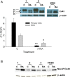
Effect of all-trans retinoic acid (ATRA) on Cx43 phosphorylation. Total cellular protein was isolated from cumulus granulosa cells (CGC) and human granulosa cell line (KGN) cells and assayed for Cx43 expression by western blotting. (A) Representative experiments of primary CGC from two patients and KGN cells treated with 10 µM ATRA (RA) or vehicle control (C) as indicated for 48 h show changes in the band intensity and distribution of non-phosphorylated (P0) and phosphorylated (P1 and P2) species of Cx43 (inset). Figure in the lower panel represents the relative intensity of the three Cx43 phosphorylation species expressed as the ratio (P1 + P2)/P0 in order to quantify the sensitivity of Cx43 dephosphorylation to ATRA treatment. Columns represent the mean (±SEM) from seven CGC patient samples (black bars) and three independent experiments with KGN cells (gray bars). Significant difference when compared with untreated controls, *P < 0.02; **P < 0.0001. (B) Representative western blot analysis using a non-phosphorylated Cx43 (Non-P Cx43)-specific antibody showing the effects of ATRA on Non-P Cx43 in CGC from three patients. Culture conditions were as in part (A). Human endometrial stromal cells (HESC) were previously determined to show up-regulation of non-phosphorylated Cx43 by ATRA (Tanmahasamut and Sidell, 2005; Wu et al., 2013) and were used as positive control cells for this effect. Quantification of six patient samples showed a mean (±SEM) fold increase of 2.31 ± 0.39 induced by ATRA (P < 0.02).
Figure 4
Figure 4
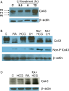
Western blot analysis showing the combined effects of all-trans retinoic acid (ATRA) and LH or hCG on Cx43 phosphorylation. (A) Cumulus granulosa cells (CGC) were cultured for up to 72 h with vehicle control or LH (2.2 IU/ml) added at different time points before the end of the culture period as indicated (representative experiment of four patient samples). Similar enhancements of Cx43 protein levels without alteration of relative intensities of the three phosphorylation species were seen in cultures treated with LH for >24 h (not shown). (B) Representative experiment of five patient samples where CGC were treated with 10 µM ATRA (RA), 80 IU/ml hCG, 2.2 IU/ml LH, or combinations as indicated for 48 h. Western blot analysis was performed for both total Cx43 and non-phosphorylated Cx43 (Non-P Cx43) using a Non-P Cx43 specific antibody. (C) Representative analysis of five independent experiments showing total Cx43 in KGN cells treated for 48 h with 80 IU/ml hCG, 10 µM ATRA (RA), or hCG + ATRA as indicated.
Figure 5
Figure 5
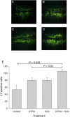
All-trans retinoic acid (ATRA) and hCG effects on gap junction intercellular communication (GJIC). Human granulosa cell line (KGN) cells were treated with (A) vehicle, (B) 10 µM ATRA, (C) 80 IU/ml hCG or (D) ATRA + hCG for 48 h, after which GJIC was assessed by the Scrape loading (SL)/dye transfer (DT) technique as described in Materials and Methods. (E) Mean (±SEM) of five determinations that quantified cell numbers showing positive dye staining.

Similar articles

Cited by

References

    1. Ackert CL, Gittens JE, O'Brien MJ, Eppig JJ, Kidder GM. Intercellular communication via connexin43 gap junctions is required for ovarian folliculogenesis in the mouse. Dev Biol 2001;233:258–270. - PubMed
    1. Alminana C, Gil MA, Cuello C, Caballero I, Roca J, Vazquez JM, Gomez E, Martinez EA. In vitro maturation of porcine oocytes with retinoids improves embryonic development. Reprod Fertil Dev 2008;20:483–489. - PubMed
    1. Borowczyk E, Johnson ML, Bilski JJ, Bilska MA, Redmer DA, Reynolds LP, Grazul-Bilska AT. Role of gap junctions in regulation of progesterone secretion by ovine luteal cells in vitro. Reproduction 2007;133:641–651. - PubMed
    1. Brown JA, Eberhardt DM, Schrick FN, Roberts MP, Godkin JD. Expression of retinol-binding protein and cellular retinol-binding protein in the bovine ovary. Mol Reprod Dev 2003;64:261–269. - PubMed
    1. Dekel N. Cellular, biochemical and molecular mechanisms regulating oocyte maturation. Mol Cell Endocrinol 2005;234:19–25. - PubMed

Publication types