

doi: 10.1128/JVI.00230-15.
Epub 2015 Apr 15.
Glycan Microheterogeneity at the PGT135 Antibody Recognition Site on HIV-1 gp120 Reveals a Molecular Mechanism for Neutralization Resistance
Affiliations
- PMID: 25878100
- PMCID: PMC4468474
- DOI: 10.1128/JVI.00230-15
Item in Clipboard
Glycan Microheterogeneity at the PGT135 Antibody Recognition Site on HIV-1 gp120 Reveals a Molecular Mechanism for Neutralization Resistance
J Virol.
2015 Jul.
Abstract
Broadly neutralizing antibodies have been isolated that bind the glycan shield of the HIV-1 envelope spike. One such antibody, PGT135, contacts the intrinsic mannose patch of gp120 at the Asn332, Asn392, and Asn386 glycosylation sites. Here, site-specific glycosylation analysis of recombinant gp120 revealed glycan microheterogeneity sufficient to explain the existence of a minor population of virions resistant to PGT135 neutralization. Target microheterogeneity and antibody glycan specificity are therefore important parameters in HIV-1 vaccine design.
Copyright © 2015, American Society for Microbiology. All Rights Reserved.
Figures
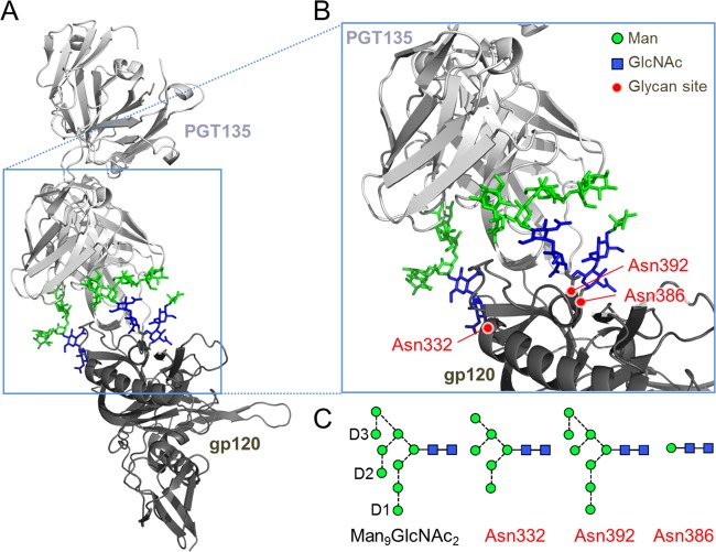
The glycan epitope of PGT135 encompasses the Asn332, Asn392, and Asn386 sites. (A) A previously reported crystal structure reveals the interaction of a PGT135 Fab domain with the Asn332 (Man6GlcNAc2), Asn392 (Man8GlcNAc2), and Asn386 (Man1GlcNAc2) glycans from a gp120JR-FL core (15). The protein moiety is depicted in a ribbon diagram, and glycans are depicted as sticks. Mannose (Man) residues are colored in green, and N-acetlyglucosamine (GlcNAc) residues are colored in blue. (B) Enlarged view of the PGT135 glycan epitope. (C) Schematic representation of a Man9GlcNAc2 glycan, with the D1 to D3 arms annotated and the glycans resolvable in the crystal structure. Glycan structures are shown according to the proposed method of Harvey et al. (40), with residues colored according to panels A and B. Images were made in PyMol using PDB code 4JM2.
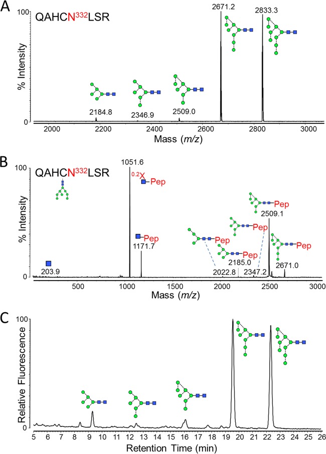
Glycans present at the Asn332 glycosylation site. Recombinant, monomeric gp120BaL was expressed in HEK 293T cells from the pHLsec vector (41) and purified by metal-affinity and size exclusion chromatography, as previously described (42). gp120 was reduced, alkylated, and digested with trypsin (Promega), before fractionation using a Jupiter C18 5-μm 250- by 4.5-mm column (300-Å pore size) and a Dionex U3000 liquid chromatography system. Fractions were collected every minute, at a flow rate of 1 ml/min, for 90 min, and then analyzed on an Autoflex Speed MALDI tandem time of flight (TOF/TOF) instrument (Bruker), operated in positive-ion mode. (A) MALDI MS of pooled fractions containing the Asn332-containing glycopeptide, QAHCNLSR. The glutamine carried a pyro-Glu modification (−17), and the cysteine was modified due to treatment with iodoacetamide (carbamidomethyl, +57). Glycan structures corresponding to the observed glycopeptide masses are indicated. (B) MALDI MS/MS fragmentation spectrum of the 2,671.2 peak, corresponding to a Man8GlcNAc2 glycopeptide. Fragment ions were observed that were characteristic of glycopeptide MALDI MS/MS fragmentation (43), including [Mpep+H + 83]+ (corresponding to 0.2X-ring cleavage of the innermost GlcNAc) and [Mpep+H + 203]+ (corresponding to Y-type cleavage of the di-N-acetylchitobiose core). Y-type fragmentation of glycosidic bonds was also observed. (C) HILIC-UPLC profile of Asn332 glycans. Glycans were released from the QAHCNLSR glycopeptides by in-solution PNGase F (QA-Bio) digestion, according to the manufacturer's instructions, and then labeled using a LudgerTag 2-AB labeling kit (Ludger Ltd., Abingdon, United Kingdom). Chromatography was performed on a Waters Acquity UPLC instrument. Glycans were assigned by comparison with known oligomannose-type glycan standards (Ludger Ltd., Abingdon, United Kingdom).
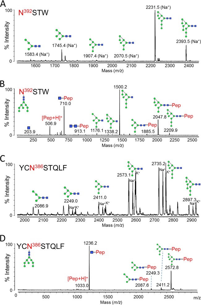
Glycans present at the Asn392 and Asn386 glycosylation sites. gp120 was digested with chymotrypsin (Promega) before RP-HPLC and MALDI analysis. (A) MALDI MS of Asn392 glycopeptides (NSTW). Sodium, [M + 23]+, and potassium, [M + 39]+, adducts were observed: both peaks were used for measuring abundances (Table 1). (B) MALDI MS/MS fragmentation spectrum of the peak corresponding to the Man8GlcNAc2 glycopeptide. Fragmentation peaks corresponded to the protonated masses. (C) MALDI MS of Asn386 glycopeptides (YCNSTQLF). The cysteine was modified due to treatment with iodoacetamide (carbamidomethyl, +57). Protonated glycopeptides, as well as sodium and potassium adducts, were detected: all were used for calculation of abundances (Table 1). (D) MALDI MS/MS fragmentation spectrum of the peak corresponding to the Man8GlcNAc2 glycopeptide. Fragmentation peaks corresponded to the protonated masses.
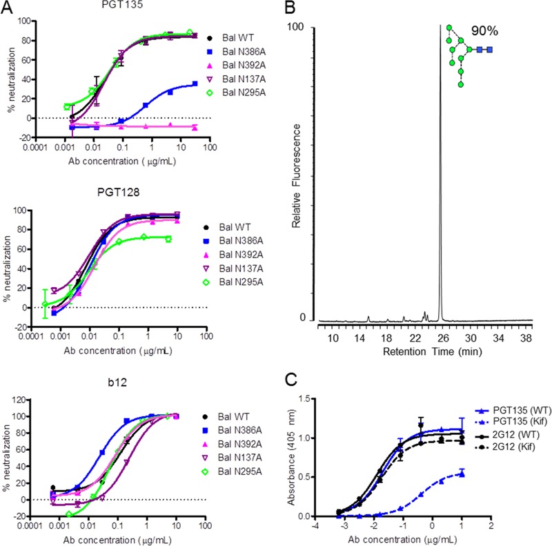
Influences of gp120 glycans on bnAb recognition. (A) Neutralization sensitivity of PGT135, PGT128, and b12 against BaL wild-type pseudovirus and glycan site mutants. bnAbs PGT128 and b12 are used as controls. To produce pseudoviruses, plasmids encoding Env were cotransfected with an Env-deficient genomic backbone plasmid (pSG3ΔEnv) in a 1:2 ratio with the transfection reagent polyethyleneimine (PEI) (1 mg/ml, 1:3 PEI-total DNA [Polysciences]) into HEK 293T cells. Pseudoviruses were harvested 72 h posttransfection. Neutralizing activity was assessed using a single-round replication pseudovirus assay with TZM-bl target cells, as described previously (2). Glycan sites N137, N295, N386, and N392 were removed using site-directed mutagenesis through an Asn-to-Ala mutation. Mutations were verified by DNA sequencing (MWG Eurofins, Germany). (B) Glycan profile of recombinant gp120 expressed in HEK 293T cells in the presence of kifunensine, analyzed as described in the legend to Fig. 2C. (C) Enzyme-linked immunosorbent assay (ELISA) data of PGT135 (blue) and 2G12 (black) binding to wild-type (continuous line) and kifunensine-treated (dashed line) gp120. ELISAs were performed as previously described (34).
Similar articles
-
Conserved Role of an N-Linked Glycan on the Surface Antigen of Human Immunodeficiency Virus Type 1 Modulating Virus Sensitivity to Broadly Neutralizing Antibodies against the Receptor and Coreceptor Binding Sites.J Virol. 2015 Oct 28;90(2):829-41. doi: 10.1128/JVI.02321-15. Print 2016 Jan 15. J Virol. 2015. PMID: 26512079 Free PMC article.
-
Structure-based, targeted deglycosylation of HIV-1 gp120 and effects on neutralization sensitivity and antibody recognition.Virology. 2003 Sep 1;313(2):387-400. doi: 10.1016/s0042-6822(03)00294-0. Virology. 2003. PMID: 12954207
-
Chinks in the armor of the HIV-1 Envelope glycan shield: Implications for immune escape from anti-glycan broadly neutralizing antibodies.Virology. 2017 Jan 15;501:12-24. doi: 10.1016/j.virol.2016.10.026. Epub 2016 Nov 13. Virology. 2017. PMID: 27846415
-
Antibody responses to the HIV-1 envelope high mannose patch.Adv Immunol. 2019;143:11-73. doi: 10.1016/bs.ai.2019.08.002. Epub 2019 Sep 11. Adv Immunol. 2019. PMID: 31607367 Free PMC article. Review.
-
GP120: target for neutralizing HIV-1 antibodies.Annu Rev Immunol. 2006;24:739-69. doi: 10.1146/annurev.immunol.24.021605.090557. Annu Rev Immunol. 2006. PMID: 16551265 Review.
Cited by
-
Global site-specific N-glycosylation analysis of HIV envelope glycoprotein.Nat Commun. 2017 Mar 28;8:14954. doi: 10.1038/ncomms14954. Nat Commun. 2017. PMID: 28348411 Free PMC article.
-
The HIV glycan shield as a target for broadly neutralizing antibodies.FEBS J. 2015 Dec;282(24):4679-91. doi: 10.1111/febs.13530. Epub 2015 Oct 23. FEBS J. 2015. PMID: 26411545 Free PMC article. Review.
-
The HIV-1 envelope glycoprotein: structure, function and interactions with neutralizing antibodies.Nat Rev Microbiol. 2025 Jul 23. doi: 10.1038/s41579-025-01206-6. Online ahead of print. Nat Rev Microbiol. 2025. PMID: 40702326 Review.
-
Structural Rearrangements Maintain the Glycan Shield of an HIV-1 Envelope Trimer After the Loss of a Glycan.Sci Rep. 2018 Oct 9;8(1):15031. doi: 10.1038/s41598-018-33390-2. Sci Rep. 2018. PMID: 30302011 Free PMC article.
-
Antibody recognition of HIV and dengue glycoproteins.Glycobiology. 2016 Aug;26(8):813-9. doi: 10.1093/glycob/cww031. Epub 2016 Mar 3. Glycobiology. 2016. PMID: 26941393 Free PMC article. Review.
References
-
- Walker LM, Phogat SK, Chan-Hui P-Y, Wagner D, Phung P, Goss JL, Wrin T, Simek MD, Fling S, Mitcham JL, Lehrman JK, Priddy FH, Olsen OA, Frey SM, Hammond PW, Kaminsky S, Zamb T, Moyle M, Koff WC, Poignard P, Burton DR. 2009. Broad and potent neutralizing antibodies from an African donor reveal a new HIV-1 vaccine target. Science 326:285–289. doi:10.1126/science.1178746. - DOI - PMC - PubMed
-
- Walker LM, Huber M, Doores KJ, Falkowska E, Pejchal R, Julien J-P, Wang S-K, Ramos A, Chan-Hui P-Y, Moyle M, Mitcham JL, Hammond PW, Olsen OA, Phung P, Fling S, Wong C-H, Phogat S, Wrin T, Simek MD, Koff WC, Wilson IA, Burton DR, Poignard P. 2011. Broad neutralization coverage of HIV by multiple highly potent antibodies. Nature 477:466–470. doi:10.1038/nature10373. - DOI - PMC - PubMed
-
- Falkowska E, Le KM, Ramos A, Doores KJ, Lee JH, Blattner C, Ramirez A, Derking R, van Gils MJ, Liang C-H, Mcbride R, von Bredow B, Shivatare SS, Wu C-Y, Chan-Hui P-Y, Liu Y, Feizi T, Zwick MB, Koff WC, Seaman MS, Swiderek K, Moore JP, Evans D, Paulson JC, Wong C-H, Ward AB, Wilson IA, Sanders RW, Poignard P, Burton DR. 2014. Broadly neutralizing HIV antibodies define a glycan-dependent epitope on the prefusion conformation of gp41 on cleaved envelope trimers. Immunity 40:657–668. doi:10.1016/j.immuni.2014.04.009. - DOI - PMC - PubMed
-
- Burton DR, Ahmed R, Barouch DH, Butera ST, Crotty S, Godzik A, Kaufmann DE, McElrath MJ, Nussenzweig MC, Pulendran B, Scanlan CN, Schief WR, Silvestri G, Streeck H, Walker BD, Walker LM, Ward AB, Wilson IA, Wyatt R. 2012. A blueprint for HIV vaccine discovery. Cell Host Microbe 12:396–407. doi:10.1016/j.chom.2012.09.008. - DOI - PMC - PubMed
Publication types
MeSH terms
Substances
Grants and funding
LinkOut - more resources
Full Text Sources
Other Literature Sources
