. 2015 Jul;89(13):6656-72.
doi: 10.1128/JVI.00571-15. Epub 2015 Apr 15.

Stable Phenotypic Changes of the Host T Cells Are Essential to the Long-Term Stability of Latent HIV-1 Infection

Affiliations

Stable Phenotypic Changes of the Host T Cells Are Essential to the Long-Term Stability of Latent HIV-1 Infection

Lillian Seu et al. J Virol. 2015 Jul.

Abstract

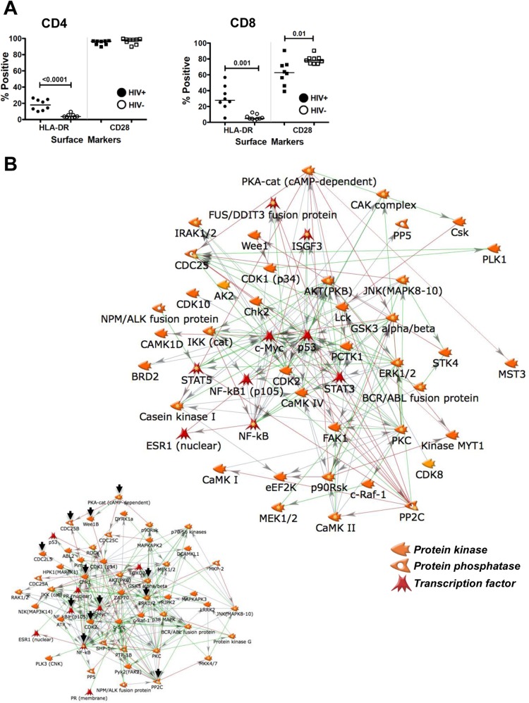
The extreme stability of the latent HIV-1 reservoir in the CD4(+) memory T cell population prevents viral eradication with current antiretroviral therapy. It has been demonstrated that homeostatic T cell proliferation and clonal expansion of latently infected T cells due to viral integration into specific genes contribute to this extraordinary reservoir stability. Nevertheless, given the constant exposure of the memory T cell population to specific antigen or bystander activation, this reservoir stability seems remarkable, unless it is assumed that latent HIV-1 resides exclusively in memory T cells that recognize rare antigens. Another explanation for the stability of the reservoir could be that the latent HIV-1 reservoir is associated with an unresponsive T cell phenotype. We demonstrate here that host cells of latent HIV-1 infection events were functionally altered in ways that are consistent with the idea of an anergic, unresponsive T cell phenotype. Manipulations that induced or mimicked an anergic T cell state promoted latent HIV-1 infection. Kinome analysis data reflected this altered host cell phenotype at a system-wide level and revealed how the stable kinase activity changes networked to stabilize latent HIV-1 infection. Protein-protein interaction networks generated from kinome data could further be used to guide targeted genetic or pharmacological manipulations that alter the stability of latent HIV-1 infection. In summary, our data demonstrate that stable changes to the signal transduction and transcription factor network of latently HIV-1 infected host cells are essential to the ability of HIV-1 to establish and maintain latent HIV-1 infection status.

Importance: The extreme stability of the latent HIV-1 reservoir allows the infection to persist for the lifetime of a patient, despite completely suppressive antiretroviral therapy. This extreme reservoir stability is somewhat surprising, since the latently HIV-1 infected CD4(+) memory T cells that form the structural basis of the viral reservoir should be exposed to cognate antigen over time. Antigen exposure would trigger a recall response and should deplete the reservoir, likely over a relatively short period. Our data demonstrate that stable and system-wide phenotypic changes to host cells are a prerequisite for the establishment and maintenance of latent HIV-1 infection events. The changes observed are consistent with an unresponsive, anergy-like T cell phenotype of latently HIV-1 infected host cells. An anergy-like, unresponsive state of the host cells of latent HIV-1 infection events would explain the stability of the HIV-1 reservoir in the face of continuous antigen exposure.


Figures

FIG 1
FIG 1
Anergy induction results in a higher level of HIV-1 latency establishment. The calcium ionophore ionomycin induces T cell anergy. Following three applications of ionomycin to Jurkat cells over a period of 7 days and a resting period of 4 days, 30 individual infection cultures each of untreated control Jurkat cells and ionomycin-treated Jurkat cells were initiated over a wide range of MOIs. (A) Active initial infection levels were determined on day 4 postinfection by using PMA-induced reactivation, with GFP as a surrogate marker for HIV-1 infection. (B) The size of the silent HIV-1 reservoir in each culture was plotted against the level of initially active infection as determined on day 3 postinfection. (C) The size of the latent HIV-1 reservoir in each culture as determined on day 14 postinfection was plotted against the level of initially active infection.
FIG 2
FIG 2
CDK2 inhibition boosts HIV-1 latency establishment. Reduced CDK2 activity has been associated with an unresponsive T cell phenotype. The data presented thus test the effect of the CDK2 inhibitor roscovitine on the establishment of latent infection. (A) Effect of the CDK2 inhibitor roscovitine (10 μM) on HIV-1 susceptibility or active de novo HIV-1 infection levels on day 4 postinfection. (B) Effect of the CDK2 inhibitor roscovitine (10 μM) on maximum HIV-1 expression levels on day 4 postinfection, plotted as a function of the active infection level. (C) Establishment of reservoirs of silent infection events (day 4 p.i.) in the presence or absence of roscovitine, plotted as a function of initial active infection levels achieved in each individual culture. (D) Establishment of latent HIV-1 infection reservoirs in control and roscovitine-treated cell cultures as determined on day 14 postinfection. Each data set represents a summary of 30 paired infection cultures generated at different MOIs.
FIG 3
FIG 3
CDK2 inhibition affects activation-induced HIV-1 reactivation. If a reduction in CDK2 activity renders T cells unresponsive, we would expect CDK2 inhibitors to reduce the ability of otherwise potent agents, such as PMA, to trigger HIV-1 reactivation. We thus tested the concentration-dependent effects of a series of specific CDK2 inhibitors in preventing PMA-induced HIV-1 reactivation in CA5 T cells. The CDK2 inhibitors used were roscovitine (A), purvalanol (B), CDK2 inhibitor III (CDK2i III) (C), and kenpaullone (D). Cells were pretreated for 2 h with the indicated concentration of the respective CDK2 inhibitor, followed by stimulation with a high dose of PMA (10 nM). HIV-1 reactivation levels were determined as the percentage of GFP-positive cells 48 h post-PMA addition. Data for at least 3 independent experiments are presented.
FIG 4
FIG 4
Effects of GRAIL (gene related to anergy in lymphocytes) and ICER (inducible cAMP early repressor) expression on activation-induced HIV-1 reactivation. High levels of GRAIL and ICER are hallmarks of T cell anergy. Latently HIV-1 infected CA5 T cells were retrovirally transduced to express increased levels of ICER or GRAIL. Two days postransduction, the parental cells and the retrovirally transduced cell populations were stimulated with PMA (10 ng/ml), and the level of HIV-1 reactivation was measured by flow cytometry using GFP as a surrogate marker of HIV-1 expression. The cells were not selected with puromycin. For many transgenic cell lines, we observe relatively fast adaptation to a new phenotype or reversion to the parental phenotype within days, which no longer represents the actual initial transgenic effect. The estimated transduction efficacy was 40 to 60%.
FIG 5
FIG 5
Latently HIV-1 infected T cells exhibit an altered, distinct kinetic NF-κB activation profile. An altered NF-κB response is a hallmark of T cell anergy. (A) Kinetic NF-κB profiles for parental Jurkat cells and latently HIV-1 infected CA5 T cells were established for a period of 360 min following PMA stimulation using TransAm assays. Kinetic profiles are shown for NF-κB p65 and NF-κB p50. (B to F) With the characteristic overshooting NF-κB peak in the latently infected T cells occurring within the first 120 min to replace the sinus wave-shaped normal NF-κB response seen in uninfected cells, we monitored the kinetic NF-κB responses in four additional latently infected T cell lines over this period. T cells latently infected with J-LAI-A, J-LAI-B, J-LAI-C, or J-LAI-E were generated using LTR chimeric viruses that would reflect the prototypical transcription factor binding site compositions of subtypes A, B, C, and E, all within the backbone of the molecular HIV-1 LAI clone. All cells were generated using the J2574 GFP reporter T cell population. We had demonstrated previously that each of these viruses establishes a different level of latency at the population level (35). Shown are NF-κB kinetic profiles for unstimulated controls (C) and following TNF-α stimulation (TNF) for the parental J2574 T cells (B), LAI-A infection (high level of latency establishment) (C), LAI-B infection (reference virus) (D), LAI-C infection (high level of latency establishment) (E), and LAI-E infection (very low level of latency establishment) (F). Independently of the latency establishment phenotype of the virus, all latently infected T cell lines exhibited the same altered kinetic NF-κB activation profile.
FIG 6
FIG 6
Kinome analysis-derived protein-protein interaction network describing phenotypic changes of CA5 T cells enabling the stability of latent HIV-1 infection. Lysates of Jurkat and latently HIV-1 infected CA5 T cells were loaded onto Kinexus kinase antibody arrays, and the expression levels and phosphorylation states of a total of 37 kinases and phosphatases were determined. Only spots with altered expression or activity in CA5 T cells relative to that in parental Jurkat cells that had a Z′ ratio of >1.2 were included. The same lysates were loaded onto PamGene chips, which revealed a total of 18 substrate peptides to be differentially phosphorylated by the kinase activity present in latently infected CA5 T cells. Using the Kinexus upstream kinase database, a total of 30 kinases were predicted to exert this phosphorylation activity. The accumulated set of kinases was uploaded on MetaCore to generate the one-step interaction network presented, which is predicted to control latent HIV-1 infection. Detailed information on the regulation of the various altered kinases can be found in Table S1 in the supplemental material.
FIG 7
FIG 7
Protein-protein interaction networks reveal synergistic drug effects. The PIN depicted in Fig. 6 revealed the convergence of PIM-1 activity and CDK2 activity. In two dose-matrix experiments, we thus tested whether the PIN prediction would translate into the ability of PIM-1 inhibitor IV–roscovitine (A) and AS601245–roscovitine (B) combinations to act in concert as inhibitors of PMA-induced HIV-1 reactivation. For this purpose, roscovitine was titrated over a concentration range from 0.01 to 10 μM, whereas the two reported PIM-1 inhibitors were cross-titrated over a concentration range from 0.01 to 10 μM. PMA-induced reactivation was then measured as the percentage of GFP-positive cells 48 h poststimulation. Cell viability was not affected by the treatment.
FIG 8
FIG 8
Host cell IκB expression levels affect HIV-1 latency establishment. On the kinase antibody arrays, IκBa was detected as the highest altered spot in latently HIV-1 infected CA5 T cells. To test for a possible influence of IκB on HIV-1 latency control, we retrovirally transduced J2574 GFP reporter T cells to express IκB. (A) Following infection of the parental J2574 cells and J2574-IκB cells over a wide MOI range, we compared the levels of HIV-1 expression in infected J2574 cells and J2574-IκB cells, under baseline and PMA-activated conditions, to demonstrate that IκB is overexpressed and physiologically active in J2574-IκB cells and that it reduces HIV-1 expression, measured using GFP mean channel fluorescence as a surrogate marker. (B) The infection levels of J2574 cells and J2574-IκB cells as a function of viral input were determined on day 3 postinfection. (C) The establishment of a silent infection reservoir in the paired infection cultures was compared on day 4 postinfection. (D) The establishment of a stable latent HIV-1 reservoir in the paired infection cultures was compared on day 14 postinfection. Each data set represents a summary of results for 30 paired infection cultures generated at different MOIs.
FIG 9
FIG 9
Dyrk1 inhibitors alter host cell control of latent HIV-1 infection. Dyrk1 was discovered as a seed node on the PamGene kinase substrate chip. (A) Dyrk1a controls the DREAM complex, which in turn controls G0 cell cycle exit and quiescence, suggesting that in this system, inhibition of Dyrk1 could alter host cells with latent HIV-1 infection to release infection events from their latent state. (B and C) The addition of Dyrk1 inhibitor INDY (B) or preINDY (C) in a concentration-dependent manner primed latent HIV-1 infection for reactivation by a second, synergistically acting activator, here bryostatin.
FIG 10
FIG 10
T cells from HIV-1 infected patients on suppressive ART exhibit a stable altered kinome signature. (A) To address whether a stably altered kinomic signature would be present in T cells from HIV-1 patients on ART, we initially immune-phenotyped PBMCs from 9 healthy controls and 8 gender-, race-, and age-matched HIV-1-infected patients for the expression of the activation marker HLA-DR and the possible loss of CD28 as an immunological senescence marker. (B) Cell lysate materials from the 9 control samples and from the 8 patient samples were pooled to produce one control sample and one HIV-1 patient sample. By this means, individual variations among donors are minimized. The samples were loaded onto Kinexus kinase antibody arrays, and expression levels and phosphorylation states of a total of 510 kinases and phosphatases were determined. Spots with altered expression or activity in HIV-1 patient samples relative to healthy-control samples that had a Z′ ratio of >1.2 were included to generate a one-step interaction network. For the purpose of better visualization, only kinases, phosphatases, and transcription factors are shown. Detailed information on the regulation of the various altered kinases can be found in Table S4 in the supplemental material. (Inset) Depiction of the equivalent CA5 T cell PIN, for easier comparability. Black arrows indicate kinases/phosphatases present in both networks.

Similar articles

Cited by

References

    1. Chun TW, Carruth L, Finzi D, Shen X, DiGiuseppe JA, Taylor H, Hermankova M, Chadwick K, Margolick J, Quinn TC, Kuo YH, Brookmeyer R, Zeiger MA, Barditch-Crovo P, Siliciano RF. 1997. Quantification of latent tissue reservoirs and total body viral load in HIV-1 infection. Nature 387:183–188. doi:10.1038/387183a0. - DOI - PubMed
    1. Chun TW, Davey RT Jr, Ostrowski M, Shawn Justement J, Engel D, Mullins JI, Fauci AS. 2000. Relationship between pre-existing viral reservoirs and the re-emergence of plasma viremia after discontinuation of highly active anti-retroviral therapy. Nat Med 6:757–761. doi:10.1038/77481. - DOI - PubMed
    1. Chun TW, Engel D, Mizell SB, Ehler LA, Fauci AS. 1998. Induction of HIV-1 replication in latently infected CD4+ T cells using a combination of cytokines. J Exp Med 188:83–91. doi:10.1084/jem.188.1.83. - DOI - PMC - PubMed
    1. Finzi D, Blankson J, Siliciano JD, Margolick JB, Chadwick K, Pierson T, Smith K, Lisziewicz J, Lori F, Flexner C, Quinn TC, Chaisson RE, Rosenberg E, Walker B, Gange S, Gallant J, Siliciano RF. 1999. Latent infection of CD4+ T cells provides a mechanism for lifelong persistence of HIV-1, even in patients on effective combination therapy. Nat Med 5:512–517. doi:10.1038/8394. - DOI - PubMed
    1. Hellerstein M, Hanley MB, Cesar D, Siler S, Papageorgopoulos C, Wieder E, Schmidt D, Hoh R, Neese R, Macallan D, Deeks S, McCune JM. 1999. Directly measured kinetics of circulating T lymphocytes in normal and HIV-1-infected humans. Nat Med 5:83–89. doi:10.1038/4772. - DOI - PubMed

Publication types

Substances