. 2015 Jul;89(13):6695-710.
doi: 10.1128/JVI.00503-15. Epub 2015 Apr 15.

The Vesicle-Forming 6K2 Protein of Turnip Mosaic Virus Interacts with the COPII Coatomer Sec24a for Viral Systemic Infection

Affiliations

The Vesicle-Forming 6K2 Protein of Turnip Mosaic Virus Interacts with the COPII Coatomer Sec24a for Viral Systemic Infection

Jun Jiang et al. J Virol. 2015 Jul.

Abstract

Positive-sense RNA viruses remodel host cell endomembranes to generate quasi-organelles known as "viral factories" to coordinate diverse viral processes, such as genome translation and replication. It is also becoming clear that enclosing viral RNA (vRNA) complexes within membranous structures is important for virus cell-to-cell spread throughout the host. In plant cells infected by turnip mosaic virus (TuMV), a member of the family Potyviridae, peripheral motile endoplasmic reticulum (ER)-derived viral vesicles are produced that carry the vRNA to plasmodesmata for delivery into adjacent noninfected cells. The viral protein 6K2 is responsible for the formation of these vesicles, but how 6K2 is involved in their biogenesis is unknown. We show here that 6K2 is associated with cellular membranes. Deletion mapping and site-directed mutagenesis experiments defined a soluble N-terminal 12-amino-acid stretch, in particular a potyviral highly conserved tryptophan residue and two lysine residues that were important for vesicle formation. When the tryptophan residue was changed into an alanine in the viral polyprotein, virus replication still took place, albeit at a reduced level, but cell-to-cell movement of the virus was abolished. Yeast (Saccharomyces cerevisiae) two-hybrid and coimmunoprecipitation experiments showed that 6K2 interacted with Sec24a, a COPII coatomer component. Appropriately, TuMV systemic movement was delayed in an Arabidopsis thaliana mutant line defective in Sec24a. Intercellular movement of TuMV replication vesicles thus requires ER export of 6K2, which is mediated by the interaction of the N-terminal domain of the viral protein with Sec24a.

Importance: Many plant viruses remodel the endoplasmic reticulum (ER) to generate vesicles that are associated with the virus replication complex. The viral protein 6K2 of turnip mosaic virus (TuMV) is known to induce ER-derived vesicles that contain vRNA as well as viral and host proteins required for vRNA synthesis. These vesicles not only sustain vRNA synthesis, they are also involved in the intercellular trafficking of vRNA. In this investigation, we found that the N-terminal soluble domain of 6K2 is required for ER export of the protein and for the formation of vesicles. ER export is not absolutely required for vRNA replication but is necessary for virus cell-to-cell movement. Furthermore, we found that 6K2 physically interacts with the COPII coatomer Sec24a and that an Arabidopsis thaliana mutant line with a defective Sec24a shows a delay in the systemic infection by TuMV.


Figures

FIG 1
FIG 1
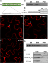
6K2 is a membrane-associated protein. (A) Schematic representation of the TuMV open reading frames, with 6K2 highlighted in red. The 6K2 amino acid sequence is shown below, with the predicted transmembrane domain shown in boldface. (B) 6K2 topology prediction using the TMHMM server. The probability values of each amino acid residue located inside (shown in blue) and outside (shown in red) are plotted against the corresponding amino acid position. (C) Immunoblot analysis of total, soluble, and membrane-associated proteins of 6K2:mCherry, mCherry, and ST-YFP. N. benthamiana plants were agroinfiltrated with 6K2:mCherry, mCherry, or ST-YFP and kept in the growth chamber for 3 days. Total proteins (S3) were extracted, and soluble proteins (S30) were separated from membrane-associated proteins (P30) by centrifugation at 30,000 × g. (D) Membrane-enriched fractions of 6K2:mCherry were treated with 1% Triton X-100, 100 mM Na2CO3, or 4 M urea for 30 min at 4°C. After centrifugation, the S30 and P30 fractions were submitted to immunoblot analysis. Proteins were separated by SDS-PAGE and analyzed by Western blotting using a rabbit serum against RFP or GFP.
FIG 2
FIG 2
The N-terminal tail of 6K2 renders cytosolic 6K1 membrane associated. (A) Protein 6K1 secondary structure prediction. The 6K1 amino acid sequence is shown below, and the predicted TMD is shown in boldface. (B and C) Confocal microscopy imaging of leaf epidermal cell of N. benthamiana expressing 6K1:mCherry (B) or mCherry (C) 3 days after agroinfiltration. (D) Fractionation of 6K1:mCherry total proteins (S3) into soluble (S30) and membrane (P30) fractions and detected by Western blotting. (E) Schematic representation of chimeric protein N2-TMD1-C1 with protein 6K1 is presented above. Black lines represent the N-terminal portion of each protein, and amino acid residues are shown. The 6K1 TMD and C-terminal tail are indicated by gray rectangles. Underlined amino acid residues VD are encoded by the incorporated SalI endonuclease restriction site. (F) Confocal microscopy imaging of leaf epidermal cell of N. benthamiana expressing N2-TMD1-C1:mCherry 3 days after agroinfiltration. (G and H) N. benthamiana leaves expressing N2-TMD1-C1:mCherry and the resulting S3, S30, and P30 were analyzed (G). P30 fractions were incubated with 1 M KCl, 100 mM Na2CO3, or 4 M urea, followed by ultracentrifugation and immunoblotting (H). All Western blots were performed with antibodies against RFP. All confocal images are optical images (1-μm thick). The nucleus is indicated by the white arrow.
FIG 3
FIG 3
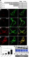
The N-terminal tail is required for ER export of 6K2. (A) Schematic representation of 6K2 and truncated 6K2Δ1–6, 6K2Δ1–12, and 6K2Δ1–18. Black lines represent the N-terminal portion of each protein, and amino acid residues are shown. The 6K2 TMD and C-terminal tail are indicated by gray rectangles. (B to E) Representative N. benthamiana epidermal cells coexpressing 6K2:mCherry (B), 6K2Δ1–6:mCherry (C), 6K2Δ1–12:mCherry (D), or 6K2Δ1–18:mCherry (E) with ER marker GFP-HDEL were imaged using confocal microscopy. These images are optical images (1-μm thick). (F) Colocalization statistical analysis between GFP-HDEL and the WT or the truncated 6K2 vesicles by calculation of the Pearson's correlation coefficient Rr values. Significant difference (Student's t tests, P < 0.001) is indicated by asterisks. Mean values ± standard deviations (SD) from three independent experiments are shown. (G) WT 6K2:mCherry and its N-terminal truncations detected by Western blotting. The bottom panels show equal loading verified by Coomassie staining. (H) Immunoblot analysis of the S3, S30, and P30 fractions of 6K2Δ1–18:mCherry. All Western blots were performed with antibodies raised against RFP.
FIG 4
FIG 4
A conserved tryptophan residue and two lysine residues are important for 6K2 ER export. (A) BLAST of predicted potyviral 6K2 N-terminal tail amino acid sequences. SMV, soybean mosaic virus; TEV, tobacco etch virus; PPV, plum pox virus; PMV, peanut mottle virus; BYMV, bean yellow mosaic virus; TuMV, turnip mosaic virus. Identical amino acid residues that are highly conserved are highlighted by black boxes, and similar amino acid residues are indicated by gray boxes. (B to D) Confocal images of N. benthamiana epidermal cells coexpressing WT 6K2 (B), 6K2K14A-K17A (C), or 6K2W15A (D) with the ER marker GFP-HDEL. These images are optical images (1 μm thick). (E) Colocalization statistical analysis between GFP-HDEL and the WT or the mutated 6K2 vesicles by calculation of the Pearson's correlation coefficient Rr values. Significant difference (Student's t tests, P < 0.001) is indicated by asterisks. Mean values ± SD from three independent experiments are shown. (F) WT 6K2:mCherry, 6K2K14A-K17A:mCherry, and 6K2W15A:mCherry detected by Western blotting with antibodies raised against RFP. The bottom panels show equal loading verified by Coomassie staining.
FIG 5
FIG 5
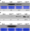
The tryptophan residue is required for TuMV systemic movement. (A) Agroinfiltrated leaves of mock-infected or TuMV-, TuMVW15A-, and TuMVVNN-infected N. benthamiana were evaluated by Western blotting 5 days later. (B) N. benthamiana protoplasts were mock transfected or transfected with p35STuMVVNN, p35STuMVW15A, and p35STuMV, and CP was detected by Western blotting 40 h after transfection. (C) The upper nonagroinfiltrated leaves from panel A were analyzed by Western blotting. The bottom panels in panels A and C show equal protein loading verified by Coomassie-staining. All immunoblotting was performed with anti-TuMV CP rabbit serum.
FIG 6
FIG 6
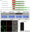
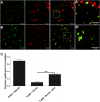
TuMVW15A replication but not virus movement is complemented by cis expression of wild-type 6K2. (A) Schematic representation of TuMV/6K2:mCherry, TuMVW15A/6K2W15A:mCherry, TuMVW15A/6K2:mCherry, TuMV/6K2:mCherry//GFP-HDEL, and TuMVW15A/6K2:mCherry//GFP-HDEL. One copy of 6K2 (wild type or mutated) between P1 and HC-Pro is shown as a red box. Endogenous 6K2 is located between the helicase and VPg, and the W15A mutation is indicated by an arrow. Gray rectangles represent the left and right borders of T-DNA, and gray arrows represent the CaMV 35S promoter. (B) Detection of CP accumulation in leaves mock infected or infected with TuMVW15A/6K2:mCherry, TuMV/6K2:mCherry, and TuMVW15A/6K2W15A:mCherry with anti-TuMV CP serum. Coomassie blue staining (bottom panel) shows equal protein loading. (C and D) Confocal images of N. benthamiana epidermal cells infected with TuMV/6K2:mCherry//GFP-HDEL (C) and TuMVW15A/6K2:mCherry//GFP-HDEL (D). Optical images (2 μm thick) of green, red, and merged colors are shown. (E) Statistical analysis of the percentage of virus cell-to-cell movement of TuMV/6K2:mCherry//GFP-HDEL and TuMVW15A/6K2:mCherry//GFP-HDEL infection foci. Significant differences (Student's t tests, P < 0.001) are indicated by asterisks. Mean values ± SD from three independent experiments are shown.
FIG 7
FIG 7
6K2 colocalizes with COPII coatomer Sec24a. (A and B) N. benthamiana cells coexpressing YFP-Sec24a (left panel) with TuMV/6K2:mCherry (middle panel) in the absence (A) and presence (B) of BFA, with merged panels shown on the right. The area in the dashed box in panels A and B is shown on the right in panels A′ and B′, respectively. (C) Colocalization statistical analysis between YFP-Sec24a and the 6K2-mCherry-tagged vesicles by calculation of the Pearson's correlation coefficient Rr values. Rr values for two colocalizing proteins, ERD2:GFP and Man49:mCherry, are given. For statistical analysis of TuMV/6K2:mCherry plus Sec24a and TuMV/6K2:mCherry plus Sec24a plus BFA colocalization, the sample number (n) is 26 for each tested combination. Significant differences (Student's t tests, 0.001 < P < 0.01) are indicated by asterisks. Mean values ± SD from three independent experiments are shown.
FIG 8
FIG 8
6K2 interacts with COPII coatomer Sec24a. (A) Yeast two-hybrid assay for protein-protein interactions of TuMV 6K2 with Sar1 and Sec24a. The transformants were plated on an SD −Leu −Trp −His plus X-Gal plus 3-AT medium. (Upper left) 6K2 plus Sar1 (LV-Cub-6K2 plus NubG-Sar1). (Upper right) 6K2 plus Sec24a (LV-Cub-6K2 plus NubG-Sec24a). (Lower left) Negative control (LV-Cub-6K2 plus NubG empty vector). (Lower right) Positive control (LV-Cub-6K2 plus NubG-6K2). (B) N. benthamiana leaves expressing combinations of mCherry and YFP, mCherry and YFP-Sec24a, YFP and 6K2:mCherry, or YFP-Sec24a and 6K2:mCherry were harvested 3 days after agroinfiltration. The cleared lysates (input) were subjected to immunopurification on a GFP-Trap resin, followed by Western blot analysis of input and immunopurified (IP) fractions using antibodies against GFP and RFP. The asterisk indicates a nonspecific or a degradation protein species recognized by the anti-GFP serum. (C) N. benthamiana leaves expressing 6K1:mCherry alone, 6K1:mCherry and YFP-Sec24a, N2-TMD1-C1:mCherry and YFP-Sec24a, and 6K2:mCherry and YFP-Sec24a were analyzed as described for panel B.
FIG 9
FIG 9
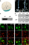
Infection of g92 Arabidopsis thaliana. (A) Yeast two-hybrid assay for protein-protein interactions of TuMV 6K2 with Sec24aR693K. The transformants were plated on an SD −Leu −Trp −His plus X-Gal plus 3-AT medium. I, negative control (LV-Cub-6K2 plus NubG empty vector); II, 6K2 plus Sec24aR693K (LV-Cub-6K2 plus NubG-Sec24aR693K); III, 6K2 plus Sec24a (LV-Cub-6K2 plus NubG-Sec24a). (B) WT and g92 A. thaliana plants were inoculated with TuMV/6K2:GFP and observed under UV light 11 days later. White arrows indicate the viral inoculation site. (C) The upper nonagroinfiltrated leaves of the WT and g92 A. thaliana plants infected with TuMV/6K2:GFP were collected at 11 dpi and analyzed for virus production by immunoblot analysis using a rabbit anti-CP serum. (D) Protoplasts were isolated from WT and g92 A. thaliana plants and were mock transfected or transfected with p35STuMVVNN and p35STuMV. Production of CP was analyzed 40 h after transfection using an anti-TuMV CP rabbit serum. Confocal images of TuMV/6K2:mCherry//GFP-HDEL-infiltrated WT (E) and g92 (F) A. thaliana epidermal cells are shown at 8 and 12 dpi. These images are three-dimensional renderings of 60 1-μm-thick slices that overlap by 0.5 μm. Scale bar, 20 μm.

Similar articles

Cited by

References

    1. den Boon JA, Ahlquist P. 2010. Organelle-like membrane compartmentalization of positive-strand RNA virus replication factories. Annu Rev Microbiol 64:241–256. doi:10.1146/annurev.micro.112408.134012. - DOI - PubMed
    1. Laliberté J-F, Sanfaçon H. 2010. Cellular remodeling during plant virus infection. Annu Rev Phytopathol 48:69–91. doi:10.1146/annurev-phyto-073009-114239. - DOI - PubMed
    1. Belov GA, Nair V, Hansen BT, Hoyt FH, Fischer ER, Ehrenfeld E. 2012. Complex dynamic development of poliovirus membranous replication complexes. J Virol 86:302–312. doi:10.1128/JVI.05937-11. - DOI - PMC - PubMed
    1. Knoops K, Kikkert M, Worm SH, Zevenhoven-Dobbe JC, van der Meer Y, Koster AJ, Mommaas AM, Snijder EJ. 2008. SARS-coronavirus replication is supported by a reticulovesicular network of modified endoplasmic reticulum. PLoS Biol 6:e226. doi:10.1371/journal.pbio.0060226. - DOI - PMC - PubMed
    1. Knoops K, Bárcena M, Limpens RWAL, Koster AJ, Mommaas AM, Snijder EJ. 2012. Ultrastructural characterization of arterivirus replication structures: reshaping the endoplasmic reticulum to accommodate viral RNA synthesis. J Virol 86:2474–2487. doi:10.1128/JVI.06677-11. - DOI - PMC - PubMed

Publication types

MeSH terms

LinkOut - more resources