. 2015 Jul;89(13):6725-45.
doi: 10.1128/JVI.03738-14. Epub 2015 Apr 15.

Immunogenic Display of Purified Chemically Cross-Linked HIV-1 Spikes

Affiliations

Immunogenic Display of Purified Chemically Cross-Linked HIV-1 Spikes

Daniel P Leaman et al. J Virol. 2015 Jul.

Abstract

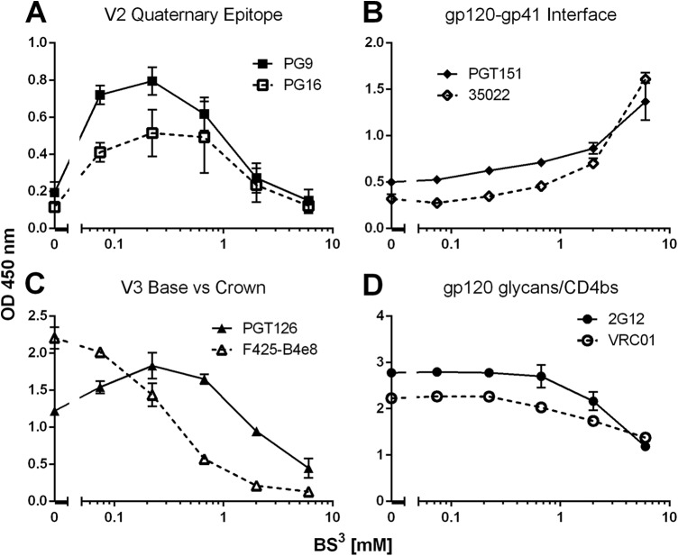
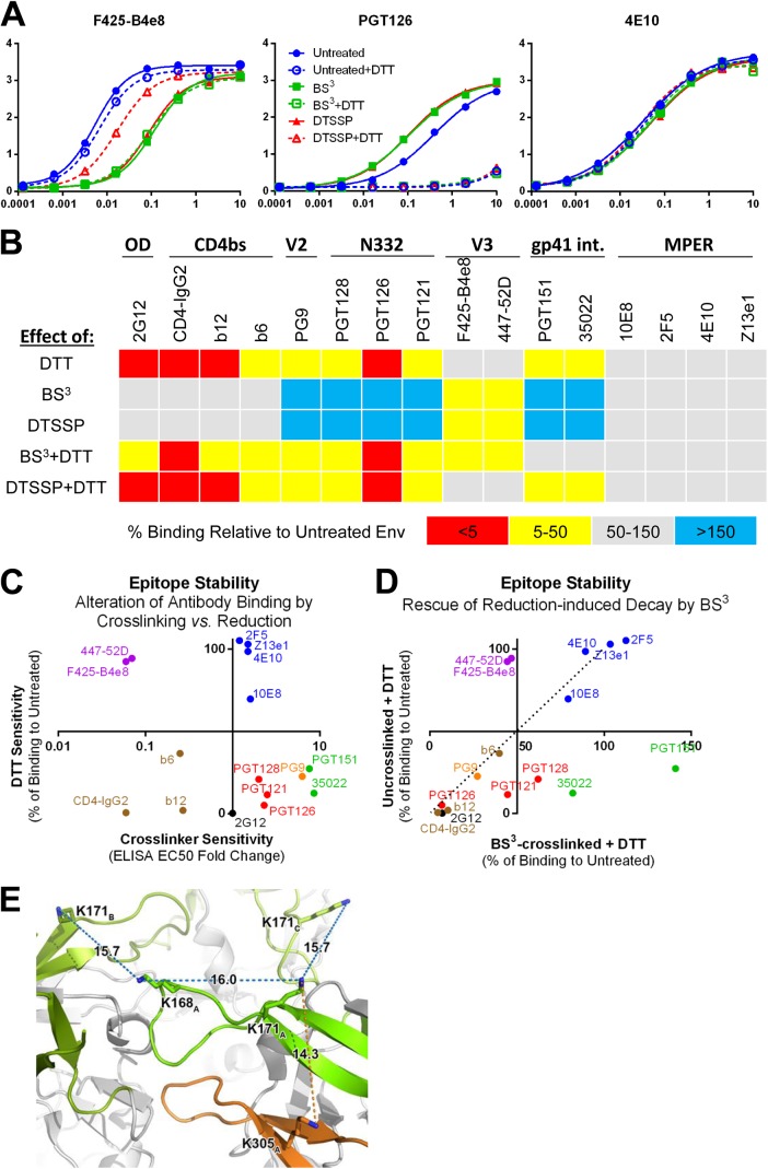
HIV-1 envelope glycoprotein (Env) spikes are prime vaccine candidates, at least in principle, but suffer from instability, molecular heterogeneity and a low copy number on virions. We anticipated that chemical cross-linking of HIV-1 would allow purification and molecular characterization of trimeric Env spikes, as well as high copy number immunization. Broadly neutralizing antibodies bound tightly to all major quaternary epitopes on cross-linked spikes. Covalent cross-linking of the trimer also stabilized broadly neutralizing epitopes, although surprisingly some individual epitopes were still somewhat sensitive to heat or reducing agent. Immunodepletion using non-neutralizing antibodies to gp120 and gp41 was an effective method for removing non-native-like Env. Cross-linked spikes, purified via an engineered C-terminal tag, were shown by negative stain EM to have well-ordered, trilobed structure. An immunization was performed comparing a boost with Env spikes on virions to spikes cross-linked and captured onto nanoparticles, each following a gp160 DNA prime. Although differences in neutralization did not reach statistical significance, cross-linked Env spikes elicited a more diverse and sporadically neutralizing antibody response against Tier 1b and 2 isolates when displayed on nanoparticles, despite attenuated binding titers to gp120 and V3 crown peptides. Our study demonstrates display of cross-linked trimeric Env spikes on nanoparticles, while showing a level of control over antigenicity, purity and density of virion-associated Env, which may have relevance for Env based vaccine strategies for HIV-1.

Importance: The envelope spike (Env) is the target of HIV-1 neutralizing antibodies, which a successful vaccine will need to elicit. However, native Env on virions is innately labile, as well as heterogeneously and sparsely displayed. We therefore stabilized Env spikes using a chemical cross-linker and removed non-native Env by immunodepletion with non-neutralizing antibodies. Fixed native spikes were recognized by all classes of known broadly neutralizing antibodies but not by non-neutralizing antibodies and displayed on nanoparticles in high copy number. An immunization experiment in rabbits revealed that cross-linking Env reduced its overall immunogenicity; however, high-copy display on nanoparticles enabled boosting of antibodies that sporadically neutralized some relatively resistant HIV-1 isolates, albeit at a low titer. This study describes the purification of stable and antigenically correct Env spikes from virions that can be used as immunogens.


Figures

FIG 1
FIG 1
Cross-linked Env trimers are highly stable. (A) Chemical structures of BS3 and DTSSP. (B) Virions were treated with 1 mM BS3 or left untreated and then incubated at either 37°C or 57°C for 1 h. Heat-treated virions were solubilized in DDM, run on BN-PAGE, and probed by Western blotting using a gp41 (4E10, 2F5, and Z13e1) antibody cocktail. (C) Untreated and BS3-cross-linked virions were boiled in Laemmli buffer with DTT for 5 min, run on SDS-PAGE, and visualized by Western blotting with an anti-gp120 (b12, 2G12, and F425-B4e8) antibody cocktail. (D) Untreated and BS3-cross-linked virions were incubated in 1% DDM detergent at 37°C for up to 4 days and then analyzed by BN-PAGE and Western blotting as described above.
FIG 2
FIG 2
Relative binding of bNAbs and non-neutralizing antibodies to BS3-cross-linked Env spikes. HIV-1 virions were either treated with 1 mM BS3 or left untreated, the virions were detergent solubilized, and Env was captured on GNL. Antibody binding was determined by ELISA and the fold change in binding (50% effective concentration [EC50]) to cross-linked Env versus un-cross-linked Env was determined (EC50 untreated/EC50 cross-linked). bNAbs used have an IC50 of <50 μg/ml against JR-FL, and non-neutralizing antibodies have an IC50 of >50 μg/ml. PG9/PG16 binding was determined using JRFL mutant E168R. PG9, PG16, PGT151, and 35O22 did not reach an EC50 against un-cross-linked Env. The results shown are averages of at least three independent experiments.
FIG 3
FIG 3
Effect of BS3 treatment on antibody recognition of virion-associated Env as a function of cross-linker concentration. Virions were treated using a 3-fold dilution series of BS3. Env was detergent solubilized and probed by bNAbs PG9 and PG16 (A), PGT151 and 35O22 (B), PGT126 and F425-B4e8 (C), and 2G12 and VRC01 (D) in a lectin-capture ELISA. The optical density (OD) at 450 nm is reported using PG9, PG16, PGT151, and 35O22 at 10 μg/ml and PGT126, F425-B4e8, 2G12, and VRC01 at 0.08 μg/ml.
FIG 4
FIG 4
Occupancy by bNAbs on cross-linked Env spikes determined using BN-PAGE gel mobility shift. (A) JR-FL virions, either BS3 cross-linked or untreated, were incubated with various antibodies, and Env was resolved using BN-PAGE. Fab fragments were used in this experiment to avoid the cross-linking of spikes that occurs with whole IgGs. Gel mobility shift with PG9 was determined using Env containing the mutation E168R that permits binding by PG9 to JR-FL. (B) Gel mobility shifts induced by antibodies were quantified using imaging software, including some antibodies not shown in panel A. The stoichiometry was calculated by measuring the distance of the gel mobility shift and comparing Fabs of known stoichiometry against un-cross-linked JR-FL (PG9 = one Fab/trimer, PGT151 = two Fabs/trimer, and PGT126 = three Fabs/trimer). Note that domain-swapped 2G12 is an F(ab′)2 and induces a gel mobility shift that is approximately twice what would be expected. Statistically significant changes were determined using an unpaired two-tailed Student t test.
FIG 5
FIG 5
Conformational epitopes on cross-linked Env spikes are relatively thermostable. (A) BS3-cross-linked Env was incubated at 57°C for 1 h and examined using BN-PAGE gel mobility shift. (B) Untreated JR-FL virions, BS3-cross-linked JR-FL virions, or soluble JR-FL SOSIP were incubated at a range of temperatures for 1 h. The Env was solubilized in 1% Empigen detergent (detergent was also added to JR-FL SOSIP for consistency), and bNAb binding was determined using a lectin-capture ELISA. All antibodies were tested at 1 μg/ml, and the percent binding at each temperature is shown relative to a control sample that was incubated at 4°C for 1 h. No binding was detected to un-cross-linked viral Env with PG16, PGT151, and 35O22, and no binding was detected to JR-FL SOSIP by 10E8, since the MPER has been removed from this construct.
FIG 6
FIG 6
The V3 crown is revealed upon cleavage of the cross-link, but bNAb epitopes on spikes are sensitive to reduction regardless of cross-linking. (A) JR-FL virions were cross-linked using BS3 or its disulfide-containing analog DTSSP. DTT breaks DTSSP linkages but does not affect BS3. Detergent-solubilized Env from virions was captured on GNL and probed in an ELISA. Full binding curves of a few representative antibodies are shown. (B) Virion-associated Env was treated as in panel A against a broader panel of bNAbs at a single concentration (1 μg/ml). The percent change in OD at 450 nm is plotted for each condition relative to untreated Env. (C) The effect of cross-linking on binding (fold change in EC50; Fig. 2) is plotted against the effect of DTT reduction (% of wild-type binding) for each antibody. Color-coding accords with class of antibody as follows: 2G12, black; CD4bs, brown; V3 crown, purple; MPER, blue; N332 supersite, red; PG9, orange; and gp120-gp41 interface, green. (D) Effect of DTT on binding to un-cross-linked Env versus BS3-cross-linked Env (% of wild-type binding) for each antibody is plotted. The dashed line denotes where antibodies would be plotted if cross-linking does not affect their binding to DTT-treated Env. Binding by antibodies plotted to the right of the dashed line is preserved when Env is cross-linked before DTT treatment, whereas binding by antibodies to the left of the line is inhibited by cross-linking. (E) Possible cross-links that could be formed at the trimer apex are shown on the crystal structure of BG505 SOSIP.664 (PDB ID 4TVP) (26). Only lysine residues that are shared between BG505 and JR-FL are shown. V1/V2 from the three gp120 protomers is shown in shades of green, and V3 of protomer A is shown in orange. The distance between the side chain primary amines of two lysine residues is labeled by the dotted line (blue, V1/V2-V1/V2; orange, V1-V3) and measured in angstroms. The subscript in the residue number indicates which gp120 protomer the residue belongs to. A combination of an intraprotomer cross-link between V3 and V1/V2 and an interprotomer cross-link between two V1/V2 regions could block accessibility to the V3 crown epitope. The measured distances are not absolute, since in solution the lysines can assume different rotameric states and the V1/V2/V3 loops can be conformationally flexible. BS3 is 11.4 Å long, and the amide bond formed upon cross-linking adds ∼1.5 Å to each end.
FIG 7
FIG 7
BS3-cross-linking stabilizes a conformation of V3 and N332 supersite on native spikes that is distinct from that on soluble gp120 and uncleaved gp140. Effects of BS3 treatment on monomeric gp120, gp140-foldon (FT) trimers, and solubilized spikes (all JR-FL) as probed by antibodies are presented as the fold change in ELISA binding (EC50 cross-linked/EC50 untreated). nb, no binding. Statistically significant changes were determined using a two-tailed t test (*, P < 0.05; **, P < 0.01; ***, P < 0.001).
FIG 8
FIG 8
Immunodepletion using non-neutralizing antibodies removes immature gp160 and other Env debris from cross-linked spike preparations. (A) A diagram demonstrating how non-native Env is removed by immunodepletion using non-neutralizing b6 and D49 antibodies. (B) Virions were cross-linked using cleavable BS3 analog, DTSSP. Detergent-solubilized non-native Env was depleted using b6, D49, or both sequentially. The DTSSP cross-link was reduced using DTT, and Env was resolved by SDS-PAGE and blotted with anti-gp120 antibodies. (C) JR-FL virions were BS3 cross-linked, solubilized in detergent, and then incubated sequentially with b6- and D49-coupled Sepharose beads. Immunodepleted and untreated samples were run side by side in a BN-PAGE gel mobility shift assay using Fabs b12 and b6. The Western blots shown were blotted with anti-gp41 antibodies. The arrow denotes the gp120-gp41 trimer. (D) Samples were analyzed for binding to a panel of neutralizing and non-neutralizing antibodies before or after being immunodepleted as described above using a GNL-capture ELISA. Shown is the average OD at 450 nm for each antibody at 10 μg/ml from at least three independent experiments. Binding by the negative-control antibody DEN3 has been subtracted.
FIG 9
FIG 9
Cross-linked virion spikes can be purified using a FLAG epitope tag. (A) A FLAG tag was genetically fused to the C terminus of full-length gp160 JR-FL Env. FLAG-tagged virions were assayed for infectivity in a single-cycle infectivity assay using TZM-bl cells (solid bar), gp120 incorporation (normalized for p24 input and measured by GNL-capture ELISA; checkered bar), and replication in MT2-CCR5ΔCT cells (p24 equivalents at day 7 relative to wild type; hatched bar). (B) Gp160 FLAG-tagged virions were analyzed using BN-PAGE mobility shift assay using Fabs b12 and b6, as well as anti-FLAG IgG. The Western blot was probed using an anti-gp41 antibody cocktail. (C) JR-FL FLAG-tagged gp160 samples were probed in an ELISA using antibody to gp120 (left panel) and p24 (right panel) before and after immunoaffinity purification. (D) Affinity-purified BS3-cross-linked JR-FL trimers were run on BN-PAGE beside recombinant JR-FL gp120 (5 μg of each protein). The purity of the proteins were analyzed using Coomassie blue staining.
FIG 10
FIG 10
Reference-free 2D class averaged EM reconstructions of BS3-cross-linked JR-FL spikes extracted from viral membrane. (A) EM image of BS3-cross-linked JR-FL Env spikes. (B) Select reference-free 2D class averages of cross-linked JR-FL Env trimers. In the enlarged image, gp120 and gp41 ectodomain regions are shaded blue, while the TM and CT domains encapsulated in a DDM micelle are shaded yellow. The white scale bar in the top left indicates 10 nm. (C) The X-ray crystal structure of the trimer ectodomain (gp120, cyan; gp41, blue; PDB ID 4TVP) (26) is shown for comparison. The predicted location of TM and CT are shown as a yellow circle.
FIG 11
FIG 11
Neutralization properties of sera from rabbits immunized using Env in a DNA prime-cross-linked spike boost format. (A) Schematic illustrating the major events in the immunization study. (B) Cross-linked spikes were captured onto 200-nm magnetic particles to generate PLNs. PLNs were analyzed by GNL-capture ELISA. Biotinylated DOPE was added at 1% of total lipid content for determination of lipid incorporation onto spike-bearing PLNs. A 5-fold dilution series of PLNs was added to GNL-coated microtiter wells and then probed using a constant concentration of anti-gp120 antibody cocktail (b12, 2G12, and F425-B4e8; left) or streptavidin (right). (C) Sera from day 0 (prebleed), day 42 (post-DNA prime), or day 98 (post-protein boost) were tested for neutralization activity against a panel of five well-characterized clade B isolates: JR-FL, JR-CSF, and ADA (tier 2); SF162 (tier 1a), and HxB2 (tier 1b). SIVmac239 and VSV-g were included as negative controls to test for nonspecific inhibition. Serum titers (IC50) are color-coded according to potency, as indicated. (D) Day 98 sera from groups A and D were tested further for neutralization activity against a reference panel of 12 clade B isolates. Serum titer IC50s are color-coded as in panel C. (E) Sera in groups A and D were compared for breadth (number of tested isolates neutralized) and potency of neutralization against SF162, HxB2, and JRCSF. The differences were not statistically significant (breadth, P = 0.19; SF162 potency, P = 0.15; HxB2 potency, P = 0.35; JRCSF potency, P = 0.36).
FIG 12
FIG 12
Neutralizing antibodies in rabbit 7547 target C3 and V5 on JR-CSF gp120. Day 98 serum from rabbit 7547 was tested for neutralization against a panel of gp120 “domain swap” pseudotyped viruses that contain substitutions of major domains from JR-FL gp120 into a JR-CSF backbone (A), major domains from JR-CSF gp120 into a JR-FL backbone (B), and V4 or V5 with substitutions to Gly/Ser residues or the mutation N301Q (C). (D) Sera 7548 and 7549 were also tested for neutralization of JR-CSF mutants V4-GGS33 and N301Q. (E) Sequence changes introduced into V4 and V5 in the mutant Envs used in panels C and D.
FIG 13
FIG 13
CD4 binding site antibodies in PLN-boosted sera 7549 and 7548 neutralize tier 1 and not tier 2 isolates. (A) Sera were incubated with TriMut core or TriMut368/370 at 10 μg/ml, or with an equivalent volume of medium, for 1 h at 37°C. Sera were then tested for neutralization of HxB2, JR-CSF, SF162, TRO.11, and SC422661. (B) Control CD4bs antibody VRC01 was tested in the assay described above. Similar results with VRC01 were observed with all tested isolates.
FIG 14
FIG 14
Binding properties of sera from immunized rabbits. (A) Binding of immune serum against a panel of antigens, including recombinant gp120 and gp41, detergent-dissociated JR-FL viral Env, purified cross-linked spikes, and BS3-cross-linked bovine serum albumin (BSA) as determined by ELISA. All Env molecules were from JR-FL. The data points correspond to individual sera from day 42 (○) and day 98 (◼), and cognate means are shown as dashed and solid lines, respectively. Statistically significant changes were determined using a two-tailed Student t test (*, P < 0.05; **, P < 0.01; ***, P < 0.001). (B) Group A and D sera were tested for binding in ELISA to the scaffold RSC3 that was designed to specifically bind neutralizing CD4bs antibodies. (C) Group A and D sera were tested for the ability to block binding by neutralizing and non-neutralizing antibodies to monomeric JR-FL gp120 or gp41. The IC50 of each serum against each antibody is shown. (D) Serum binding to gp120 V3 and gp41 DSL peptides was determined by using ELISA. Statistical significance was determined as in panel A. (E) Relationship between binding of day 98 sera (EC50) to V3 peptide versus neutralization (IC50) of SF162 virus.

Similar articles

Cited by

References

    1. Mascola JR, Montefiori DC. 2010. The role of antibodies in HIV vaccines. Annu Rev Immunol 28:413–444. doi:10.1146/annurev-immunol-030409-101256. - DOI - PubMed
    1. Walker LM, Burton DR. 2010. Rational antibody-based HIV-1 vaccine design: current approaches and future directions. Curr Opin Immunol 22:358–366. doi:10.1016/j.coi.2010.02.012. - DOI - PMC - PubMed
    1. Stephenson KE, Barouch DH. 2013. A global approach to HIV-1 vaccine development. Immunol Rev 254:295–304. doi:10.1111/imr.12073. - DOI - PMC - PubMed
    1. Wyatt R, Sodroski J. 1998. The HIV-1 envelope glycoproteins: fusogens, antigens, and immunogens. Science 280:1884–1888. doi:10.1126/science.280.5371.1884. - DOI - PubMed
    1. Zwick MB, Burton DR. 2007. HIV-1 neutralization: mechanisms and relevance to vaccine design. Curr HIV Res 5:608–624. doi:10.2174/157016207782418443. - DOI - PubMed

Publication types

MeSH terms

LinkOut - more resources