. 2015 May 22;290(21):13354-71.
doi: 10.1074/jbc.M115.638098. Epub 2015 Apr 15.

Heterogeneous Nuclear Ribonucleoprotein C Proteins Interact with the Human Papillomavirus Type 16 (HPV16) Early 3'-Untranslated Region and Alleviate Suppression of HPV16 Late L1 mRNA Splicing

Affiliations

Heterogeneous Nuclear Ribonucleoprotein C Proteins Interact with the Human Papillomavirus Type 16 (HPV16) Early 3'-Untranslated Region and Alleviate Suppression of HPV16 Late L1 mRNA Splicing

Soniya Dhanjal et al. J Biol Chem. .

Abstract

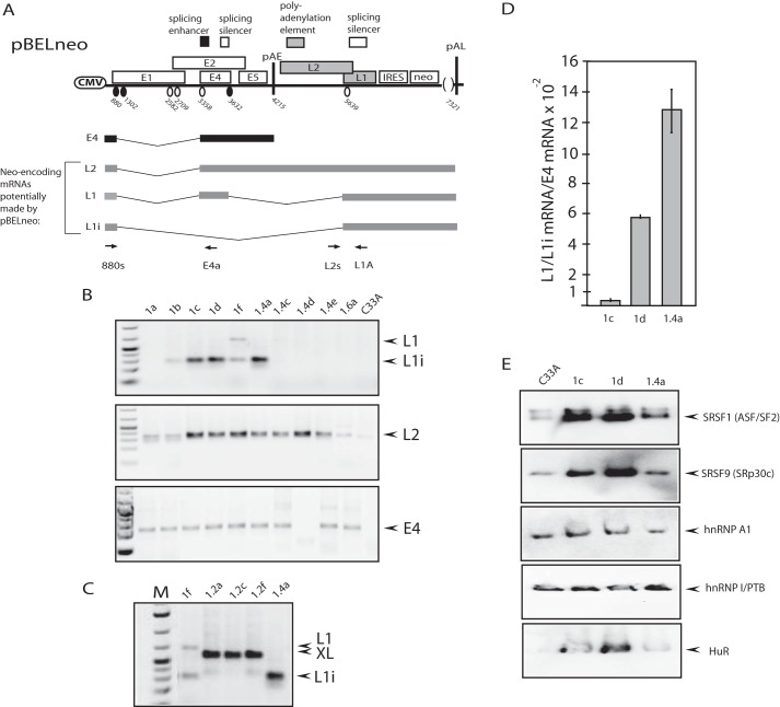
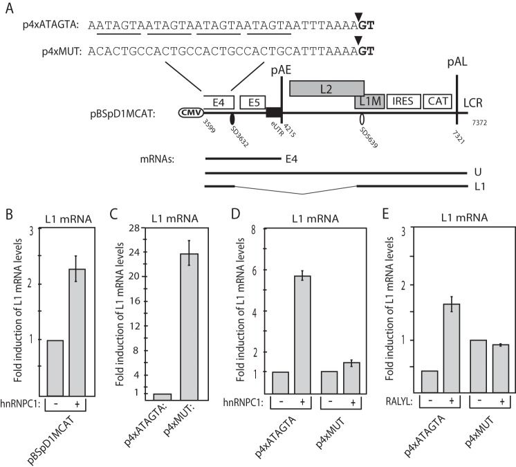
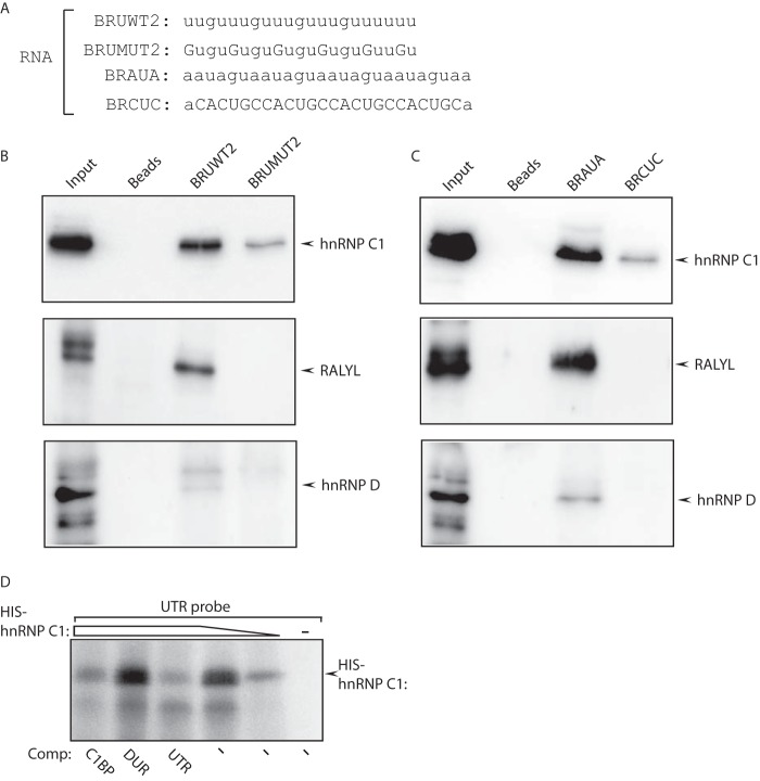
In order to identify cellular factors that regulate human papillomavirus type 16 (HPV16) gene expression, cervical cancer cells permissive for HPV16 late gene expression were identified and characterized. These cells either contained a novel spliced variant of the L1 mRNAs that bypassed the suppressed HPV16 late, 5'-splice site SD3632; produced elevated levels of RNA-binding proteins SRSF1 (ASF/SF2), SRSF9 (SRp30c), and HuR that are known to regulate HPV16 late gene expression; or were shown by a gene expression array analysis to overexpress the RALYL RNA-binding protein of the heterogeneous nuclear ribonucleoprotein C (hnRNP C) family. Overexpression of RALYL or hnRNP C1 induced HPV16 late gene expression from HPV16 subgenomic plasmids and from episomal forms of the full-length HPV16 genome. This induction was dependent on the HPV16 early untranslated region. Binding of hnRNP C1 to the HPV16 early, untranslated region activated HPV16 late 5'-splice site SD3632 and resulted in production of HPV16 L1 mRNAs. Our results suggested that hnRNP C1 controls HPV16 late gene expression.

Keywords: DNA viruses; HPV; RNA processing; RNA splicing; RNA-binding protein; tumor virus.


Figures

FIGURE 1.
FIGURE 1.
A, schematic representation of the HPV16 genome and subgenomic HPV16 expression plasmids. The early and late viral promoters p97 and p670 and the long control region (LCR) are indicated. Numbers indicate nucleotide positions of 5′- (filled circles) and 3′-splice sites (open circles) or the early and late poly(A) sites pAE and pAL, respectively. The major late mRNAs are indicated (15). A schematic representation of the pBELneo and pTEx4Mneo plasmids is shown. The human CMV, the poliovirus IRES, and the neomycin resistance gene (neo) are indicated. Locations of mutations that inactivate the splicing enhancer downstream of SA3358 and the splicing silencers downstream of SA5639 are shown (24, 29). The structures of the E4 mRNAs produced by pBELneo and the E4 and L1i mRNAs produced by pTEx4Mneo are displayed below the plasmids. B, graphs display the number of neomycin-resistant colonies observed at 14 days posttransfection of pBELneo or pTEx4Mneo into C33A or HaCaT cells following selection in 1 mg/ml G418. Three different transfection reagents were used: Turbofect (T), Fugene 6 (F), and Lipofectamine (L).
FIGURE 2.
FIGURE 2.
A, schematic representation of the HPV16 subgenomic expression plasmid pBELneo. Numbers indicate the nucleotide positions of 5′- (filled circles) and 3′-splice sites (open circles) or the early and late poly(A) sites pAE and pAL, respectively, and the borders of deletions. B, RT-PCR with primers 880S and L1A (L1 and L1i mRNAs), L2S and L1A (L2 mRNAs), and 880S and E4a (E4 mRNAs) on cDNA of cytoplasmic RNA extracted from the various cell clones indicated at the top of the gels, as well as from the C33A control cells transfected with pRSVneo. C, RT-PCR with primers 880S and L1A (L1 and L1i mRNAs) on cDNA of cytoplasmic RNA extracted from clones indicated at the top of the gel. The alternatively spliced XL1 mRNA is indicated (for structure of the XL1 mRNA, see Fig. 3A). D, ratio of L1 mRNAs over E4 mRNA levels determined by RT-qPCR with primers 880S and L1A or E4a, respectively. RNA from the three cell clones 1c, 1d, and 1.4a was analyzed. E, Western blot analysis of cell extracts from the pBELneo-transfected and G418-selected cell clones indicated at the top of the gels as well as from the C33A control cells transfected with pRSVneo and selected in G418, with antibodies to SRSF1, SRSF9, hnRNP A1, hnRNP I, or HuR. Error bars, S.D.
FIGURE 3.
FIGURE 3.
A, schematic representation of the HPV16 genome. The early and late viral promoters p97 and p670 are indicated. Numbers indicate the nucleotide positions of 5′- (filled circles) and 3′-splice sites (open circles) or the early and late poly(A) sites pAE and pAL, respectively. LCR, long control region. HPV16 L2 and L1 mRNAs are indicated, as is the novel, alternatively spliced XL1 mRNA. Note that the E1 AUG followed by part of the E4 ORF is fused in frame with a short sequence from the L2 region (but not in the L2 reading frame) and the entire L1 ORF, creating an mRNA with the potential to produce an E4-L1 fusion protein. An mRNA with similar structure identified in HPV31-infected cells is indicated (mRNA E1E4*) (15, 16, 39). The E4 and L1 ORFs are not in frame on the HPV31 mRNA. B, sequences immediately upstream of the HPV16 cryptic 5′-splice site SD3519 are shown and aligned with sequences encompassing the downstream cryptic 3′-splice site SA4980. The splice sites are underlined (the first or last dinucleotides of the intron). The seven nucleotides immediately upstream of SD3519 are 100% homologues to seven nucleotides encompassing SA4980. C, Western blot analysis of extracts from cells transfected with cDNA plasmids expressing either the L1 ORF or the XL1 ORF.
FIGURE 4.
FIGURE 4.
A, Western blot analysis of cell extracts from the pBELneo-transfected and G418-selected cell clone 1.4a and the C33A control cells transfected with pRSVneo and selected in G418 with antibodies to RALYL or actin. B, RT-PCR with primers RALYLS and RALYLA (which detect spliced RALYL mRNAs on cDNA from cytoplasmic RNA extracted from various cell lines indicated at the top of the gel). C, DAPI staining and immunofluorescence staining of C33A cells with anti-RALYL antibody and a composite image.
FIGURE 5.
FIGURE 5.
A, schematic representation of the HPV16 genome and subgenomic HPV16 expression plasmids. The early and late viral promoters p97 and p670 and the long control region (LCR) are indicated. Numbers indicate nucleotide positions of 5′- (filled circles) and 3′-splice sites (open circles) or the early and late poly(A) sites pAE and pAL, respectively. The major late mRNAs are indicated. A schematic representation of the pBELsLuc and pBELMsLuc plasmids is shown. The human CMV, the poliovirus IRES, and the sLuc gene are indicated. Locations of mutations that inactivate the splicing silencers downstream of SA5639 are shown (29). B, Western blot analysis with antibodies to RALYL or actin of cell extracts from C33A cells cotransfected with pBELsLuc and serially diluted pCRALYL. C, sLuc enzyme activity produced by 293T, C33A, HeLa, or HFK cells transfected with pBELsLuc (28) in the absence or presence of serially diluted pCRALYL plasmid. Mean values and S.D. (error bars) are shown. D, sLuc enzyme activity produced by 293T, C33A, HeLa, or HFK cells transfected with pBELMsLuc (28) in the absence or presence of serially diluted pCRALYL plasmid. Mean values and S.D. are shown. E, sLuc enzyme activity produced by C33A cells transfected with pBELsLuc (28) and pCRALYL or the various RALYL deletion mutants indicated to the right. Mean values and S.D. are shown. The indicated RNA-binding domains RNP-1 and -2 are predicted, and RNA-binding assays have not been performed. F, RT-PCR on cytoplasmic RNA extracted from C33A cells transfected with pBELsLuc and empty CMV promoter plasmid (pCL086) (32) or pCRALYL. RT-PCR was performed with primers 880S and E4a to detect spliced E4 mRNAs, with 880S and L1A to detect L1 and L1I mRNAs, and with L2S and L1A to detect L2 mRNAs. Because the latter did not span a splice junction, RT-PCR was performed in the absence (−RT) or presence of RT (+RT). G, RT-qPCR on the HPV16 L1 and L2 mRNAs produced by pBELsLuc cotransfected with either empty vector (pC) or pCRALYL.
FIGURE 6.
FIGURE 6.
A, schematic representation of genomic HPV16 plasmid pHPV16ANSL (25). LoxP sites and HPV16 early (p97) and late (p670) promoters and early (pAE) and late (pAL) poly(A) signals are indicated. The arrows denote positions of PCR primers 16S and 16A. The cassette encoding the Rous sarcoma virus long terminal repeat promoter driving the neomycin resistance gene, followed by the simian virus 40 poly(A) signal, is indicated in black. The effect of the cre recombinase on these plasmids is illustrated, as described previously (25). L1 and L2, late HPV16 genes L1 and L2. B, sLuc activity produced by HeLa cells or HFK cells transfected with pHPV16ANSL in the absence or presence of the RALYL expression plasmid pCRALYL. Error bars, S.D.
FIGURE 7.
FIGURE 7.
A, sLuc enzyme activity produced by C33A cells transfected with subgenomic HPV16 plasmid pBELMsLuc (28) in the absence or presence of serially diluted pCRALYL plasmid. Mean values and S.D. (error bars) are shown. B, sLuc enzyme activity produced by C33A cells transfected with pBELsLuc or pBELMsLuc (28) in the absence or presence of CMV promoter-driven plasmid pCRALY, pCRALYL, or pChnRNPC1. Mean values and S.D. are shown. C, Western blot analysis with antibodies to RALY, hnRNP C1, or actin of cell extracts from C33A cells cotransfected with pBELMsLuc and serially diluted pCRALY or pChnRNPC1. Note that also endogenous hnRNP C1 protein is detected by the hnRNP C1 antibody. D, sLuc enzyme activity produced by the previously described C33A2 reporter cell line containing stably integrated copies of the pBELsLuc plasmid (28) and transiently transfected with empty plasmid pCL086 (32) or with RALY or hnRNP C1 expression plasmid pCRALY or pChnRNPC1. E, RT-PCR on cytoplasmic RNA extracted from the C33A2 reporter cell line transfected with empty plasmid pCL086, pCRALYL, or pChnRNPC1. RT-PCR was performed with primers 880S and E4a to detect spliced E4 mRNAs and with 880S and L1A to detect L1 and L1i mRNAs. F, -fold induction of HPV16 L1, L2, or E4 mRNAs in the C33A2 reporter cell line (28) transfected with pCRALYL or pChnRNPC1 over reporter cell C33A2 transfected with empty vector (pCL086) (32). RT-qPCR was performed with primers 880S and E4a to detect spliced E4 mRNAs, with 880S and L1A to detect L1 and L1i mRNAs, and with L2S and L1A to detect L2 mRNAs. Error bars, S.D.
FIGURE 8.
FIGURE 8.
A, schematic representation of the subgenomic HPV16 plasmid pBEL (29). The 5′- (filled circles) and 3′-splice sites (open circles) and the early and late poly(A) sites pAE and pAL are indicated. pBEL is driven by the human CMV promoter (29). An enlargement of the region upstream of pAE indicates the deletion of the entire early UTR in plasmid pBELDUTR (18). The nucleotide positions of the borders are indicated. Nucleotide positions refer to the HPV16R sequence. B, RT-PCR on total RNA extracted from the C33A cells transfected with pBEL (29) or pBELDUTR in the absence or presence of plasmids expressing RALY, RALYL, or hnRNP C1. RT-PCR was performed with primers 880S and E4a to detect spliced E4 mRNAs and with 880S and L1A to detect L1 and L1i mRNAs. *, a primer-dimer band. C, RT-PCR on cytoplasmic RNA extracted from the 293T cells transfected with pBEL (29) or pBELDUTR (18) in the absence or presence of plasmids expressing RALYL or hnRNP C1. RT-PCR was performed with primers 880S and E4a to detect spliced E4 mRNAs, with 880S and L1A to detect L1 and L1i mRNAs, and with ACTINS and ACTINA to monitor spliced actin mRNA. RT-PCR was performed in the absence (−RT) or presence (+RT) of reverse transcriptase. D, RT-qPCR on the HPV16 E4 and L1 mRNAs produced in C33A cells transfected with pBEL (29) or pBELDUTR (18) in the absence or presence of pCRALYL or pChnRNPC1. E, RT-PCR on nuclear (N) and cytoplasmic (C) RNA extracted from C33A cells transfected with pBEL in the absence or presence of plasmid expressing hnRNP C1. RT-PCR was performed with primers 880S and E4a to detect spliced E4 mRNAs, with 880S and L1A to detect L1 and L1i mRNAs, with actins and actina to monitor spliced actin mRNA, and with ACTINS-1 and ACTINA to detect unspliced actin pre-mRNA. F, RT-PCR on nuclear (N) RNA extracted from C33A cells transfected with pBEL (29) or pBELDUTR (18) in the absence or presence of plasmid expressing hnRNP C1. RT-PCR was performed with primers 880S and E4a to detect spliced E4 mRNAs, with 880S and L1A to detect L1 and L1i mRNAs, with actins and actina to monitor spliced actin mRNA, and with ACTINS-1 and ACTINA to detect unspliced actin pre-mRNA. Error bars, S.D.
FIGURE 9.
FIGURE 9.
A, schematic representation of the subgenomic HPV16 plasmid p4xATAGTA, p4xMUT, and pBSpD1MCAT (28). The 5′- (filled circles) and 3′-splice sites (open circles) and the early and late poly(A) sites pAE and pAL are indicated. pBEL is driven by the human CMV promoter. The four copies of the wild type active or mutant inactive motifs of the splicing silencer inserted upstream SD3632 are shown. SD3632 is indicated by a filled triangle. mRNAs produced by the plasmids are indicated. Nucleotide positions refer to the HPV16R sequence. B, RT-qPCR on the HPV16 L1 mRNAs produced in C33A cells transfected with pBSpD1MCAT (28) in the absence or presence of pChnRNPC1. C, RT-qPCR on the HPV16 L1 mRNAs produced in C33A cells transfected with p4xATAGTA or p4xMUT (28). D, RT-qPCR on the HPV16 L1 mRNAs produced in C33A cells transfected with p4xATAGTA or p4xMUT (28) in the absence or presence of pChnRNPC1 or RALYL (E). Error bars, S.D.
FIGURE 10.
FIGURE 10.
A, RT-PCR on HPV16 E4 mRNAs with primers 880S and E4a on cDNA synthesized from in vivo UV cross-linked, RNA-protein complexes immunoprecipitated with monospecific antibodies to the c-Myc tag on RALYL or mouse IgG. Cells were transfected with pBEL in the absence or presence of pCRALYL. B, RT-qPCR on the HPV16 E4 mRNAs immunoprecipitated with antibodies to the c-Myc tag on RALYL or mouse IgG using primers 880S and E4a. C, RT-PCR on HPV16 E4 mRNAs with primers 880S and E4a on cDNA synthesized from in vivo UV-cross-linked RNA-protein complexes immunoprecipitated with monospecific antibodies to the c-Myc tag on RALYL or mouse IgG. Cells where transfected with pBEL (29) or pBELDUTR (18) in the absence or presence of pCRALYL. D, RT-PCR on HPV16 E4 mRNAs with primers 880S and E4a on cDNA synthesized from in vivo UV-cross-linked RNA-protein complexes immunoprecipitated with IgG or monospecific antibodies to hnRNP C1. Cells were transfected with pBEL (29) in the absence or presence of pChnRNPC1. RT-PCR was also performed on the input cellular extract prior to immunoprecipitation, and 2% of the PCR was loaded on the gel. E, RT-PCR on HPV16 E4 mRNAs with primers 880S and E4a on cDNA synthesized from in vivo UV cross-linked, RNA-protein complexes immunoprecipitated with hnRNP C1 antibody. Cells were transfected with pBEL (29) or pBELDUTR (18) in the presence of pChnRNPC1. RT-PCR was also performed on the input cellular extract prior to immunoprecipitation, and 10% of the PCR-reaction was loaded on the gel. F, RT-PCR on HPV16 E4 mRNAs with primers 880S and E4a on cDNA synthesized from in vivo UV cross-linked, RNA-protein complexes immunoprecipitated with hnRNP C1 antibody. Cells were transfected with empty plasmid (pCL086), pBEL (29), or pBELDUTR (18) in the absence or presence of pChnRNPC1. RT-PCR was also performed on the input cellular extract prior to immunoprecipitation, and 10% was loaded on the gel. G, RT-qPCR on HPV16 E4 mRNAs detected by immunoprecipitation with anti-hnRNP C1 antibody in pBEL- or pBELDUTR-transfected cells in the absence or presence of cotransfected pChnRNPC1 plasmid. Error bars, S.D.
FIGURE 11.
FIGURE 11.
A, sequences of the RNA oligonucleotides used for protein pull-down experiments. BRUWT2 and BRUMUT2 represent the wild type and mutant U-rich region of the HPV16 early untranslated region, respectively, whereas RNA oligonucleotides BRAUA and BRCUC contain five copies of the wild type (AUAGUA) or mutant RNA (CACUGC) motif in the splicing silencer upstream of HPV16 SD3632, respectively. B, Western blot analysis of proteins pulled down by RNA oligonucleotides BRUWT2 and BRUMUT2 using antibodies to hnRNP C1, RALYL, or hnRNP D. C, Western blot analysis of proteins pulled down by RNA oligonucleotides BRAUA and BRCUC using antibodies to hnRNP C1, RALYL, or hnRNP D. D, UV-cross-linking of radiolabeled HPV16 early UTR RNA to recombinant His-tagged hnRNP C1 protein in the absence or presence of competitor RNA. C1BP, RNA consisting four copies of hnRNP C1 binding site AUUUUUA; DUTR, HPV16 early UTR with a deletion that removes the U-rich region of the UTR; UTR, full-length HPV16 early UTR.

Similar articles

Cited by

References

    1. Walboomers J. M., Jacobs M. V., Manos M. M., Bosch F. X., Kummer J. A., Shah K. V., Snijders P. J., Peto J., Meijer C. J., Muñoz N. (1999) Human papillomavirus is a necessary cause of invasive cervical cancer worldwide. J. Pathol. 189, 12–19 - PubMed
    1. Bouvard V., Baan R., Straif K., Grosse Y., Secretan B., El Ghissassi F., Benbrahim-Tallaa L., Guha N., Freeman C., Galichet L., Cogliano V., and WHO International Agency for Research on Cancer Monograph Working Group (2009) A review of human carcinogens: part B: biological agents. Lancet Oncol. 10, 321–322 - PubMed
    1. Bosch F. X., Lorincz A., Muñoz N., Meijer C. J., Shah K. V. (2002) The causal relation between human papillomavirus and cervical cancer. J. Clin. Pathol. 55, 244–265 - PMC - PubMed
    1. zur Hausen H. (2002) Papillomaviruses and cancer: from basic studies to clinical application. Nat. Rev. Cancer 2, 342–350 - PubMed
    1. Howley P. M., Lowy D. R. (2006) in Virology, 5th Ed (Knipe D. M., Howley P. M., eds) pp 2299–2354, Lippincott, Philadelphia

Publication types

MeSH terms