. 2015 Apr 15;35(15):6038-50.
doi: 10.1523/JNEUROSCI.3716-14.2015.

Non-aggregating tau phosphorylation by cyclin-dependent kinase 5 contributes to motor neuron degeneration in spinal muscular atrophy

Affiliations

Non-aggregating tau phosphorylation by cyclin-dependent kinase 5 contributes to motor neuron degeneration in spinal muscular atrophy

Nimrod Miller et al. J Neurosci. .

Abstract

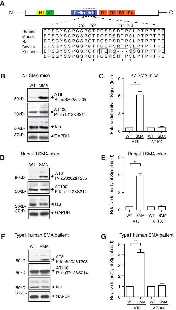
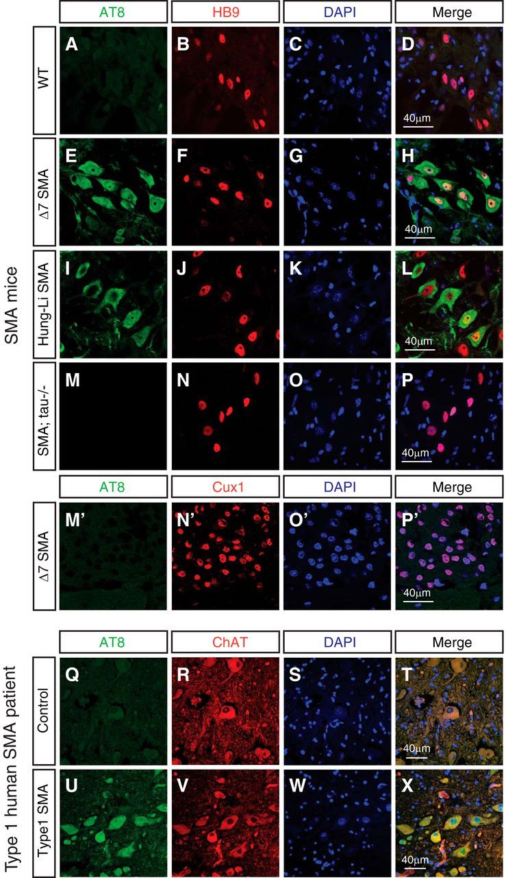
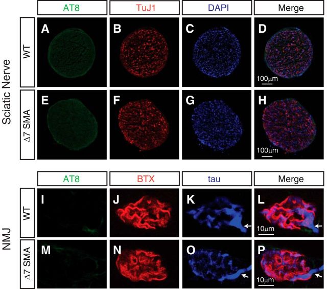
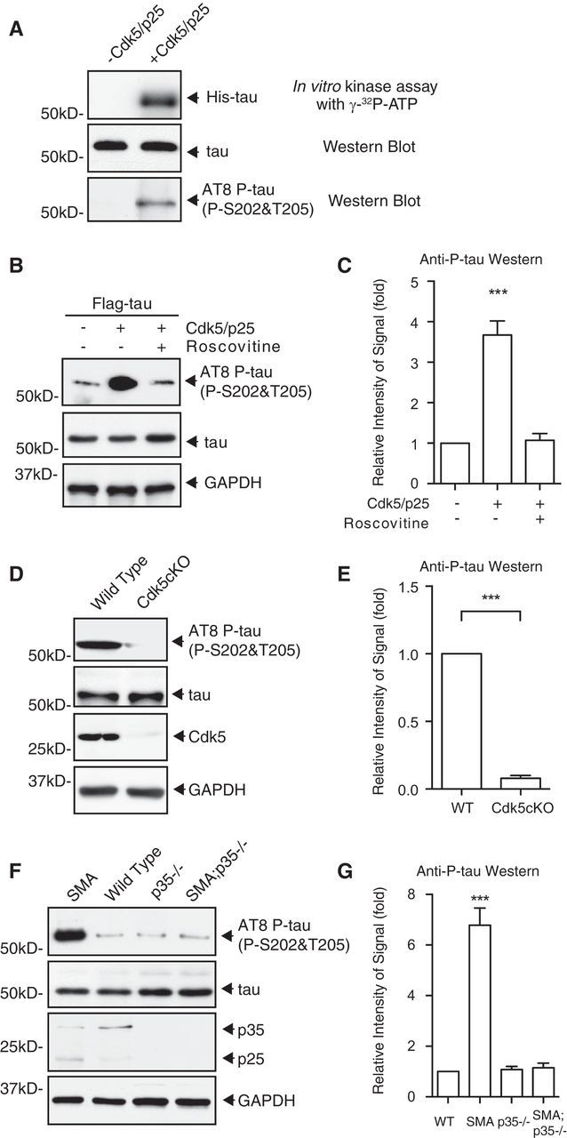
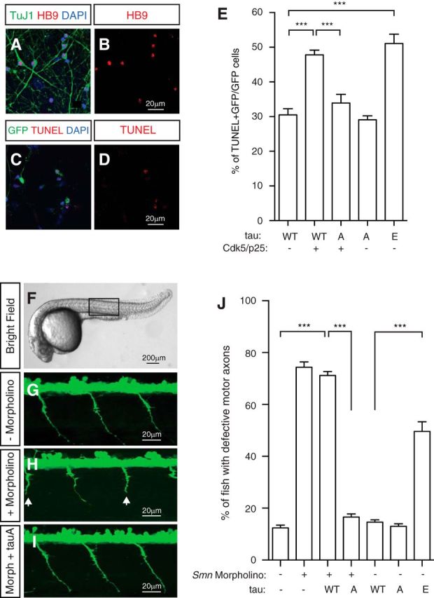
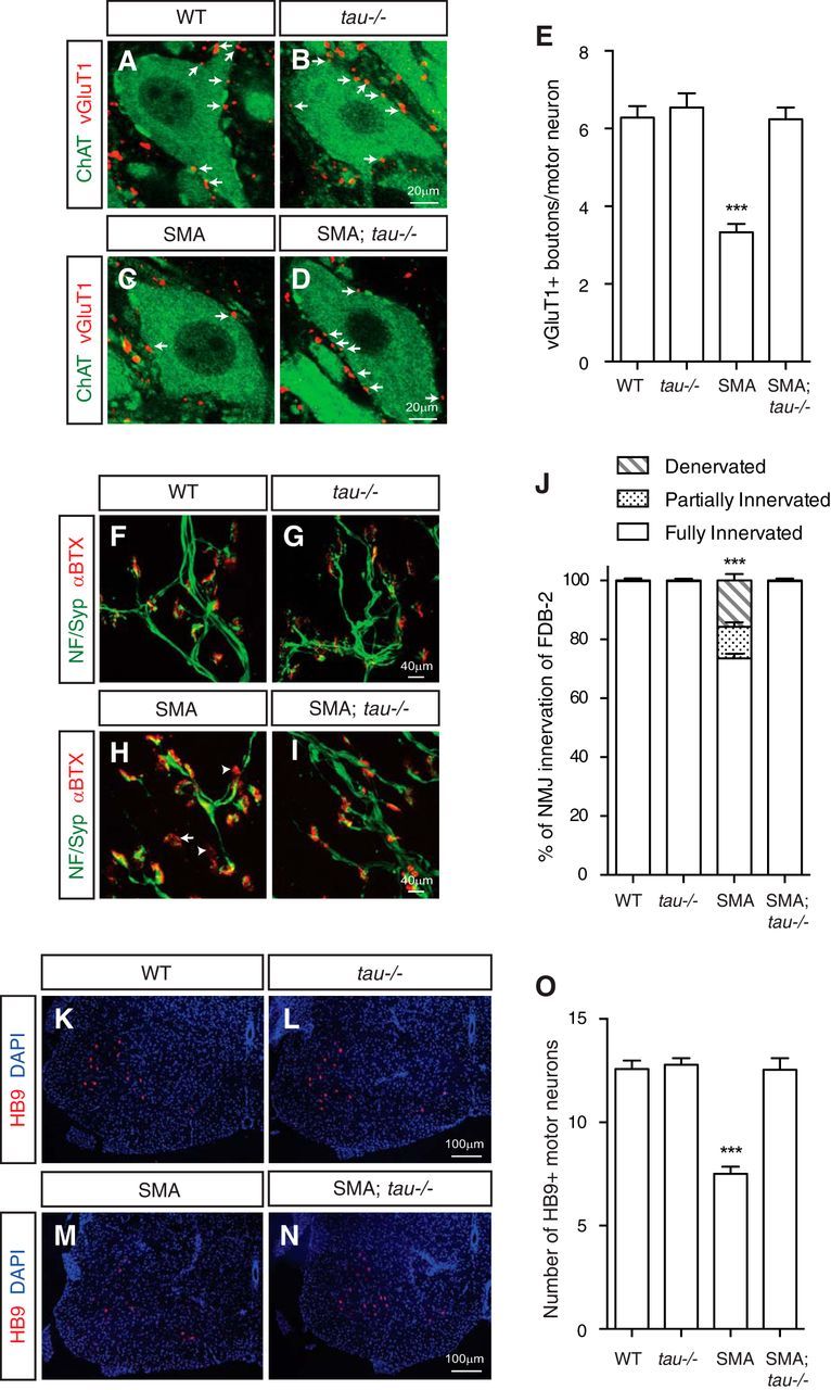
Mechanisms underlying motor neuron degeneration in spinal muscular atrophy (SMA), the leading inherited cause of infant mortality, remain largely unknown. Many studies have established the importance of hyperphosphorylation of the microtubule-associated protein tau in various neurodegenerative disorders, including Alzheimer's and Parkinson's diseases. However, tau phosphorylation in SMA pathogenesis has yet to be investigated. Here we show that tau phosphorylation on serine 202 (S202) and threonine 205 (T205) is increased significantly in SMA motor neurons using two SMA mouse models and human SMA patient spinal cord samples. Interestingly, phosphorylated tau does not form aggregates in motor neurons or neuromuscular junctions (NMJs), even at late stages of SMA disease, distinguishing it from other tauopathies. Hyperphosphorylation of tau on S202 and T205 is mediated by cyclin-dependent kinase 5 (Cdk5) in SMA disease condition, because tau phosphorylation at these sites is significantly reduced in Cdk5 knock-out mice; genetic knock-out of Cdk5 activating subunit p35 in an SMA mouse model also leads to reduced tau phosphorylation on S202 and T205 in the SMA;p35(-/-) compound mutant mice. In addition, expression of the phosphorylation-deficient tauS202A,T205A mutant alleviates motor neuron defects in a zebrafish SMA model in vivo and mouse motor neuron degeneration in culture, whereas expression of phosphorylation-mimetic tauS202E,T205E promotes motor neuron defects. More importantly, genetic knock-out of tau in SMA mice rescues synapse stripping on motor neurons, NMJ denervation, and motor neuron degeneration in vivo. Altogether, our findings suggest a novel mechanism for SMA pathogenesis in which hyperphosphorylation of non-aggregating tau by Cdk5 contributes to motor neuron degeneration.

Keywords: Cdk5; SMA; motor neuron; neurodegeneration; tau.


Figures

Figure 1.
Figure 1.
Microtubule-associated protein tau is hyperphosphorylated in SMA mouse models and human SMA patients. A, Functional domains and conserved phosphorylation sites of tau. Phosphorylated S202 and T205 recognized by the AT-8 antibody and phosphorylated T212 and S214 recognized by the AT-100 antibody are located in the PR domain of tau. Amino acid sequence containing S202 and T205 is conserved across species. N1, N2, N-Terminal regions of tau; R1, R2, R3, R4, microtubule-binding repeat sequences; N, N terminus; C, C terminus. Numbering is based on the longest human tau isoform with 441 residues. B, C, Western blot analysis of tau phosphorylation using spinal cord lysates from Δ7 SMA mice (Smn−/−;SMN2tg/tg;SMNΔ7tg/tg) at P9. Phosphorylation of tau on S202 and T205 as recognized by AT-8, but not on T212 and S214 as recognized by AT-100, is increased in mice affected by SMA. Data are quantified by densitometry from four independent experiments and are mean ± SEM. **p < 0.01, Student's t test. D–E, Western blot analysis of tau phosphorylation using spinal cord lysates from Hung-Li SMA mice (Smn−/−;SMN2Hungtg/tg) at P9. The phosphorylation of tau on S202 and T205, but not on T212 and S214, is increased in the Hung-Li SMA mice. Data are quantified by densitometry from four independent experiments and are mean ± SEM. **p < 0.01, Student's t test. F, G, Western blot analysis of tau phosphorylation in type I human SMA patient spinal cord lysates and age-matched control samples. The phosphorylation of tau on S202 and T205, but not on T212 and S214, is upregulated in humans affected by SMA. Data are quantified from three independent experiments and are mean ± SEM. **p < 0.01, Student's t test.
Figure 2.
Figure 2.
Increased phosphorylation of tau on S202 and T205 occurs specifically in motor neurons affected by SMA without forming aggregates. A–P′, Immunostaining of WT, SMA, and tau−/−;SMA compound mutant mouse spinal cords and cortices with the AT-8 antibody recognizing tau phosphorylated on S202 and T205. Spinal cord sections from P9 WT mice (A–D), Δ7 SMA mice (E–H), Hung-Li SMA mice (I–L), and tau−/−;SMA compound mutant mice (M–P) are stained with AT-8 (green) and the motor neuron-specific HB9 antibody (red). The phosphorylation of tau on S202 and T205 is increased in spinal motor neurons in both SMA mouse models (A–L) but not in the tau−/−;SMA mice (M–P). Immunostaining of cortex sections from P9 Δ7 SMA mice (M′–P′) with AT-8 (green) and the layer II/III cortical neuronal marker Cux1 (red) shows minimal tau hyperphosphorylation detected in layer II/III cortical neurons. Data shown are representative images from eight sets of mice in four independent experiments. Q–X, Immunostaining of spinal cord sections from type I human SMA patients (U–X) and age-matched control samples (Q–T) with the AT-8 (phospho-tauS202&T205) antibody. Tau phosphorylated on S202 and T205 (green) is increased in ChAT-positive (red) motor neurons affected in SMA patients. Data shown are representative images from four sets of samples in four independent experiments.
Figure 3.
Figure 3.
Phosphorylation of tau on S202 and T205 is not increased in the sciatic nerve or NMJs of SMA mice. A–H, Immunostaining of WT and SMA mouse sciatic nerves with the AT-8 antibody recognizing tau phosphorylated on S202 and T205. Sciatic nerve cross-sections from P9 WT mice (A–D) and Δ7 SMA mice (E–H) are stained with the AT-8 (green) antibody and TuJ1 (red) antibody recognizing neuronal β-III tubulin. The phosphorylation of tau on S202 and T205 is at similarly low levels in the sciatic nerves of SMA and control mice. I–P, Immunostaining of NMJs of Δ7 SMA mice at P12 with antibodies recognizing tau and phosphorylated tau (AT-8). At NMJs of the EDL muscle, little phosphorylation on S202 and T205 (I, M, green) or aggregates of phosphorylated tau is detected in either WT or SMA mice at the end stage of SMA pathology (P12). AChRs labeled by BTX (J, N, red) show poor organization in SMA mice (N) compared with WT mice (J). Arrows highlight the junction point at which motor axons enter NMJs (K–P).
Figure 4.
Figure 4.
Cdk5 mediates the increased phosphorylation of tau on S202 and T205 in SMA disease conditions. A, Recombinant tau can be phosphorylated directly by Cdk5 on S202 and T205 in vitro. Autoradiograph showing that His-tagged tau purified from bacteria is phosphorylated by recombinant Cdk5/p25 in a reconstitution kinase assay with [γ-32P]ATP. Western blot analysis with the AT-8 antibody reveals that tau is phosphorylated on S202 and T205 by Cdk5/p25 in the in vitro kinase assay. B, C, Western blot analysis of tau phosphorylation on S202 and T205 by Cdk5 in motor neurons. Expression of Cdk5 together with its activating subunit p25 in cultured mouse motor neurons leads to increased phosphorylation of WT tau on S202 and T205 as recognized by the AT-8 antibody but not the tauS202A,T205A mutant. The Cdk5 inhibitor roscovitine reduces the phosphorylation of tau on S202 and T205. Data shown in C are quantified by densitometry from four independent experiments and are mean ± SEM. ***p < 0.0001, one-way ANOVA with Tukey's HSD post hoc analysis. D, E, Western blot analysis of tau phosphorylation on S202 and T205 in neuronal tissue-specific Cdk5 knock-out mice (Cdk5F/F;Nestin–Cre or Cdk5KO). The phosphorylation of tau on S202 and T205 recognized by the AT-8 antibody is reduced significantly in spinal cord lysates of Cdk5 neuronal tissue-specific knock-out mice. Data shown in E are quantified by densitometry from four independent experiments and are mean ± SEM. ***p < 0.0001, Student's t test. F, G, Western blot analysis of tau phosphorylation on S202 and T205 in p35−/−;Smn−/−;SMN2Hungtg/tg or p35−/−;SMA compound mutant mice. The increased phosphorylation of tau on S202 and T205 is significantly rescued by genetic knock-out of Cdk5 activating subunit p35 in Hung-Li SMA mice. Data shown in G are quantified by densitometry from four independent experiments and are mean ± SEM. ***p < 0.0001, one-way ANOVA with Tukey's HSD post hoc analysis.
Figure 5.
Figure 5.
Phosphorylation of tau on S202 and T205 is critical for motor neuron functions in vitro and in vivo. A–E, Increased tau phosphorylation by Cdk5 promotes degeneration of mouse spinal cord neurons. Cultured mouse spinal cord neurons (A, B) can be recognized by antibodies to the motor neuron marker HB9 (A, B, red) and the pan-neuronal marker TuJ1 (A, green). TUNEL staining (C, D, red) shows degeneration induced by expressing tau constructs together with Cdk5/p25 and GFP. Expression of WT tau, but not the phosphorylation-deficient tauS202A,T205A mutant, together with Cdk5/p25 or expressing the phosphorylation mimetic tauS202E,T205E mutant leads to significantly increased neurodegeneration. Quantified results (E) are from five independent experiments and 6116 neurons and are mean ± SEM. ***p < 0.0001, Student's t test. F–J, Phosphorylation-deficient tauS202A,T205A mutant rescues motor neuron defects in a zebrafish SMA model in vivo. Antisense MO knockdown of Smn in Tg(olig2:egfp) transgenic zebrafish leads to truncations (white arrows in H) or abnormal branching of motor axons. G–I correspond to regions similar to the boxed area in F. Motor neuron defects can be rescued by the expression of RNA encoding the phosphorylation-deficient mutant tauS202A,T205A (J). Expression of tauS202E,T205E leads to significantly increased motor axon defects in zebrafish (J). Data are from five independent experiments and 572 fish and are mean ± SEM. ***p < 0.0001, Student's t test.
Figure 6.
Figure 6.
Genetic knock-out of tau rescues synaptic and NMJ defects and motor neuron degeneration in SMA mice. A–E, Immunostaining of excitatory glutamatergic synapse formation on spinal cord motor neurons. Colocalization of the glutamatergic synapse marker VGluT1 (A–D, red) with the motor neuron marker ChAT (A–D, green) shows that excitatory synaptic boutons (arrows) on motor neurons are reduced significantly in SMA mice compared with WT littermates. The synaptic defect is rescued to the WT level by genetic knock-out of tau in SMA mice. Quantification in E is from three to five mice from each genotype and are mean ± SEM. ***p < 0.0001, one-way ANOVA with Tukey's HSD post hoc analysis. F–J, Staining of motor neuron axon innervation of NMJs. Colocalization of presynaptic nerve terminals marked by immunostaining with anti-neurofilament (NF) and anti-synaptophysin (Syp) antibodies (F–I, green) with postsynaptic AChRs labeled by BTX (F–I, red) shows increased denervation (arrowheads) and partial innervation (arrow) of NMJs on the FDB-2 muscle of SMA. The NMJ innervation defect is rescued to the WT level by genetic knock-out of tau in SMA mice. Quantification in J is from >100 NMJs on the FDB-2 muscle from each mouse. Four mice for each genotype were used. Denervated, partially innervated, and fully innervated NMJs were analyzed separately. Results are mean ± SEM. ***p < 0.0001, one-way ANOVA with Tukey's HSD post hoc analysis. K–O, Immunostaining of mouse spinal cord sections with the motor neuron marker HB9 to quantify motor neuron degeneration. Lumbar level spinal cord sections of P9 SMA mice show a near 40% reduction of HB9-positive (K–N, red) motor neurons compared with WT littermates. Genetic knock-out of tau in SMA (tau−/−;SMA compound mutant) mice rescues motor neuron degeneration to the WT level (O). Quantification in O is from 10 to 15 sections of four mice from each genotype in four independent experiments and are mean ± SEM. ***p < 0.0001, one-way ANOVA with Tukey's HSD post hoc analysis.

Similar articles

Cited by

References

    1. Akten B, Kye MJ, Hao le T, Wertz MH, Singh S, Nie D, Huang J, Merianda TT, Twiss JL, Beattie CE, Steen JA, Sahin M. Interaction of survival of motor neuron (SMN) and HuD proteins with mRNA cpg15 rescues motor neuron axonal deficits. Proc Natl Acad Sci U S A. 2011;108:10337–10342. doi: 10.1073/pnas.1104928108. - DOI - PMC - PubMed
    1. Bailey RM, Covy JP, Melrose HL, Rousseau L, Watkinson R, Knight J, Miles S, Farrer MJ, Dickson DW, Giasson BI, Lewis J. LRRK2 phosphorylates novel tau epitopes and promotes tauopathy. Acta Neuropathologica. 2013;126:809–827. doi: 10.1007/s00401-013-1188-4. - DOI - PMC - PubMed
    1. Baumann K, Mandelkow EM, Biernat J, Piwnica-Worms H, Mandelkow E. Abnormal Alzheimer-like phosphorylation of tau-protein by cyclin-dependent kinases cdk2 and cdk5. FEBS Lett. 1993;336:417–424. doi: 10.1016/0014-5793(93)80849-P. - DOI - PubMed
    1. Bevan AK, Hutchinson KR, Foust KD, Braun L, McGovern VL, Schmelzer L, Ward JG, Petruska JC, Lucchesi PA, Burghes AH, Kaspar BK. Early heart failure in the SMNDelta7 model of spinal muscular atrophy and correction by postnatal scAAV9-SMN delivery. Hum Mol Genet. 2010;19:3895–3905. doi: 10.1093/hmg/ddq300. - DOI - PMC - PubMed
    1. Biernat J, Gustke N, Drewes G, Mandelkow EM, Mandelkow E. Phosphorylation of Ser262 strongly reduces binding of tau to microtubules: distinction between PHF-like immunoreactivity and microtubule binding. Neuron. 1993;11:153–163. doi: 10.1016/0896-6273(93)90279-Z. - DOI - PubMed

Publication types

MeSH terms