. 2015 Apr 15;35(15):6093-106.
doi: 10.1523/JNEUROSCI.3412-14.2015.

A new CRB1 rat mutation links Müller glial cells to retinal telangiectasia

Affiliations

A new CRB1 rat mutation links Müller glial cells to retinal telangiectasia

Min Zhao et al. J Neurosci. .

Abstract

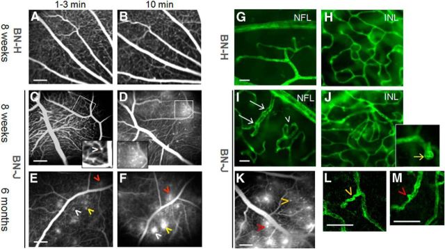
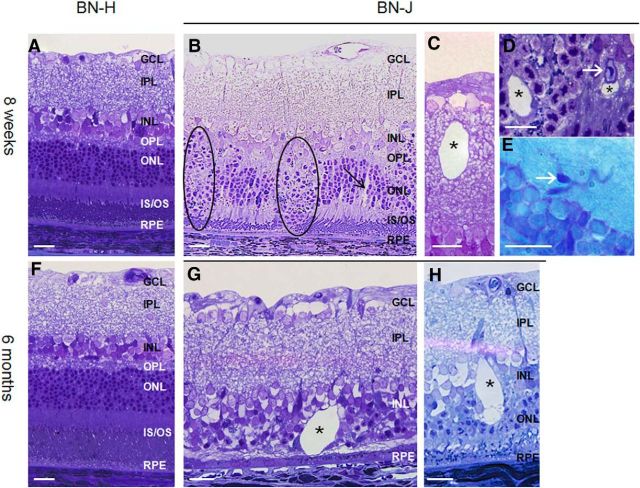
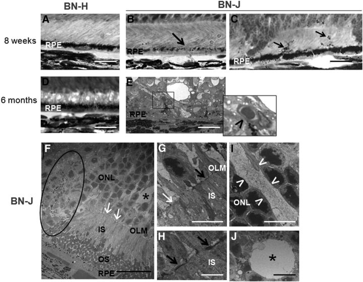
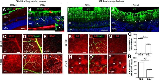
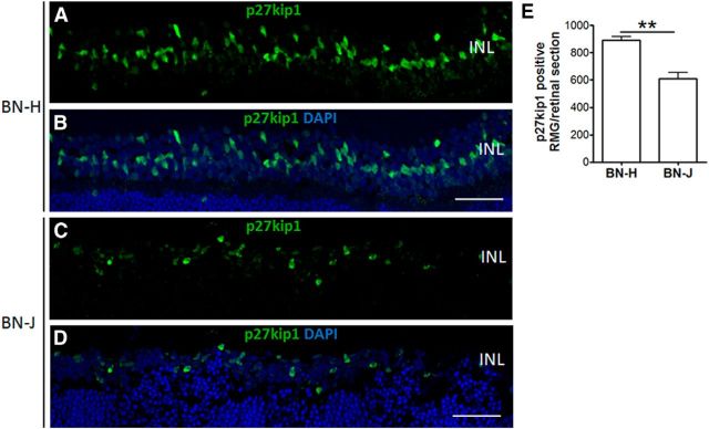
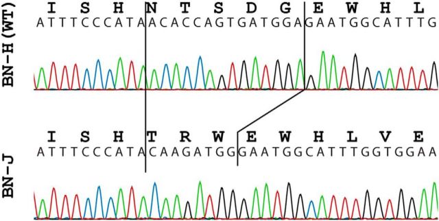
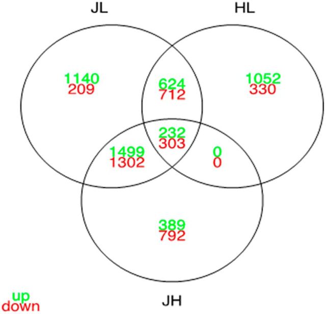
We have identified and characterized a spontaneous Brown Norway from Janvier rat strain (BN-J) presenting a progressive retinal degeneration associated with early retinal telangiectasia, neuronal alterations, and loss of retinal Müller glial cells resembling human macular telangiectasia type 2 (MacTel 2), which is a retinal disease of unknown cause. Genetic analyses showed that the BN-J phenotype results from an autosomal recessive indel novel mutation in the Crb1 gene, causing dislocalization of the protein from the retinal Müller glia (RMG)/photoreceptor cell junction. The transcriptomic analyses of primary RMG cultures allowed identification of the dysregulated pathways in BN-J rats compared with wild-type BN rats. Among those pathways, TGF-β and Kit Receptor Signaling, MAPK Cascade, Growth Factors and Inflammatory Pathways, G-Protein Signaling Pathways, Regulation of Actin Cytoskeleton, and Cardiovascular Signaling were found. Potential molecular targets linking RMG/photoreceptor interaction with the development of retinal telangiectasia are identified. This model can help us to better understand the physiopathologic mechanisms of MacTel 2 and other retinal diseases associated with telangiectasia.

Keywords: adherens junction; disease model; genetics; microcirculation; retinal blood vessels; retinal degeneration.


Figures

Figure 1.
Figure 1.
Vascular abnormalities in BN-J rats. Shown is in vivo fluorescein angiography of retinal vessels of BN-H and BN-J rats (AF). Normal retinal vessels of BN-H rat at early (1–3 min, A) and late phase (10 min, B) of the angiographic sequence. Eight-week-old BN-J rat exhibits subtle capillary dilation hardly detected in the early phase (1–3 min) of angiography (C, arrowhead in the inset) that becomes more visible with leakage at later time point of 10 min (D and inset). At 6 months of age, similar but leakier capillary ectasia are observed (E, F, arrowheads, the same color indicates the same spot). Hyperfluorent leaking dots are observed at 1–3 min and their size increases at 10 min. Scale bar, 200 μm. Confocal imaging of lectin-stained retinal vessels on flat-mounted retinas from BN-H and BN-J rats is shown (GJ, L, M). Normal retinal vascular network (green) at the nerve fiber layer (NFL) and in the deep plexus at the INL from BN-H rat (G, H). In the BN-J rat retina, irregular vascular diameter (white arrows) and increased tortuosity (arrowhead) are observed at the NFL level (I). In the INL, disorganized capillary plexus is observed (J), together with multiple capillary telangiectasia (inset, yellow arrow). Images of a lectin-labeled flat-mounted retina of BN-J rat are linked to their corresponding angiographic pattern (K). Higher magnifications of the lectin-labeled vessels show that leaky telangiectasia (K) correspond to capillary tortuousness (L) and focal capillary ectasia (M). Arrowheads of the same color indicate the same spot. Scale bars: GJ, 20 μm; K, 200 μm; L, M, 50 μm.
Figure 2.
Figure 2.
Retinal morphology of BN-H and BN-J rats at 8 weeks and 6 months. Compared with the normally developed retina of BN-H rat at 8 weeks (A), the retina of the BN-J rat shows focal disorganization of the outer retinal layers (B, dark circles), where segments are not formed and nuclei of photoreceptors dive toward the retinal pigment epithelium (RPE). In areas where segments are present, swollen RMG cells can be observed (B, black arrow). Cysts (asterisks) can be found in both the inner (C) and the outer (D) retina. Telangiectasia are also identified on histological sections (D, E, white arrow). At 6 months, the BN-H rat retina is unchanged (F), whereas the retina of the BN-J rat shows variable degrees of degeneration. Photoreceptors have totally disappeared in some areas (G) and cysts are more abundant with irregular shapes (G, H, asterisks). GCL, Ganglion cell layer; IPL, inner plexiform layer; OPL, outer plexiform layer; IS/OS; inner and outer segments of photoreceptors. Scale bar, 20 μm.
Figure 3.
Figure 3.
Outer retinal alterations in BN-J rats. AE, Histological sections of the outer retina. FJ, TEM images. Contrasting with the heavy pigments located in the apical side of retinal pigment epithelium (RPE) in BN-H retina (A), melanosomes are poorly formed in BN-J rat at 8 weeks, even in areas where the segments have formed (B, black arrow) and pigments migrate in the photoreceptor segment layer (C, black arrows). At 6 months, the outer retina of BN-H rat does not change (D), whereas abnormal vessels are observed between RPE cells and the degenerated retina of BN-J rat (E, inset and black arrowhead), potentially corresponding to neovascularization. TEM analysis allows detection of more subtle changes in BN-J retina such as focal decrease in junction structures (F, G, white arrows) at the outer limiting membrane (OLM), alternating with normal OLM structures (G, H, black arrows). Abrupt disorganization of retinal layers is observed (F, dark circle). Swollen RMG cells (I, in between the white arrowheads) are identified in the ONL and cysts (F, J, asterisks) are surrounded by a membrane-like structure, suggesting intracellular swollen. IS, Inner segments of photoreceptors; OS, outer segments of photoreceptors. Scale bars: AE, 20 μm; F, 25 μm; G, I, J, 10 μm; H, 2 μm.
Figure 4.
Figure 4.
Postnatal retinal development morphology of BN-H and BN-J rats. From P1 to P8, the neuronal layers are segmented into inner neuroblastic (INbL) and outer neuroblastic layers (ONbL) both in BN-H (A, B) and in BN-J (D, E) rats. However, from P8 to P15, whereas inner and outer segments (IS and OS) elongate normally in the BN-H retina (C), focal areas without segment elongation and persistent neuroblastic nuclei (circled areas) are observed in the BN-J retina (F). Dilated capillaries can be observed in the INL of BN-J retina (F, arrow). GCL, Ganglion cell layer. Scale bar, 20 μm.
Figure 5.
Figure 5.
Immunohistochemistry of retinal neurons of BN-J rats. Different neuronal types are immunostained with specific markers in the BN-H retina: Cone arrestin stains the entire cone photoreceptors, including outer segments and synaptic bodies (A), rhodopsin stains the outer segments (OS) of rod photoreceptors (F), PKC-α labels the bipolar cells (I), and synaptophysin labels synaptic connections between retinal neurons (M). B, F, J and N are merged images with DAPI (blue). In the BN-J retina, cone segments are shorter or even absent (C, arrowhead) and cones are missing in cystic formations (C, asterisk) and nuclei of some cones without segments are displaced (C, arrow). Cell count shows significant reduction of cone cells in BN-J rats (E). Rods are absent in disorganized area (G, asterisk) and their segments are shorter in other regions (G, arrows), suggesting segment elongation disruption. Quantification of rhodopsin-positive surface shows significant decrease in rod outer segment areas in BN-J rats (H). In disorganized areas, nuclei of bipolar cell are internally displaced (K, circle) and neuronal synapses are disrupted in the outer plexiform layer (OPL, O, arrows). D, G, L and P are merged images with DAPI. GCL, Ganglion cell layer; IPL, inner plexiform layer. Scale bars: AD, F, G, MP, 50 μm; IL, 20 μm. For E and H, n = 5 rats per strain; **p < 0.01.
Figure 6.
Figure 6.
RMG morphologic alterations in sectioned and flat-mounted BN-J retinas. GFAP stains RMG end feet and astrocytes on BN-H retinal cross-section (A). On flat-mounted retinas, GFAP staining is surrounding vessels (labeled with lectin) in the nerve fiber layer (NFL, C, D) and, in RMG apices, in the outer limiting membrane (OLM, E). In the BN-J retina, activated RMG cells extend up to the subretinal space in disorganized areas (B, filled arrows). Activated RMG cells surround vessels in the INL (B, top inset, open arrow) and form the border of cysts (B, bottom inset, asterisk). On flat-mounted BN-J retinas, GFAP is enhanced in the RMG end feet in the NFL (F, G) and extends up to the OLM, where swollen apices (H, filled arrows) and disorganization of RMG (H) are observed. In the BN-H retina, GS stains the RMG from their end feet to processes around the vessels (labeled with lectin) and to their apices (retinal section, I, and flat-mounted retina, KM). In the BN-J retina, GS immunoreactivity is enhanced in hypertrophic RMG and reduced in the surrounding areas, as observed in both retinal section (J, between the arrowheads) and flat-mounted retina (NP, asterisks), suggesting focal loss of RMG cells. GS-positive and CRALBP (another RMG marker)-positive RMG cells are significantly decreased in BN-J rat retinas (Q, R). GCL, Ganglion cell layer; IPL, inner plexiform layer. Scale bars: A, B, I, J, 50 μm; C, D, F, G, K, L, N, O, 100 μm; E, H, M, P, 50 μm. For Q and R, n = 5 rats per strain; **p < 0.01.
Figure 7.
Figure 7.
Loss of Müller glial cells in BN-J rat retinas. RMG cells were immunostained with p27kip1, an RMG nuclear marker. P27kip1-positive nuclei in the INL are reduced in the BN-J rat retina (C) compared with BN-H rat retina (A). B and D are merged images with DAPI. Scale bar, 50 μm. Cell count of p27kip1-positive RMG shows significant decrease in BN-J rat retinas (E). n = 7 rats for BN-H and 6 rats for BN-J; **p < 0.01.
Figure 8.
Figure 8.
Early retinal function alterations in BN-J rats. ERG was performed on 3-week-old BN-H and BN-J rats. Although global ERG responses show a trend but not significant reduction in a-wave amplitude (A), the b-wave amplitude is significantly decreased (B), suggesting postreceptoral disturbance of the visual signal in the inner retina. Scotopic ERG shows a significant reduction in a-wave (C) and b-wave (D) amplitudes from 0.1 to 3 cd · s/m2, suggesting intense rod visual pathway dysfunction. n = 5 rats for BN-H and n = 4 for BN-J. *p < 0.05.
Figure 9.
Figure 9.
Electropherograms of part of crb1 exon 6 in BN-H and BN-J rats. The insertion–deletion in the BN-J sequence is indicated by solid black lines.
Figure 10.
Figure 10.
CRB1 immunolocalization in BN-H and BN-J retinas. In the BN-H retina, CRB1 localizes in the subapical region above the level of outer limiting membrane (OLM) labeled with GS (AC, open arrows in the left inset). Higher magnification shows colocalization of CRB1 with GS in the microvilli of RMG cells (arrowheads in the right inset). CRB1 stains also the inner segments (IS) of photoreceptors (A, D). Double staining with PNA (E) shows colocalization of CRB1 with cone IS (F, arrowheads in the inset). In the BN-J retina, CRB1 loses its localization in the subapical region (GI, inset), OLM is even disrupted in disorganized areas (H, filled arrow). In organized areas, CRB1 remains in the photoreceptor IS (J). Double staining with PNA (K) shows colocalization with cone IS (L, arrowhead in the inset). Scale bar, 20 μm.
Figure 11.
Figure 11.
Crossing of the differential expression analyses of BN-J, BN-H, and Lewis P17 primary RMG cells. Venn diagram shows the number of overexpressed (in green) and underexpressed genes (in red) that are shared and unique for each comparison.

References

    1. Abukawa H, Tomi M, Kiyokawa J, Hori S, Kondo T, Terasaki T, Hosoya K. Modulation of retinal capillary endothelial cells by Muller glial cell-derived factors. Mol Vis. 2009;15:451–457. - PMC - PubMed
    1. Alves CH, Pellissier LP, Wijnholds J. The CRB1 and adherens junction complex protiens in retinal development and maintenance. Prog Retin Eye Res. 2014;40:35–52. doi: 10.1016/j.preteyeres.2014.01.001. - DOI - PubMed
    1. Behzadian MA, Wang XL, Windsor LJ, Ghaly N, Caldwell RB. TGF-beta increases retinal endothelial cell permeability by increasing MMP-9: possible role of glial cells in endothelial barrier function. Invest Ophthalmol Vis Sci. 2001;42:853–859. - PubMed
    1. Bringmann A, Pannicke T, Grosche J, Francke M, Wiedemann P, Skatchkov SN, Osborne NN, Reichenbach A. Muller cells in the healthy and diseased retina. Prog Retin Eye Res. 2006;25:397–424. doi: 10.1016/j.preteyeres.2006.05.003. - DOI - PubMed
    1. Charbel Issa P, Finger RP, Kruse K, Baumüller S, Scholl HP, Holz FG. Monthly ranibizumab for nonproliferative macular telangiectasia type 2: a 12-month prospective study. Am J Ophthalmol. 2011;151:876–886.e1. - PubMed

Publication types

MeSH terms

Substances

LinkOut - more resources