. 2015 Apr 15;35(15):6221-30.
doi: 10.1523/JNEUROSCI.2552-14.2015.

Tau-dependent Kv4.2 depletion and dendritic hyperexcitability in a mouse model of Alzheimer's disease

Affiliations

Tau-dependent Kv4.2 depletion and dendritic hyperexcitability in a mouse model of Alzheimer's disease

Alicia M Hall et al. J Neurosci. .

Abstract

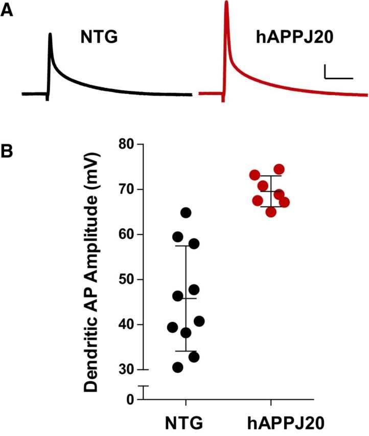
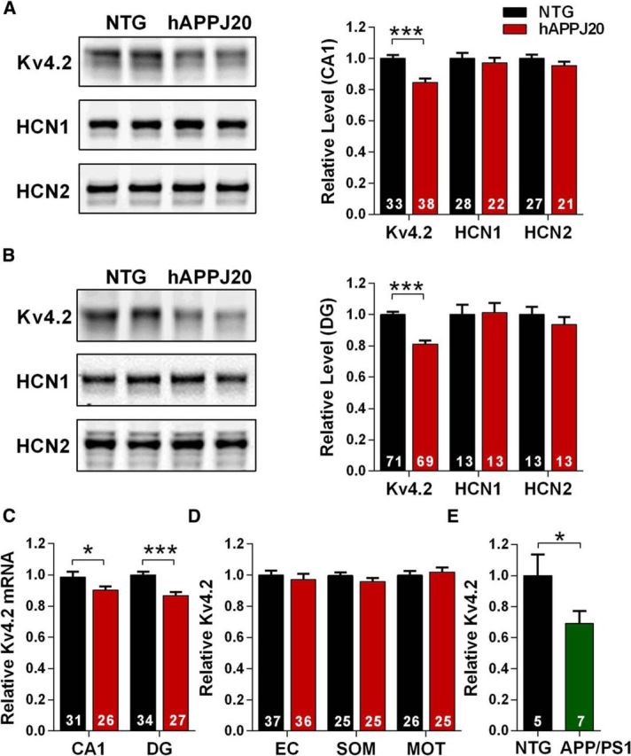
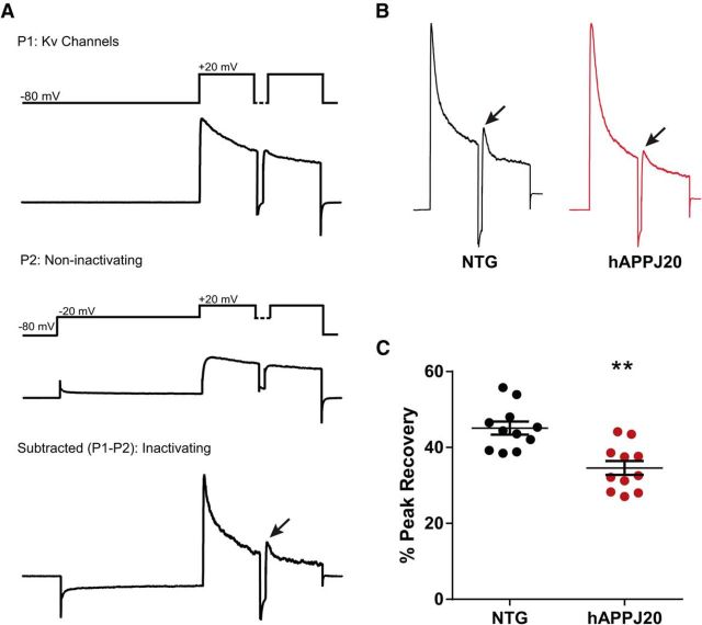
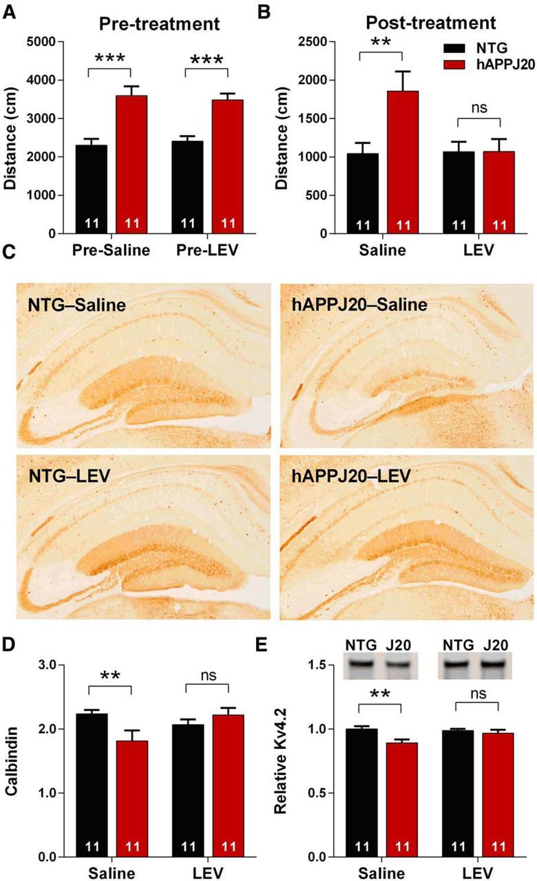
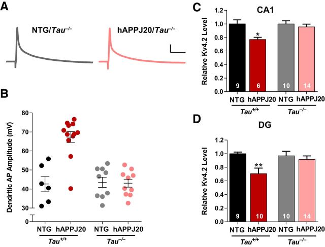
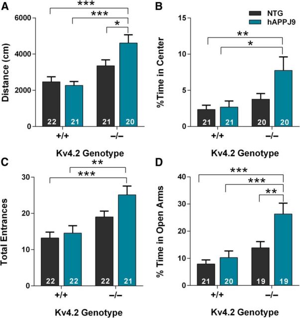
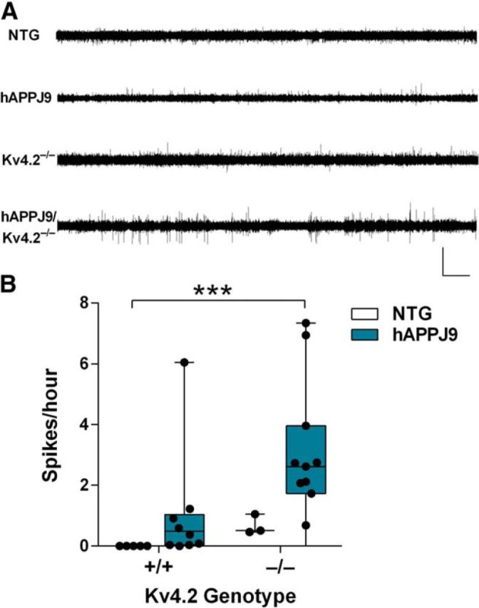
Neuronal hyperexcitability occurs early in the pathogenesis of Alzheimer's disease (AD) and contributes to network dysfunction in AD patients. In other disorders with neuronal hyperexcitability, dysfunction in the dendrites often contributes, but dendritic excitability has not been directly examined in AD models. We used dendritic patch-clamp recordings to measure dendritic excitability in the CA1 region of the hippocampus. We found that dendrites, more so than somata, of hippocampal neurons were hyperexcitable in mice overexpressing Aβ. This dendritic hyperexcitability was associated with depletion of Kv4.2, a dendritic potassium channel important for regulating dendritic excitability and synaptic plasticity. The antiepileptic drug, levetiracetam, blocked Kv4.2 depletion. Tau was required, as crossing with tau knock-out mice also prevented both Kv4.2 depletion and dendritic hyperexcitability. Dendritic hyperexcitability induced by Kv4.2 deficiency exacerbated behavioral deficits and increased epileptiform activity in hAPP mice. We conclude that increased dendritic excitability, associated with changes in dendritic ion channels including Kv4.2, may contribute to neuronal dysfunction in early stages AD.

Keywords: Alzheimer; Kv4.2; amyloid-beta; dendrites; excitability; tau.


Figures

Figure 1.
Figure 1.
Increased dendritic excitability in hAPPJ20 mice. A, Representative back-propagating dendritic APs recorded in apical dendrites of CA1 pyramidal neurons in NTG and hAPPJ20 mice. Calibration: 10 mV, 12.5 ms. B, Dendritic AP amplitude was increased by 52 ± 3% in hAPPJ20 mice, indicating increased dendritic excitability (age 3 months; p < 0.0001, t test).
Figure 2.
Figure 2.
Kv4.2 is depleted in hippocampal regions in hAPPJ20 mice. A, Immunoblots for dendritic ion channels in area CA1. Kv4.2 was decreased in hAPPJ20 mice (ANOVA: hAPP × channel interaction, p < 0.05). ***p < 0.0001 (post hoc test; age 4.5–7 months). B, Immunoblots for dendritic ion channels in dentate gyrus. Kv4.2 was decreased in hAPPJ20 mice (ANOVA: hAPP × channel interaction, p < 0.05). ***p < 0.0001 (post hoc test; age 4.5–7 months). C, Kv4.2 mRNA levels were decreased in area CA1 and DG of hAPPJ20 mice. *p < 0.05 (one-tailed t test). ***p < 0.0001 (one-tailed t test; age 4.5–7 months). D, Kv4.2 levels were unchanged in cortex in hAPPJ20 mice. EC, Entorhinal cortex; SOM, somatosensory cortex; MOT, motor cortex (age 4.5–7 months). E, Kv4.2 was depleted in the hippocampus of hAPPswe/PS1dE9 mice by immunoblot. *p < 0.05 (t test; age 6 months).
Figure 3.
Figure 3.
Decreased IA in hAPPJ20 mice. A, Protocol used to isolate Kv4-mediated component of IA, based on Chen et al. (2006) and Rüschenschmidt et al. (2006). The first pass (P1) activates voltage-gated potassium channels (recordings are performed under conditions that block sodium, calcium, and calcium-activated potassium channels, plus glutamate and GABA receptors). The second pass (P2) includes a depolarizing step to inactivate fast-inactivating (A-type) channels. The subtraction (P1–P2) represents IA, the fast-inactivating potassium current. Each pass includes two pulses to distinguish Kv1- and Kv4-mediated currents, which both contribute to IA. Kv4 currents recover from inactivation an order of magnitude more quickly than Kv1 currents, so the peak current after the second pulse represents Kv4-mediated current, when Kv1 has not yet recovered from inactivation. Thus, the peak recovery between the first and second pulses (arrow) in the P1–P2 subtraction trace represents Kv4-mediated IA (Chen et al., 2006). B, Subtraction (P1–P2) traces for NTG and hAPPJ20 mice, showing decreased Kv4-mediated IA (arrows). C, Quantification of data in B showing decreased Kv4-mediated IA in hAPPJ20 mice (N = 11 cells per genotype; age 4–5 months). **p < 0.01 (t test).
Figure 4.
Figure 4.
Levetiracetam blocks Kv4.2 depletion in hAPPJ20 mice. A, hAPPJ20 mice showed increased ambulatory distance in open field when pretested before LEV treatment (age 4–5 months, p < 0.0001, t test). B, After 18 d of LEV treatment, hAPPJ20 mice showed normal ambulatory distance (ANOVA: hAPP × treatment interaction, p < 0.05, **p < 0.01 on post hoc test). C, Representative images of calbindin immunohistochemistry after 28 d of LEV treatment. D, Quantification of calbindin immunoreactivity after 28 d of LEV treatment, expressed as the ratio of DG to CA1 immunoreactivity as in Palop et al. (2011). hAPPJ20 mice treated with LEV showed normal levels of calbindin (ANOVA: hAPP × treatment interaction, p < 0.05, **p < 0.01 on post hoc test; age 5–6 months). E, Quantification of Kv4.2 after LEV treatment for 28 d. LEV blocked the Kv4.2 depletion in the hAPPJ20 mice. Representative blots and quantification of Kv4.2 in the DG of hAPPJ20 mice (ANOVA: hAPP effect, p < 0.05, **p < 0.01 on post hoc test; age 5–6 months).
Figure 5.
Figure 5.
Tau is required for Aβ-induced dendritic hyperexcitability and loss of Kv4.2 in hAPPJ20 mice. A, Representative back-propagating dendritic APs recorded in apical dendrites of CA1 pyramidal neurons from Tau−/− mice. Calibration: 10 mV, 12.5 ms. B, Dendritic AP amplitude was increased in hAPPJ20/Tau+/+ mice, but not in hAPPJ20/Tau−/− mice (ANOVA: hAPP × tau interaction, p < 0.001; on post hoc tests, hAPPJ20/Tau+/+ differed from all other groups, p < 0.0001; age 6.5–8.5 months). C, Tau reduction blocked the loss of Kv4.2 in area CA1 (ANOVA: p < 0.02; *p < 0.05 on post hoc tests, hAPP/Tau+/+ differed from other groups; age 4.5–7 months). D, Tau reduction blocked the loss of Kv4.2 in the dentate gyrus (ANOVA: p < 0.01; **p < 0.01 on post hoc tests, hAPP/Tau+/+ differed from other groups, p < 0. 005; age 4.5–7 months).
Figure 6.
Figure 6.
Kv4.2 deficiency exacerbates behavioral abnormalities in hAPPJ9 mice. A, In the open field, hAPPJ9/Kv4.2−/− mice showed increased ambulatory distance, an abnormality seen in hAPPJ20 mice (ANOVA: hAPP × Kv4.2 interaction, p < 0.05; *p < 0.05 and ***p < 0.0001 on post hoc test; age 5–7 months). B, In the open field, hAPPJ9/Kv4.2−/− mice showed increased time in the center, an abnormality seen in hAPPJ20 mice (ANOVA: Kv4.2 effect, p < 0.01; *p < 0.05 and **p < 0.01 on post hoc test; age 5–7 months). C, In the elevated plus maze, hAPPJ9/Kv4.2−/− mice showed increased total entrances, an abnormality seen in hAPPJ20 mice (ANOVA: Kv4.2 effect, p < 0.0001; **p < 0.01 and ***p < 0.001 on post hoc test; age 5–7 months). D, In elevated plus maze, hAPPJ9/Kv4.2−/− mice showed increased time in the open arms, an abnormality seen in hAPPJ20 mice (ANOVA: hAPP effect p < 0.0001; Kv4.2 effect p < 0.001; **p < 0.01 and ***p < 0.001 on post hoc test; age 5–7 months).
Figure 7.
Figure 7.
Kv4.2 deficiency exacerbates epileptiform activity in hAPPJ9 mice. A, Representative EEG recordings showing more frequent spikes in hAPPJ9/Kv4.2−/− mice compared with other genotypes. Calibration: 7 mV, 15 min. B, Quantification of epileptiform spikes on EEG showing a significant increase only in hAPPJ9/Kv4.2−/− mice (Kruskal–Wallis test, p < 0.005). ***p < 0.001 (post hoc test; age 10–12 months).

References

    1. Andrásfalvy BK, Makara JK, Johnston D, Magee JC. Altered synaptic and non-synaptic properties of CA1 pyramidal neurons in Kv4.2 knockout mice. J Physiol. 2008;586:3881–3892. doi: 10.1113/jphysiol.2008.154336. - DOI - PMC - PubMed
    1. Aronica E, Boer K, Doorn KJ, Zurolo E, Spliet WG, van Rijen PC, Baayen JC, Gorter JA, Jeromin A. Expression and localization of voltage dependent potassium channel Kv4.2 in epilepsy associated focal lesions. Neurobiol Dis. 2009;36:81–95. doi: 10.1016/j.nbd.2009.06.016. - DOI - PubMed
    1. Bernard C, Anderson A, Becker A, Poolos NP, Beck H, Johnston D. Acquired dendritic channelopathy in temporal lobe epilepsy. Science. 2004;305:532–535. doi: 10.1126/science.1097065. - DOI - PubMed
    1. Brager DH, Johnston D. Channelopathies and dendritic dysfunction in fragile X syndrome. Brain Res Bull. 2014;103:11–17. doi: 10.1016/j.brainresbull.2014.01.002. - DOI - PMC - PubMed
    1. Busche MA, Chen X, Henning HA, Reichwald J, Staufenbiel M, Sakmann B, Konnerth A. Critical role of soluble amyloid-β for early hippocampal hyperactivity in a mouse model of Alzheimer's disease. Proc Natl Acad Sci U S A. 2012;109:8740–8745. doi: 10.1073/pnas.1206171109. - DOI - PMC - PubMed

Publication types

MeSH terms