

Comparison of special stains for keratin with routine hematoxylin and eosin stain
- PMID: 25878469
- PMCID: PMC4385717
Comparison of special stains for keratin with routine hematoxylin and eosin stain
Abstract
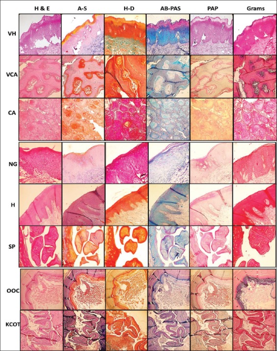
Background: Keratins are the most abundant proteins and are characteristic findings in many epithelial pathologies, making it a diagnostically important marker, both histopathologically and immunohistochemically. Since, immunohistochemistry is an expensive diagnostic tool, special stains to detect the degree of keratinization could serve as a faster and economic option. The aim of the present study was to compare the efficacy of special stains for keratin with standard hematoxylin and eosin stain (H and E). Objectives include: (i) To subject the diagnosed cases of keratin disorders to the selected special stains: Ayoub-shklar method, Dane-Herman method, Alcian blue -periodic acid Schiff 's (PAS), rapid papanicolaou (PAP) and Gram's stain. (ii) To compare the staining specificity and staining intensity of special stains with respect to routine hematoxylin and eosin (H and E) stain. (iii) To compare the efficacy of special stains to routine H and E stain in identification of the type of keratin present in the selected cases.
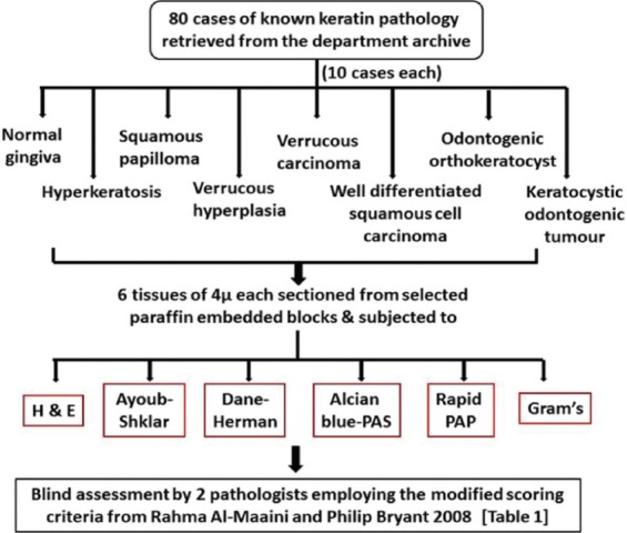
Materials and methods: A total of 80 cases of known pathology for keratin were retrieved from the department archive, which included 10 each of normal gingiva, hyperkeratosis, squamous papilloma, verrucous hyperplasia, verrucous carcinoma, well-differentiated squamous cell carcinoma, orthokeratinized odontogenic cyst and keratocystic odontogenic tumors. Six sections of 4 µ each from the paraffin blocks were made, stained with H and E and the special stains and these were evaluated by 2 pathologists based on the modified scoring criteria from Rahma Al-Maaini and Philip Bryant 2008.
Results: The results were tabulated using Chi square and kappa statistics. The statistical values for identification of the type of keratinization was insignificant showing that ortho and parakeratinized epithelia could be correctly identified by both H and E as well as all the special stains. Furthermore, all the special stains showed a positive result and statistical significance (P < 0.001) with respect to the staining of keratin.
Conclusion: To conclude, though the special stains distinctly stained the keratin with a higher intensity, H and E proves to be overall better stain with respect to specificity.
Keywords: Ayoub-Shklar; Dane-Herman; Keratins; PAP; alcian blue-periodic acid schiff; gram.
Conflict of interest statement
Figures
References
-
- Nanci A. Ten Cate's Oral Histology: Development, Structure, and Function. 8th ed. Missouri, USA: Mosby Elsevier; 2012.
-
- Bancroft JD, Gamble M. Theory and Practice of Histological Techniques. 6th ed. Philadelphia: Churchill Livingstone Elsevier; 2008.
LinkOut - more resources
Full Text Sources
Research Materials