

Neurons in the hippocampal CA1 region, but not the dentate gyrus, are susceptible to oxidative stress in rats with streptozotocin-induced type 1 diabetes
- PMID: 25878595
- PMCID: PMC4396109
- DOI: 10.4103/1673-5374.153695
Neurons in the hippocampal CA1 region, but not the dentate gyrus, are susceptible to oxidative stress in rats with streptozotocin-induced type 1 diabetes
Abstract
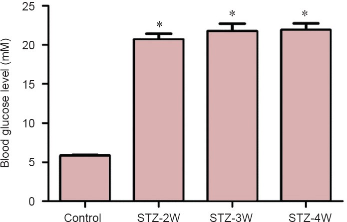
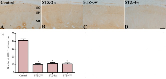
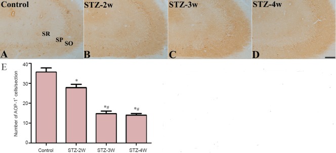
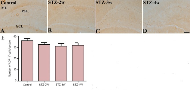
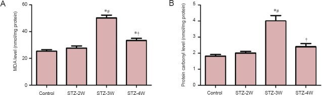
In this study, we investigated the effects of streptozotocin-induced type 1 diabetes on antioxidant-like protein-1 immunoreactivity, protein carbonyl levels, and malondialdehyde formation, a marker for lipid peroxidation, in the hippocampus. For this study, streptozotocin (75 mg/kg) was intraperitoneally injected into adult rats to induce type 1 diabetes. The three experimental parameters were determined at 2, 3, 4 weeks after streptozotocin treatment. Fasting blood glucose levels significantly increased by 20.7-21.9 mM after streptozotocin treatment. The number of antioxidant-like protein-1 immunoreactive neurons significantly decreased in the hippocampal CA1 region, but not the dentate gyrus, 3 weeks after streptozotocin treatment compared to the control group. Malondialdehyde and protein carbonyl levels, which are modified by oxidative stress, significantly increased with a peak at 3 weeks after malondialdehyde treatment, and then decreased 4 weeks after malondialdehyde treatment. These results suggest that neurons in the hippocampal CA1 region, but not the dentate gyrus, are susceptible to oxidative stress 3 weeks after malondialdehyde treatment.
Keywords: dentate gyrus; hippocampus; lipid peroxidation; malondialdehyde; nerve regeneration; neural regeneration; neurons; type 1 diabetes.
Conflict of interest statement
Figures
Similar articles
-
Electrophysiology and immunohistochemistry in the hippocampal ca1 and the dentate gyrus of rats chronically exposed to 1-bromopropane, a substitute for specific chlorofluorocarbons.Neuroscience. 2004;124(3):593-603. doi: 10.1016/j.neuroscience.2003.12.025. Neuroscience. 2004. PMID: 14980730
-
Piperine restores streptozotocin-induced cognitive impairments: Insights into oxidative balance in cerebrospinal fluid and hippocampus.Behav Brain Res. 2018 Jan 30;337:131-138. doi: 10.1016/j.bbr.2017.09.031. Epub 2017 Sep 20. Behav Brain Res. 2018. PMID: 28939403
-
Reduced hippocampal cell differentiation in the subgranular zone of the dentate gyrus in a rat model of type II diabetes.Neurochem Res. 2008 Mar;33(3):394-400. doi: 10.1007/s11064-007-9440-8. Epub 2007 Aug 22. Neurochem Res. 2008. PMID: 17712629
-
Cerebrolysin reverses hippocampal neural atrophy in a mice model of diabetes mellitus type 1.Synapse. 2015 Jun;69(6):326-35. doi: 10.1002/syn.21819. Synapse. 2015. PMID: 25851531
-
Dynamic changes of behaviors, dentate gyrus neurogenesis and hippocampal miR-124 expression in rats with depression induced by chronic unpredictable mild stress.Neural Regen Res. 2020 Jun;15(6):1150-1159. doi: 10.4103/1673-5374.270414. Neural Regen Res. 2020. PMID: 31823896 Free PMC article.
Cited by
-
Protective effect of Tat fused HPCA protein on neuronal cell death caused by ischemic injury.Heliyon. 2023 Dec 16;10(1):e23488. doi: 10.1016/j.heliyon.2023.e23488. eCollection 2024 Jan 15. Heliyon. 2023. PMID: 38192804 Free PMC article.
-
The Reciprocal Causation of the ASK1-JNK1/2 Pathway and Endoplasmic Reticulum Stress in Diabetes-Induced Cognitive Decline.Front Cell Dev Biol. 2020 Jul 17;8:602. doi: 10.3389/fcell.2020.00602. eCollection 2020. Front Cell Dev Biol. 2020. PMID: 32766246 Free PMC article.
-
Stress injuries and autophagy in mouse hippocampus after chronic cold exposure.Neural Regen Res. 2017 Mar;12(3):440-446. doi: 10.4103/1673-5374.202932. Neural Regen Res. 2017. PMID: 28469659 Free PMC article.
-
Early Life Vitamin C Deficiency Does Not Alter Morphology of Hippocampal CA1 Pyramidal Neurons or Markers of Synaptic Plasticity in a Guinea Pig Model.Nutrients. 2018 Jun 8;10(6):749. doi: 10.3390/nu10060749. Nutrients. 2018. PMID: 29890692 Free PMC article.
References
-
- Biessels GJ, Kamal A, Ramakers GM, Urban IJ, Spruijt BM, Erkelens DW, Gispen WH. Place learning and hippocampal synaptic plasticity in streptozotocin-induced diabetic rats. Diabetes. 1996;45:1259–1266. - PubMed
-
- Biessels GJ, Kamal A, Urban IJ, Spruijt BM, Erkelens DW, Gispen WH. Water maze learning and hippocampal synaptic plasticity in streptozotocin-diabetic rats: effects of insulin treatment. Brain Res. 1998;800:125–135. - PubMed
-
- Bonnefont-Rousselot D, Bastard JP, Jaudon MC, Delattre J. Consequences of the diabetic status on the oxidant/antioxidant balance. Diabetes Metab. 2000;26:163–176. - PubMed
-
- Chae HZ, Chung SJ, Rhee SG. Thioredoxin-dependent peroxide reductase from yeast. J Biol Chem. 1994;269:27670–27678. - PubMed
LinkOut - more resources
Full Text Sources
Other Literature Sources
Research Materials
Miscellaneous