. 2015 Apr 12;11(1):14.
doi: 10.1186/s13223-015-0081-1. eCollection 2015.

Therapeutic potential of anti-IL-6 therapies for granulocytic airway inflammation in asthma

Affiliations

Therapeutic potential of anti-IL-6 therapies for granulocytic airway inflammation in asthma

Derek K Chu et al. Allergy Asthma Clin Immunol. .

Abstract

Background: Determining the cellular and molecular phenotypes of inflammation in asthma can identify patient populations that may best benefit from targeted therapies. Although elevated IL-6 and polymorphisms in IL-6 signalling are associated with lung dysfunction in asthma, it remains unknown if elevated IL-6 levels are associated with a specific cellular inflammatory phenotype, and how IL-6 blockade might impact such inflammatory responses.

Methods: Patients undergoing exacerbations of asthma were phenotyped according to their airway inflammatory characteristics (normal cell count, eosinophilic, neutrophilic, mixed granulocytic), sputum cytokine profiles, and lung function. Mice were exposed to the common allergen, house dust-mite (HDM), in the presence or absence of endogenous IL-6. The intensity and nature of lung inflammation, and levels of pro-granulocytic cytokines and chemokines under these conditions were analyzed.

Results: Elevated IL-6 was associated with a lower FEV1 in patients with mixed eosinophilic-neutrophilic bronchitis. In mice, allergen exposure increased lung IL-6 and IL-6 was produced by dendritic cells and alveolar macrophages. Loss-of-function of IL-6 signalling (knockout or antibody-mediated neutralization) abrogated elevations of eosinophil and neutrophil recruiting cytokines/chemokines and allergen-induced airway inflammation in mice.

Conclusions: We demonstrate the association of pleiotropic cellular airway inflammation with IL-6 using human and animal data. These data suggest that exacerbations of asthma, particularly those with a combined eosinophilic and neutrophilic bronchitis, may respond to therapies targeting the IL-6 pathway and therefore, provide a rational basis for initiation of clinical trials to evaluate this.

Keywords: Airway inflammation; Allergy; Asthma; Bronchitis; Eosinophil; Granulocyte; House dust-mite (HDM); IL-6; IL-6R; Neutrophil.


Figures
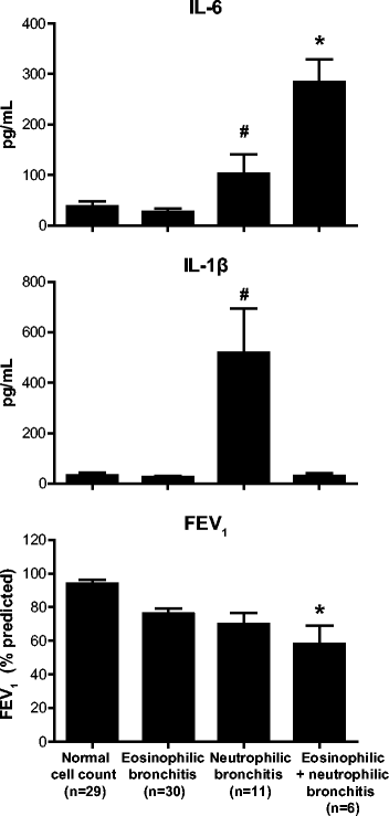
Figure 1
Figure 1
IL-6 associates with eosinophilic-neutrophilic granulocytic inflammation and worse pulmonary function in patients with asthma. Sputum IL-6 levels (upper panel) were highest and FEV1 (% predicted) (lower panel) was lowest in patients with asthma who had a combined eosinophilic and neutrophilic bronchitis compared to patients with asthma who had normal cell counts in sputum, an eosinophilic bronchitis (>3%) or a neutrophilic bronchitis (total cell count >106/g and neutrophils >65%). IL-1β was elevated only in the neutrophilic bronchitis group (middle panel). Mean + SEM. Sample size shown in lower panel. *p < 0.05 vs Neutrophilic bronchitis and eosinophilic bronchitis groups. #p < 0.05 vs eosinophilic bronchitis group.
Figure 2
Figure 2
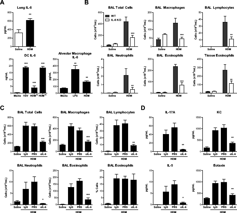
IL-6 is produced by allergen-stimulated mononuclear phagocytes and mediates allergic airway inflammation through eosinophil/neutrophil chemokines/cytokines. WT or IL-6 KO mice received 25 μg HDM i.n. daily for 10d, with or without 50 μg anti-IL-6 (αIL-6) or control IgG i.p. on d0, 3, 5 and 7. (A) IL-6 by ELISA in lung homogenates, or cultures of dendritic cells or alveolar macrophages. (B, C) Cell counts from bronchoalveolar lavage (BAL) or digested lung tissue quantified by hemocytometer manual counting with Turks, followed by differential cell counting of Hema 3-stained cytospins or flow cytometry. (D) Cytokines in lung tissue homogenates quantified by Luminex. Mean + SEM, n = 3-5 mice per group per experiment, 2-3 independent experiments performed. *p < 0.05 compared to saline, media, WT HDM, or HDM + IgG control groups. **p < 0.01. ***p < 0.001.

References

    1. Nair P. Update on clinical inflammometry for the management of airway diseases. Can Respir J. 2013;20(2):117–120. - PMC - PubMed
    1. Dasgupta A, Neighbour H, Nair P. Targeted therapy of bronchitis in obstructive airway diseases. Pharmacol Ther. 2013;140(3):213–222. doi: 10.1016/j.pharmthera.2013.07.001. - DOI - PubMed
    1. Nair P, Dasgupta A, Brightling CE, Chung KF. How to diagnose and phenotype asthma. Clin Chest Med. 2012;33(3):445–457. doi: 10.1016/j.ccm.2012.05.003. - DOI - PubMed
    1. Nair P, Pizzichini MM, Kjarsgaard M, Inman MD, Efthimiadis A, Pizzichini E, et al. Mepolizumab for prednisone-dependent asthma with sputum eosinophilia. N Engl J Med. 2009;360(10):985–993. doi: 10.1056/NEJMoa0805435. - DOI - PubMed
    1. Rincon M. Interleukin-6: from an inflammatory marker to a target for inflammatory diseases. Trends Immunol. 2012;33(11):571–577. doi: 10.1016/j.it.2012.07.003. - DOI - PubMed